. 1999 Jul;19(7):4757-65.
doi: 10.1128/MCB.19.7.4757.

Inhibition of double-stranded RNA- and tumor necrosis factor alpha-mediated apoptosis by tetratricopeptide repeat protein and cochaperone P58(IPK)

Affiliations

Inhibition of double-stranded RNA- and tumor necrosis factor alpha-mediated apoptosis by tetratricopeptide repeat protein and cochaperone P58(IPK)

N M Tang et al. Mol Cell Biol. 1999 Jul.

Abstract

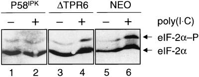
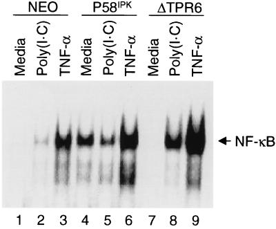
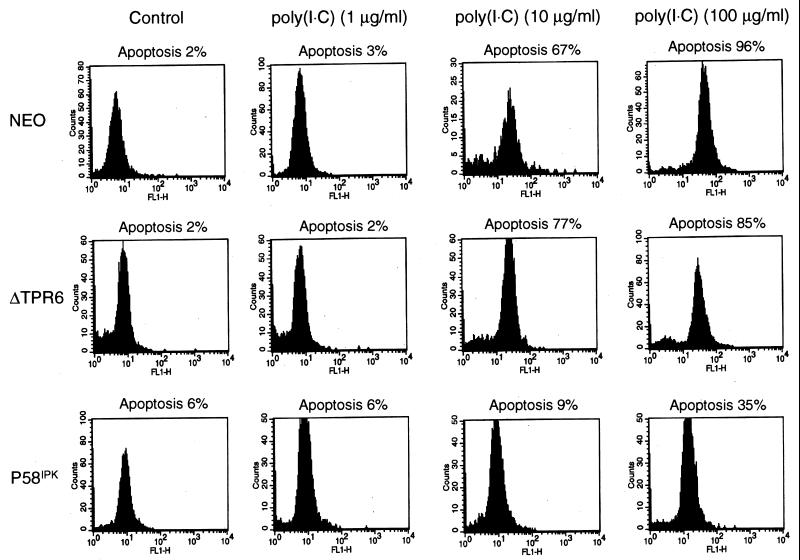
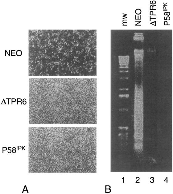
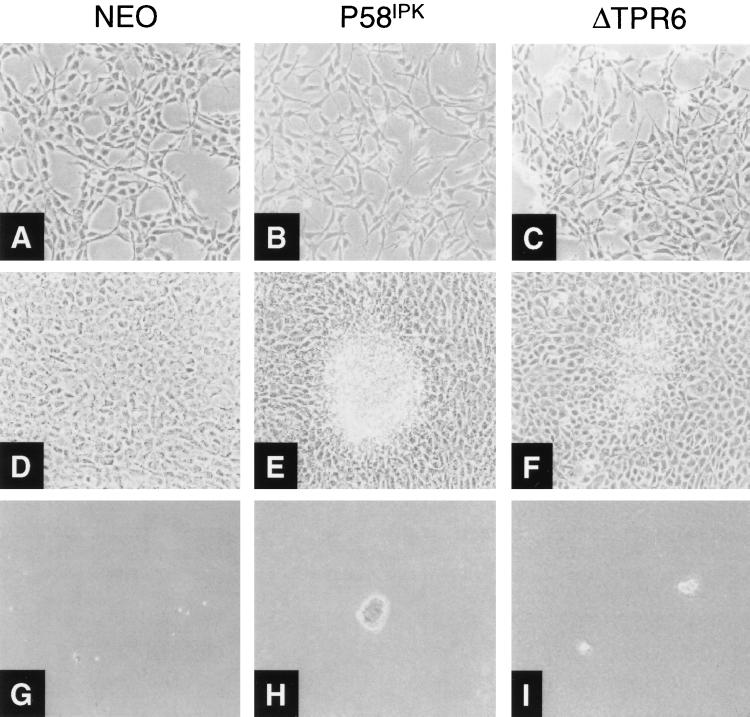
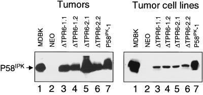
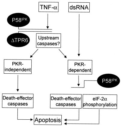
P58(IPK) is a tetratricopeptide repeat-containing cochaperone that is involved in stress-activated cellular pathways and that inhibits the activity of protein kinase PKR, a primary mediator of the antiviral and antiproliferative properties of interferon. To gain better insight into the molecular actions of P58(IPK), we generated NIH 3T3 cell lines expressing either wild-type P58(IPK) or a P58(IPK) deletion mutant, DeltaTPR6, that does not bind to or inhibit PKR. When treated with double-stranded RNA (dsRNA), DeltaTPR6-expressing cells exhibited a significant increase in eukaryotic initiation factor 2alpha phosphorylation and NF-kappaB activation, indicating a functional PKR. In contrast, both of these PKR-dependent events were blocked by the overexpression of wild-type P58(IPK). In addition, the P58(IPK) cell line, but not the DeltaTPR6 cell line, was resistant to dsRNA-induced apoptosis. Together, these findings demonstrate that P58(IPK) regulates dsRNA signaling pathways by inhibiting multiple PKR-dependent functions. In contrast, both the P58(IPK) and DeltaTPR6 cell lines were resistant to tumor necrosis factor alpha-induced apoptosis, suggesting that P58(IPK) may function as a more general suppressor of programmed cell death independently of its PKR-inhibitory properties. In accordance with this hypothesis, although PKR remained active in DeltaTPR6-expressing cells, the DeltaTPR6 cell line displayed a transformed phenotype and was tumorigenic in nude mice. Thus, the antiapoptotic function of P58(IPK) may be an important factor in its ability to malignantly transform cells.


Figures

FIG. 1
FIG. 1
Bovine P58IPK or ΔTPR6 is efficiently produced in stably transfected NIH 3T3 cell lines. Immunoblot analysis of P58IPK and ΔTPR6 protein production was performed. Protein extracts (100 μg) from wild-type P58IPK (P58IPK-1 and P58IPK-2) and ΔTPR6 (ΔTPR6-1 and ΔTPR6-2) cell lines were analyzed with a polyclonal antibody that recognizes bovine P58IPK (lane 1, Madin-Darby bovine kidney cells) but not the endogenous P58IPK protein present in murine cells (lane 2, NEO). Since the deletion in ΔTPR6 is small, the ΔTPR6 protein migrates at approximately the same position as P58IPK in the 12% acrylamide gel shown. Gradient gels (10 to 20% acrylamide) were therefore used to distinguish between the 58-kDa wild-type P58IPK and the 54-kDa ΔTPR6 protein (not shown).
FIG. 2
FIG. 2
PKR phosphorylates eIF-2α in ΔTPR6-expressing cells. An analysis of the steady-state level of eIF-2α phosphorylation in cultured cell lines was performed. Extracts were prepared from P58IPK-1, ΔTPR6-1, or NEO cells in mid-log phase, and 20 μg of each extract was subjected to vertical-slab isoelectric focusing. The resolved proteins were transferred to a nitrocellulose membrane, and the blot was probed with an eIF-2α monoclonal antibody. The ratio of phosphorylated to unphosphorylated eIF-2α in each lane was determined by scanning laser densitometry. A change in the ratio in response to treatment with poly(I · C) is indicative of PKR activity. The ratio of phosphorylated to unphosphorylated eIF-2α increased 1.3-fold in the P58IPK cell line and 4.5-fold in the ΔTPR6 and NEO cell lines. Phosphorylated (P) and unphosphorylated forms of eIF-2α are indicated by arrows.
FIG. 3
FIG. 3
P58IPK, but not ΔTPR6, inhibits the dsRNA-induced activation of NF-κB. NF-κB DNA-binding activities in NEO, P58IPK-1, and ΔTPR6-1 cell lines are shown. Cells were treated with medium alone, medium containing poly(I · C) (100 μg/ml), or medium containing TNF-α (10 ng/ml). NF-κB activation was detected in an EMSA using a 32P-labeled PRDII oligonucleotide probe. NF-κB–PRDII complexes are indicated by the arrow.
FIG. 4
FIG. 4
P58IPK, but not ΔTPR6, mediates resistance to dsRNA-induced apoptosis. An analysis of dsRNA-induced apoptosis in NEO, ΔTPR6-1, and P58IPK-1 cell lines was performed. Cells were treated with poly(I · C) (1, 10, or 100 μg/ml) for 16 h. Apoptosis-induced DNA fragmentation was detected by the labeling of DNA strand breaks with fluorescein dUTP and deoxynucleotidyltransferase. The percentage of apoptotic cells was quantified by flow cytometry as described in Materials and Methods.
FIG. 5
FIG. 5
P58IPK and ΔTPR6 mediate resistance to TNF-α-induced apoptosis. The NEO, ΔTPR6-1, and P58IPK-1 cell lines were treated with TNF-α for 16 h and examined for cell viability and apoptosis-induced DNA fragmentation as described in Materials and Methods. (A) Morphologic characteristics of cell monolayers 16 h post-TNF-α treatment. (B) Electrophoretic analysis of DNA prepared from TNF-α-treated cells.
FIG. 6
FIG. 6
ΔTPR6-expressing cells exhibit a transformed phenotype. Morphologic and growth characteristics of the NEO, P58IPK-1, and ΔTPR6-1 cell lines are shown. Cell lines were plated at 2 × 10⁴ cells per 100-mm-diameter dish in DMEM containing 10% FBS and 400 μg of G418 per ml. (A through C) Morphologic characteristics of mid-log-phase cells. In contrast to NEO cells, P58IPK and ΔTPR6 cell lines exhibited spindle-shaped morphology and increased refractivity. (D through F) Cells maintained in culture 4 days after they reached confluency, demonstrating transformed foci on P58IPK and ΔTPR6 cell monolayers. (G through I) Growth in soft agar. Anchorage-independent growth was observed in P58IPK and ΔTPR6 cell lines. Magnification, ×100.
FIG. 7
FIG. 7
Bovine P58IPK or ΔTPR6 is produced in tumors and tumor cell lines. Immunoblot analysis of P58IPK and ΔTPR6 protein production was performed. (Left) Four tumors from mice injected with ΔTPR6 cell lines (two from mice injected with ΔTPR6-1-expressing cells, designated ΔTPR6-1.1 and ΔTPR6-1.2, and two from mice injected with ΔTPR6-2-expressing cells, designated ΔTPR6-2.1 and ΔTPR6-2.2) and one tumor from a mouse injected with the P58IPK-1 cell line (P58IPK-1) were analyzed by using the anti-P58IPK monoclonal antibody, 2F8 (5). (Right) Cell lines derived from ΔTPR6-expressing and P58IPK-overexpressing tumors were analyzed by immunoblotting using the P58IPK polyclonal antibody. In each panel, cell extracts prepared from MDBK cells were used as a source of bovine P58IPK protein to serve as a positive control (lane 1). Cell extracts prepared from the NEO cell line were analyzed in parallel as a negative control (lane 2).
FIG. 8
FIG. 8
Model of P58IPK suppression of apoptosis. PKR has been implicated as an essential component of the apoptotic pathway that is induced by dsRNA, and inhibition of PKR by overexpression of P58IPK results in resistance to dsRNA-induced apoptosis. Consistent with the inability of ΔTPR6 to inhibit PKR-dependent functions, ΔTPR6-expressing cells undergo apoptosis in response to dsRNA treatment. In contrast, both the P58IPK and ΔTPR6 cell lines were resistant to TNF-α-induced apoptosis. We propose that P58IPK (and ΔTPR6) can also act in a PKR-independent fashion to regulate the TNF-α pathway. This regulation most likely occurs at a point in the pathway that is upstream of PKR. Suppression of apoptosis may be a primary mechanism by which overexpression of P58IPK induces malignant transformation. See Discussion for additional details.

Similar articles

Cited by

References

    1. Abraham N, Stojdl D F, Duncan P I, Méthot N, Ishii T, Dubé M, Vanderhyden B C, Atkins H L, Gray D A, McBurney M W, Koromilas A E, Brown E G, Sonenberg N, Bell J C. Characterization of transgenic mice with targeted disruption of the catalytic domain of the double-stranded RNA-dependent protein kinase, PKR. J Biol Chem. 1999;274:5953–5962. - PubMed
    1. Ausubel F M, Brent R, Kingston R E, Moore D D, Seidman J G, Smith J A, Struhl K. Current protocols in molecular biology. New York, N.Y: John Wiley & Sons, Inc.; 1988.
    1. Balachandran S, Kim C N, Yeh W-C, Mak T W, Barber G N. Activation of the dsRNA-dependent protein kinase, PKR, induces apoptosis through FADD-mediated death signaling. EMBO J. 1998;17:6888–6902. - PMC - PubMed
    1. Barber G N, Jagus R, Meurs E F, Hovanessian A G, Katze M G. Molecular mechanisms responsible for malignant transformation by regulatory and catalytic domain variants of the interferon-induced enzyme RNA-dependent protein kinase. J Biol Chem. 1995;270:17423–17428. - PubMed
    1. Barber G N, Thompson S, Lee T G, Strom T, Jagus R, Darveau A, Katze M G. The 58-kilodalton inhibitor of the interferon-induced double-stranded RNA-activated protein kinase is a tetratricopeptide repeat protein with oncogenic properties. Proc Natl Acad Sci USA. 1994;91:4278–4282. - PMC - PubMed

Publication types

MeSH terms

LinkOut - more resources