. 1999 Jul;19(7):4832-42.
doi: 10.1128/MCB.19.7.4832.

Use of a recombination reporter insert to define meiotic recombination domains on chromosome III of Saccharomyces cerevisiae

Affiliations

Use of a recombination reporter insert to define meiotic recombination domains on chromosome III of Saccharomyces cerevisiae

V Borde et al. Mol Cell Biol. 1999 Jul.

Abstract

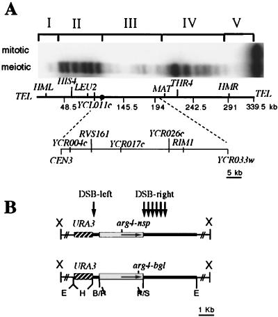
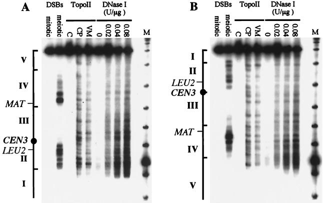
In Saccharomyces cerevisiae, meiotic recombination is initiated by DNA double-strand breaks (DSBs). DSBs usually occur in intergenic regions that display nuclease hypersensitivity in digests of chromatin. DSBs are distributed nonuniformly across chromosomes; on chromosome III, DSBs are concentrated in two "hot" regions, one in each chromosome arm. DSBs occur rarely in regions within about 40 kb of each telomere and in an 80-kb region in the center of the chromosome, just to the right of the centromere. We used recombination reporter inserts containing arg4 mutant alleles to show that the "cold" properties of the central DSB-deficient region are imposed on DNA inserted in the region. Cold region inserts display DSB and recombination frequencies that are substantially less than those seen with similar inserts in flanking hot regions. This occurs without apparent change in chromatin structure, as the same pattern and level of DNase I hypersensitivity is seen in chromatin of hot and cold region inserts. These data are consistent with the suggestion that features of higher-order chromosome structure or chromosome dynamics act in a target sequence-independent manner to control where recombination events initiate during meiosis.


Figures

FIG. 1
FIG. 1
Structure of the recombination construct and insert locations. (A) DSBs on chromosome III. Breaks were mapped by pulsed-field gel electrophoresis of DNA prepared from MJL2305 just before (mitotic) or 6 h after (meiotic) transfer to sporulation medium. The probe used was a PCR fragment from the left end of chromosome III (nt 15838 to 16857). Positions of DSB regions I to V (3) are indicated above the autoradiogram; locations of the cold region URA3-arg4 inserts used in this study are indicated on the map of chromosome III below the autoradiogram. (B) Structure of inserts. Plasmids contain pBR322 sequences (thick line), a 1.2-kb HindIII URA3 fragment (hatched box), and a 3.3-kb PstI arg4 fragment (open box), containing either the arg4-nsp or arg4-bgl allele. Thin lines represent flanking genomic sequences used for integration, with × indicating the restriction site used for integration. Vertical arrows indicate the approximate location of DSBs seen in all inserts.
FIG. 2
FIG. 2
Chromatin structure and topoisomerase II cleavage of mitotic chromosome III. Mitotic and meiotic rad50S samples are from MJL2305, 0 and 6 h, respectively, after transfer to sporulation medium. Chromatin was prepared from exponentially growing cells of strain MJL1578. For topoisomerase II (TopoII) cleavage sites, chromatin was incubated with 1% dimethyl sulfoxide (C), 100 μM CP-115,953 (CP), or 100 μM VM26 (VM); for DNase I-sensitive sites, chromatin was incubated with the indicated concentration of DNase I (U/μg of DNA). DNA was displayed on a pulsed-field gel (see Materials and Methods), and the resulting filter was probed with a CHA1 probe (chromosome III nt 15838 to 16857) (A) or a YCR098c probe (nt 296511 to 297070) (B). Lanes M contain bacteriophage λ DNA concatemers plus a HindIII digest of bacteriophage λ. The hot or cold DSB regions I to V are indicated alongside each panel.
FIG. 3
FIG. 3
DSBs in inserts at different locations on chromosome III detected by using pBR322 sequences as probes. (A) DSBs between URA3 and arg4 sequences (DSB-left). DNA was digested with PstI (P), which cuts in pBR322 and in URA3, and probed with a PstI-AlwNI fragment from pBR322. DSB-left is indicated by a solid arrow, and the ARG4 promoter (prom.) is indicated by a dotted arrow. Mitotic DNA is from MJL2105. Meiotic DNAs are, from left to right: MJL2105, MJL2144, MJL2237, MJL1185, MJL2139, MJL2106, MJL1176, MJL1170, MJL2324, and MJL2326. (B) DSBs in the pBR322 sequence downstream of the arg4 fragment (DSB-right). DNA was cut with StuI (St) and a locus-specific enzyme: RSV161, SacII; YCR004c, AflII; YCR017c, AflII; CHA1, XhoI; YCR026c, AflII; RIM1, SphI; MAT, XbaI; YCL011c, AflII; LEU2, AflII; and HIS4, Bpu1102I. The probe used was a HindIII-BamHI pBR322 fragment. DSB-right is indicated by solid arrows and the ARG4 promoter is indicated by a dotted arrow. Mitotic DNA is from MJL2105. Meiotic DNAs are, from left to right: MJL2105, MJL2144, MJL2237, MJL2139, MJL1185, MJL1176, MJL2324, MJL2326, and MJL1170.
FIG. 4
FIG. 4
Frequencies of DSBs and recombination within URA3-arg4 inserts. Chromosome III DSB hot and cold regions are indicated. The thick horizontal lines denote a physical map of chromosome III, with 50-kb intervals indicated by vertical hatches and the centromere marked by a filled circle. Bars in the top panel indicate the frequency of Arg+ recombinants within each insert (hatched bars); bars in the bottom panel indicate the frequencies of both DSB-left (shaded) and DSBs-right (white) for inserts at the same location. Recombination data for inserts at MAT, CHA1, LEU2, and HIS4 are from a previous study (67).
FIG. 5
FIG. 5
DNase I-hypersensitive sites in the URA3-arg4 insert at HIS4 and RVS161. (A) Location of the URA3-arg4 insert on each chromosome III of strain MJL2418. (B) DSBs and DNase I-hypersensitive sites in inserts. DNA was cut with both AflII and XbaI, and the same filter was probed successively with an RVS161 probe (nt 128743 to 129300; left) and a HIS4 probe (nt 65967 to 66522; right). For DNase I, chromatin from MJL2418 was prepared at indicated times after transfer to sporulation medium and incubated with DNase I. Lanes: 1, 4, and 6, no DNase I; 2 and 5: 0.8 U of DNase I/μg of DNA; 3, 0.8 U of DNase I/μg of DNA; 7, 2 U of DNase I/μg of DNA. For rad50S DSBs, DNA from MJL2420 0 h (lanes 9) or 6 h (lanes 8) after transfer to sporulation medium. (C) Quantitative comparison of DNase I digestion profiles of URA3-arg4 inserts located at either RVS161 (thin lines) or HIS4 (thick lines). Densitometric profiles of lanes 3 (0 h), 5 (2 h), and 7 (4 h) are superimposed.
FIG. 6
FIG. 6
Crossing over within cold region III. For each insert, genetic distances are presented above the line, and crossover densities are given below. The location of each URA3-arg4 insert used for measuring genetic distances, as well as the position of CEN3, is shown on the chromosome III map.

Similar articles

Cited by

References

    1. Alani E, Padmore R, Kleckner N. Analysis of wild-type and rad50 mutants of yeast suggests an intimate relationship between meiotic chromosome synapsis and recombination. Cell. 1990;61:419–436. - PubMed
    1. Baker B S, Carpenter A T, Esposito M S, Esposito R E, Sandler L. The genetic control of meiosis. Annu Rev Genet. 1976;10:53–134. - PubMed
    1. Baudat F, Nicolas A. Clustering of meiotic double-strand breaks on yeast chromosome III. Proc Natl Acad Sci USA. 1997;94:5213–5218. - PMC - PubMed
    1. Becker D M, Guarente L. High-efficiency transformation of yeast by electroporation. Methods Enzymol. 1991;194:182–187. - PubMed
    1. Bickel S E, Wyman D W, Miyazaki W Y, Moore D P, Orr-Weaver T L. Identification of ORD, a Drosophila protein essential for sister chromatid cohesion. EMBO J. 1996;15:1451–1459. - PMC - PubMed

LinkOut - more resources