. 1999 Jul;19(7):4855-65.
doi: 10.1128/MCB.19.7.4855.

Repressive effect of Sp1 on the C/EBPalpha gene promoter: role in adipocyte differentiation

Affiliations

Repressive effect of Sp1 on the C/EBPalpha gene promoter: role in adipocyte differentiation

Q Q Tang et al. Mol Cell Biol. 1999 Jul.

Abstract

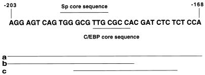
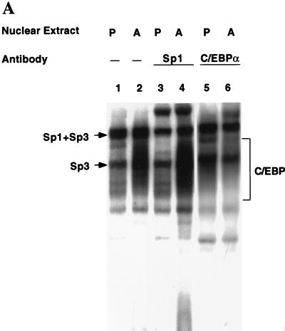
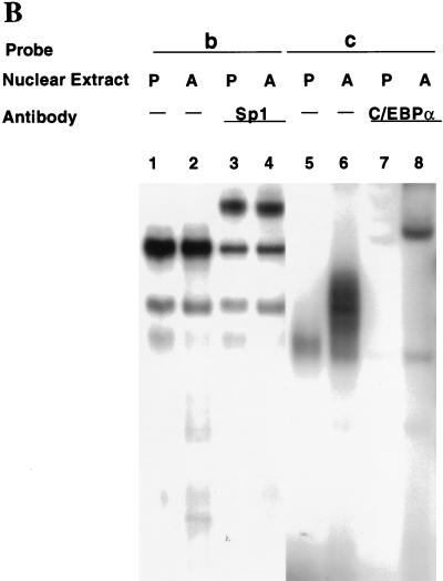
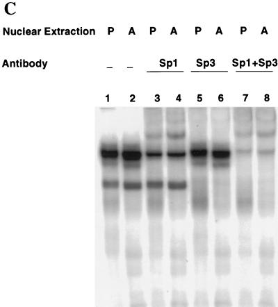
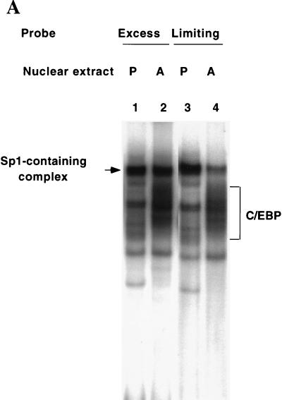
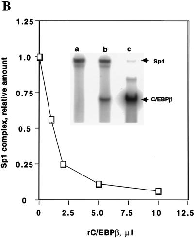
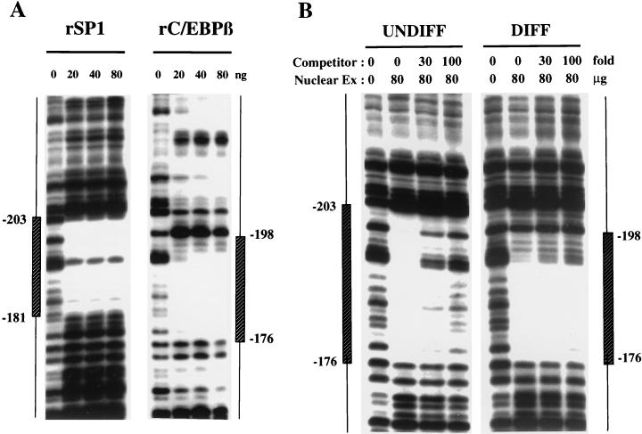
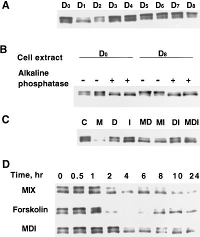
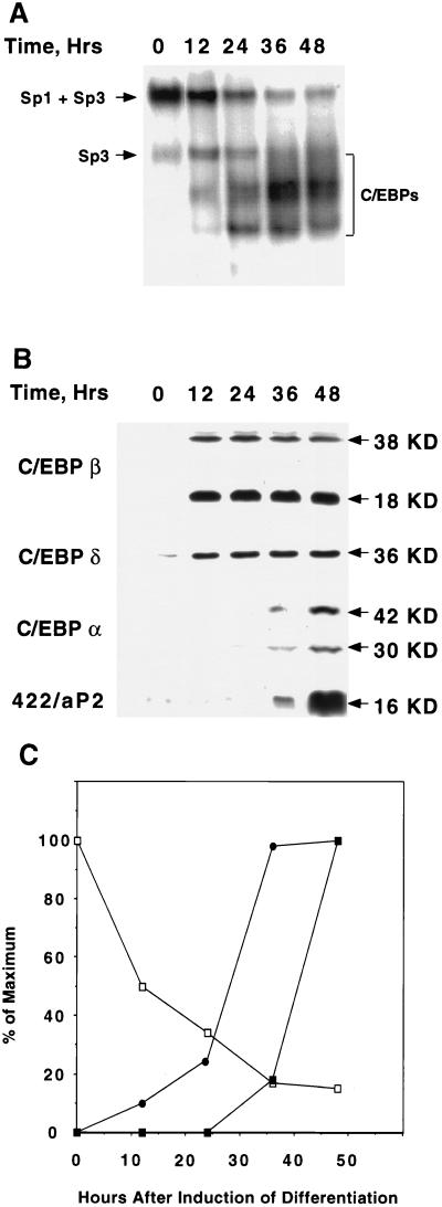
Expression of C/EBPalpha is required for differentiation of 3T3-L1 preadipocytes into adipocytes. Previous investigations indicated that transcription of the C/EBPalpha gene is sequentially activated during differentiation, initially by C/EBPbeta and C/EBPdelta and later by C/EBPalpha (autoactivation). These events are mediated by a C/EBP regulatory element in the promoter of the C/EBPalpha gene. This article presents evidence that members of the Sp family, notably Sp1, act repressively on the C/EBPalpha promoter prior to the induction of differentiation. Sp1 was shown to bind to a GC box at the 5' end of the C/EBP regulatory element in the C/EBPalpha promoter and, in so doing, to competitively prevent binding to and transactivation of the promoter by the C/EBPs. One of the differentiation inducers methylisobutylxanthine (a cAMP phosphodiesterase inhibitor) or Forskolin, both of which increase the cellular cAMP level, causes down-regulation of Sp1. This decrease in Sp1 level early in the differentiation program appears to facilitate access of C/EBPbeta and/or C/EBPdelta to the C/EBP regulatory element and, thereby, derepression of the C/EBPalpha gene.


Figures

FIG. 1
FIG. 1
Nucleotide sequences in the C/EBPα gene promoter protected from DNaseI digestion (footprinted) by nuclear proteins from 3T3-L1 preadipocytes and adipocytes. Nuclear extracts were prepared from 3T3-L1 cells either maintained in the undifferentiated state (UNDIFF) as preadipocytes or induced to differentiate into adipocytes (DIFF). A 204-bp SmaI-StyI fragment (nt −348 to −144) of the C/EBPα gene promoter was incubated with increasing amounts (20 to 80 μg of protein) of nuclear extract and then subjected to digestion with DNaseI. The footprinted regions are indicated by vertical hatched boxes indicating the number of nucleotides from the transcriptional start site (determined by a sequencing gel run in parallel).
FIG. 2
FIG. 2
Nucleotide sequence encompassing the C/EBP and Sp binding sites in the C/EBP α gene promoter. The consensus Sp core binding site and the consensus C/EBP core sequence for members of the C/EBP family are indicated. Lines labeled a, b, and c indicate the lengths of the labeled oligonucleotide probes used in the EMSAs.
FIG. 3
FIG. 3
EMSA of oligonucleotides corresponding to the Sp and/or C/EBP binding sites in the C/EBPα gene promoter. (A) EMSA of preadipocyte (P) and adipocyte (A) nuclear extract and 32P-labeled oligonucleotide a (nt −203 to −168; see Fig. 2) encompassing both the Sp and C/EBP binding sites. In lanes 3 and 4, nuclear extracts were supershifted with antibody directed against Sp1 and in lanes 5 and 6 extracts were supershifted with antibody directed against C/EBPα. (B) EMSA of preadipocyte (P) and adipocyte (A) nuclear extract and 32P-labeled oligonucleotide b (nt −203 to −180, which encompass the Sp core binding site; Fig. 2) or oligonucleotide c (nt −191 to −172, which encompass the C/EBP binding site; Fig. 2). In lanes 3 and 4, nuclear extracts were supershifted with antibody directed against Sp1 and in lanes 7 and 8 the extracts were supershifted with antibody directed against C/EBPα. (C) EMSA of preadipocyte (P) and adipocyte (A) nuclear extract and 32P-labeled oligonucleotide b (nt −203 to −180, which encompass the Sp core binding site; Fig. 2). In lanes 3 and 4, nuclear extracts were supershifted with antibody directed against Sp1, in lanes 5 and 6, extracts were supershifted with antibody directed against Sp3, and in lanes 7 and 8, extracts were supershi with antibody directed against both Sp1 and Sp3. In all cases, a 100-fold excess of unlabeled oligonucleotide probes a, b, and c effectively competed away virtually all detectable protein–32P-labeled oligonucleotide complexes on the gels shown above (results not shown).
FIG. 3
FIG. 3
EMSA of oligonucleotides corresponding to the Sp and/or C/EBP binding sites in the C/EBPα gene promoter. (A) EMSA of preadipocyte (P) and adipocyte (A) nuclear extract and 32P-labeled oligonucleotide a (nt −203 to −168; see Fig. 2) encompassing both the Sp and C/EBP binding sites. In lanes 3 and 4, nuclear extracts were supershifted with antibody directed against Sp1 and in lanes 5 and 6 extracts were supershifted with antibody directed against C/EBPα. (B) EMSA of preadipocyte (P) and adipocyte (A) nuclear extract and 32P-labeled oligonucleotide b (nt −203 to −180, which encompass the Sp core binding site; Fig. 2) or oligonucleotide c (nt −191 to −172, which encompass the C/EBP binding site; Fig. 2). In lanes 3 and 4, nuclear extracts were supershifted with antibody directed against Sp1 and in lanes 7 and 8 the extracts were supershifted with antibody directed against C/EBPα. (C) EMSA of preadipocyte (P) and adipocyte (A) nuclear extract and 32P-labeled oligonucleotide b (nt −203 to −180, which encompass the Sp core binding site; Fig. 2). In lanes 3 and 4, nuclear extracts were supershifted with antibody directed against Sp1, in lanes 5 and 6, extracts were supershifted with antibody directed against Sp3, and in lanes 7 and 8, extracts were supershi with antibody directed against both Sp1 and Sp3. In all cases, a 100-fold excess of unlabeled oligonucleotide probes a, b, and c effectively competed away virtually all detectable protein–32P-labeled oligonucleotide complexes on the gels shown above (results not shown).
FIG. 4
FIG. 4
Bindings of C/EBPα, C/EBPβ, and Sp1 at the C/EBP and Sp sites in the C/EBPα gene promoter are mutually exclusive. (A) EMSA of preadipocyte (P) and adipocyte (A) nuclear extract and differing amounts (the standard level, 0.25 ng or the “limiting” level, 0.008 ng) of 32P-labeled oligonucleotide a (nt −203 to −168; see Fig. 2) which encompasses both the Sp and C/EBP binding sites. Exposure of film for lanes 1 and 2 was 12 h and for lanes 3 and 4 exposure was 96 h. (B) Effect of rC/EBPβ on the binding of rSp1 to oligonucleotide a. EMSA was conducted with a limiting amount (see above) of probe a, the amount of rSp1 was held constant (10 ng), and the amount of C/EBPβ (8.25 ng per μl) was increased as shown. The inset shows the autoradiograms for gels, corresponding to the 0, 1, and 10 μl levels of rC/EBPβ. (C) Effect of rSp1 on the binding of rC/EBPβ to oligonucleotide a. EMSA was conducted with a limiting amount (see above) of probe a, the amount of C/EBPβ was held constant (8.25 ng) and the amount of rSp1 (10 ng per μl) was increased as shown. The inset shows the autoradiograms for gels corresponding to the 0-, 1-, and 10-μl levels of rSp1 added.
FIG. 4
FIG. 4
Bindings of C/EBPα, C/EBPβ, and Sp1 at the C/EBP and Sp sites in the C/EBPα gene promoter are mutually exclusive. (A) EMSA of preadipocyte (P) and adipocyte (A) nuclear extract and differing amounts (the standard level, 0.25 ng or the “limiting” level, 0.008 ng) of 32P-labeled oligonucleotide a (nt −203 to −168; see Fig. 2) which encompasses both the Sp and C/EBP binding sites. Exposure of film for lanes 1 and 2 was 12 h and for lanes 3 and 4 exposure was 96 h. (B) Effect of rC/EBPβ on the binding of rSp1 to oligonucleotide a. EMSA was conducted with a limiting amount (see above) of probe a, the amount of rSp1 was held constant (10 ng), and the amount of C/EBPβ (8.25 ng per μl) was increased as shown. The inset shows the autoradiograms for gels, corresponding to the 0, 1, and 10 μl levels of rC/EBPβ. (C) Effect of rSp1 on the binding of rC/EBPβ to oligonucleotide a. EMSA was conducted with a limiting amount (see above) of probe a, the amount of C/EBPβ was held constant (8.25 ng) and the amount of rSp1 (10 ng per μl) was increased as shown. The inset shows the autoradiograms for gels corresponding to the 0-, 1-, and 10-μl levels of rSp1 added.
FIG. 5
FIG. 5
DNaseI footprint analysis of the C/EBPα proximal promoter. (A) DNaseI footprint of a segment (nt −348 to −144; cut with SmaI and StyI) of the 5′ flanking region of the C/EBPα gene was subjected to digestion with DNaseI in the presence of increasing amounts (20 to 80 ng) of rSp1 or rC/EBPβ. The footprinted regions are indicated by vertical boxes indicating the number of nucleotides from the transcriptional start site (determined by a sequencing gel run in parallel). (B) DNaseI footprint of a segment (nt −348 to −144) of the 5′ flanking region of the C/EBPα gene was subjected to digestion with DNaseI in the presence or absence of nuclear extract (80 μg of protein) from undifferentiated (UNDIFF) or differentiated (DIFF) 3T3-L1 cells and increasing amounts of unlabeled oligonucleotide corresponding to the Sp binding site (nt −203 to −180) in the C/EBPα promoter.
FIG. 6
FIG. 6
Effect of Sp1 on transactivation by C/EBPβ or -α mediated by the C/EBPα or obese gene promoters. (A) A C/EBPα promoter-luciferase reporter gene construct (2 μg) containing 343 bp of 5′ flanking sequence and 125 bp of 5′ untranslated sequence of the promoter was cotransfected into 3T3-L1 preadipocytes with a C/EBPβ or C/EBPα expression vector (2 μg) and increasing amounts of an Sp1 expression vector. After 48 h, luciferase assays with cell lysates were conducted. (B) An obese gene promoter-luciferase reporter gene construct (2 μg) containing 700 bp of 5′ flanking sequence of the promoter (containing a C/EBP regulatory element at nt −55) was cotransfected into 3T3-L1 preadipocytes with a C/EBPα expression vector (2 μg) and increasing amounts of an Sp1 expression vector. After 48 h, luciferase assays with cell lysates were conducted.
FIG. 7
FIG. 7
Effect of mutation of the Sp core element in the C/EBPα gene promoter on binding and inhibition of transactivation by Sp1. (A) An EMSA was performed with oligonucleotide a and a-mut (mutated in the Sp binding site; see Materials and Methods) with preadipocyte (P) or adipocyte (A) nuclear extract. A 100-fold excess of unlabeled a-mut was added during the binding reaction in lanes 3 and 4. Antibody against C/EBPα was used for the supershift experiments (lanes 7 and 8). (B) C/EBPα promoter-luciferase reporter gene constructs (2 μg) containing 343 bp of 5′ flanking sequence and 125 bp of 5′ untranslated sequence either mutated in the Sp binding site (■) or the C/EBP binding site (●) (see Materials and Methods) were cotransfected into 3T3-L1 preadipocytes with a C/EBPα expression vector (2 μg) and increasing amounts of an Sp1 expression vector. After 48 h, luciferase assays with cell lysates were conducted. (C) C/EBPα promoter-reported constructs (wild types or constructs mutated in the Sp site as in panel B) were cotransfected into 3T3-L1 preadipocytes with an Sp1 expression vector (1 μg) without or with different amounts of a C/EBPα expression vector. After 48 h, luciferase assays with cell lysates were conducted. (D) Effect of mutating the C/EBP and Sp binding sites in the C/EBPα promoter on reporter gene expression following induction of differentiation. Wild-type (wt) promoter-luciferase, C/EBP site-mutated promoter-luciferase (C/EBP Mut), and Sp site mutated promoter-luciferase (Sp Mut) constructs were transfected into D0 postconfluent 3T3-L1 preadipocytes. Twenty-four hours later, the cells were induced to differentiate by using the standard protocol, and after an additional 24 h, the cells were lysed and luciferase assays with the cell lysates were performed.
FIG. 8
FIG. 8
Effect of adipocyte differentiation inducers on the level and phosphorylation state of Sp1. (A) Two-day postconfluent 3T3-L1 preadipocytes were induced to differentiate into adipocytes by using the standard differentiation protocol described in Materials and Methods. At different times (subscripts indicate to the number of days following induction of differentiation) after the induction of differentiation, cell lysates were subjected to SDS-PAGE and then Western blotted with anti-Sp1 antibody. (B) Cell lysates on day 0 and Day 8 were subjected to dephosphorylation by alkaline phosphatase treatment (see Materials and Methods). Following dephosphorylation, cell lysates were subjected to SDS-PAGE and Western blotting with anti-Sp1 antibody. (C) Two-day postconfluent 3T3-L1 preadipocytes were exposed to individual components of the differentiation inducer mixture either alone or in combination. After 24 h, cell lysates were analyzed as described for panel A. M, MIX; D, DEX; I, INS. (D) Two-day postconfluent 3T3-L1 preadipocytes were exposed to MIX, Forskolin, or MDI for the times indicated, after which cell lysates were analyzed as described for panel in A.
FIG. 9
FIG. 9
Changes in Sp1/Sp3 binding activity and expression of C/EBPβ, -δ, and -α following induction of differentiation. (A) An EMSA was performed with a limiting amount (0.004 ng) of an oligonucleotide (probe a) containing the Sp and C/EBP binding sites in the proximal C/EBPα gene promoter. An EMSA was performed with nuclear extracts from 3T3-L1 cells at 12, 24, 36, and 48 h after induction of differentiation. (B) Western blot analyses for C/EBPβ, -δ, and -α, and 422/aP2 using the cell lysates as described for panel A. (C) Changes in the binding activity of Sp1 + Sp3 (□) and C/EBPβ and -δ (●) and the expression of C/EBPα (■) at 12, 24, 36, and 48 h after induction of differentiation. Data are from panels A and B above.

References

    1. Bernlohr D A, Bolanowski M A, Kelly T J, Lane M D. Evidence for an increase in transcription of specific mRNAs during differentiation of 3T3-L1 preadipocytes. J Biol Chem. 1985;260:5563–5567. - PubMed
    1. Cao Z, Umek R M, McKnight S L. Regulated expression of three C/EBP isoforms during adipose conversion of 3T3-L1 cells. Genes Dev. 1991;5:1538–1552. - PubMed
    1. Cheneval D, Christy R J, Geiman D, Cornelius P, Lane M D. Cell-free transcription directed by the 422 adipose P2 gene promoter: activation by the CCAAT/enhancer binding protein. Proc Natl Acad Sci USA. 1991;88:8465–8469. - PMC - PubMed
    1. Christy R J, Kaestner K H, Geiman D E, Lane M D. CCAAT/enhancer binding protein gene promoter: binding of nuclear factors during differentiation of 3T3-L1 preadipocytes. Proc Natl Acad Sci USA. 1991;88:2593–2597. - PMC - PubMed
    1. Christy R J, Yang V W, Ntambi J M, Geiman D E, Landschulz W H, Friedman A D, Nakabeppu Y, Kelly T J, Lane M D. Differentiation-induced gene expression in 3T3-L1 preadipocytes: CCAAT/enhancer binding protein interacts with and activates the promoters of two adipocyte-specific genes. Genes Dev. 1989;3:1323–1335. - PubMed

Publication types

MeSH terms

LinkOut - more resources