. 1999 Jul;19(7):4918-26.
doi: 10.1128/MCB.19.7.4918.

Homeoproteins CDP and SATB1 interact: potential for tissue-specific regulation

Affiliations

Homeoproteins CDP and SATB1 interact: potential for tissue-specific regulation

J Liu et al. Mol Cell Biol. 1999 Jul.

Abstract

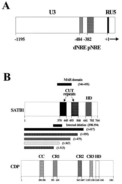
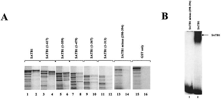
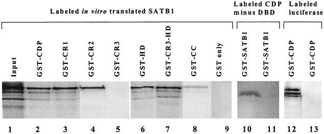
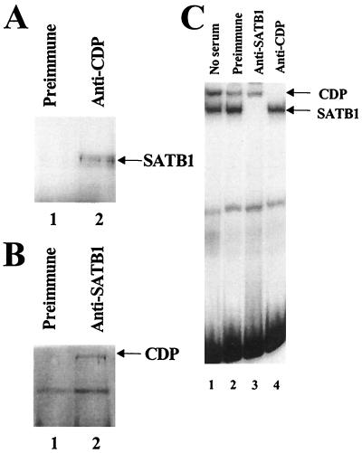
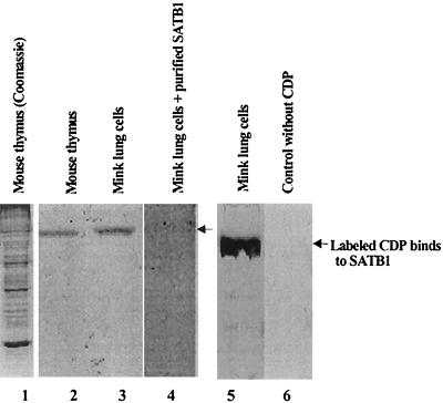
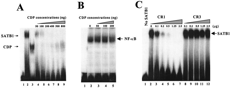
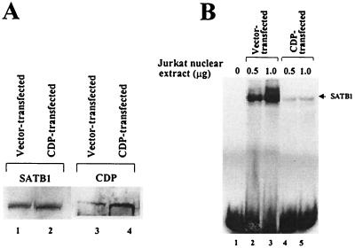
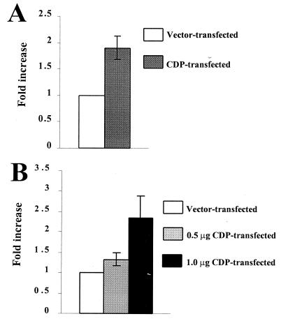
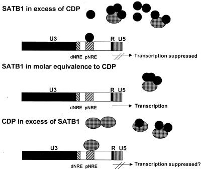
Homeoproteins are known to participate in development and cell type specification. The homeoproteins CCAAT displacement protein (CDP) and special AT-rich sequence binding protein 1 (SATB1) have been shown to bind to nuclear matrix-associated regions and to act as repressors of many cellular genes. Moreover, binding of SATB1 to the mouse mammary tumor virus (MMTV) promoter region dramatically affects the tissue-specific transcription of this retrovirus. Because protein-protein interactions are a common means of regulating homeoprotein function, we tested whether SATB1 and CDP interact in vivo and in vitro. SATB1 interacted with CDP through its DNA-binding domain, as demonstrated by glutathione S-transferase (GST) pull-down assays. GST pull-down assays also showed that CDP associated with SATB1 through three of its four DNA-binding domains (CR1, CR2, and the homeodomain). SATB1-specific antisera, but not preimmune sera, precipitated CDP from nuclear extracts, and CDP-specific antisera precipitated SATB1 from the same extracts. Far-Western blotting detected interaction of SATB1 and CDP in several different tissue extracts. Association of purified SATB1 and CDP in vitro resulted in the inability of each protein to bind to DNA in gel retardation assays. CDP overexpression in cultured T cells led to a loss of detectable SATB1 binding to the MMTV promoter region, as measured by gel shift experiments. CDP overexpression also elevated MMTV long terminal repeat reporter gene activity in transient-transfection assays, a result consistent with neutralization of the SATB1 repressor function in T cells. SATB1 is very abundant in certain tissues, particularly thymus, whereas CDP is relatively ubiquitous, except in certain terminally differentiated cell types. Because of the tissue and cell type distribution of SATB1 and CDP, we propose that the SATB1-to-CDP ratio in different tissues is a novel mechanism for homeoproteins to control gene expression and differentiation in mammals.


Figures

FIG. 1
FIG. 1
(A) The MMTV LTR, divided into U3, R, and U5 regions. The U3 region contains the transcription regulatory signals for the standard MMTV promoter that allows RNA initiation at the U3-R border. The approximate positions of the promoter-proximal (pNRE) and promoter-distal (dNRE) negative elements, as determined by reporter gene expression in transient-transfection assays, are shown. The probes used in this study include a 120-bp fragment that spans the pNRE and an oligonucleotide probe that includes an inverted repeat at the 3′ end of the 120-bp probe (see Materials and Methods). (B) Domain structures of SATB1 and CDP. The cut domains CR1, CR2, and CR3 and the homeodomain (HD) in CDP all can bind DNA independently, whereas the coiled-coil (CC) domain cannot (2, 18). An internal MAR domain, including the CR domains, contains the DNA-binding domain of SATB1 (40). Numbers indicate amino acid positions. CDP (the human protein) is known in other species as Cut (Drosophila), Clox (dogs), and Cux (mice) (1, 8, 53). In this paper, we use CDP to refer to proteins from all species.
FIG. 2
FIG. 2
SATB1 and CDP interaction in vitro. (A) Mapping of the SATB1 region necessary for CDP association. GST-CDP (CR2-Cterm) was immobilized on glutathione-agarose beads and incubated with 35S-labeled in vitro-translated full-length SATB1 (lanes 1 and 2), SATB1 mutants with C-terminal truncations (lanes 3 to 12), an internal-deletion mutant (lanes 13 and 14), or GST alone (lanes 15 and 16). After affinity chromatography, bound proteins were resolved on SDS–10% polyacrylamide gels (even-numbered lanes) and compared with input labeled protein (odd-numbered lanes). (B) Lack of DNA binding of internally deleted SATB1. Wild-type SATB1 and an internal-deletion mutant were purified and used for gel shift assays with a labeled 120-bp proximal NRE probe prior to electrophoresis on a 4% nondenaturing polyacrylamide gel. The 120-bp NRE probe contains two binding sites for CDP and two binding sites for SATB1 (59). Numbers indicate amino acid positions.
FIG. 3
FIG. 3
Binding of different CDP domains to SATB1. GST-CDP (CR2-Cterm), GST-CR1, GST-CR2, GST-CR3, GST-HD, GST-CR3 plus HD, GST-coiled-coil (GST-CC), or GST only (11) (lanes 2 to 9) were incubated with in vitro-translated labeled SATB1 (lane 1), as described for Fig. 2A. As controls, labeled in vitro-translated CDP containing the spacer region between CR1 and CR2 (lanes 10 and 11) or firefly luciferase (lanes 12 and 13) also was incubated with a GST–full-length SATB1 fusion (lane 11) or a GST-CDP (CR2-Cterm) fusion (lane 13). Lanes 1, 10, and 12 show the input radioactively labeled protein.
FIG. 4
FIG. 4
SATB1 and CDP interact in vivo. (A) Coimmunoprecipitation of SATB1 with CDP-specific antibody. Jurkat T-cell lysate was incubated with an insoluble Staphylococcus aureus suspension to eliminate nonspecific binding prior to being immunoprecipitated with preimmune (lane 1) or anti-CDP (lane 2) serum. Immunoprecipitates were resolved on an SDS–8% polyacrylamide gel, transferred to nitrocellulose, and incubated with SATB1-specific serum, followed by development as suggested by the instructions with the Amersham ECL kit. (B) Coimmunoprecipitation of CDP with SATB1-specific antibody. Western blotting was performed as described for panel A, except that immunoprecipitations were performed with preimmune (lane 1) or anti-SATB1 (lane 2) serum prior to incubation of nitrocellulose transfers with anti-CDP. (C) Specificity of anti-SATB1 and anti-CDP sera. Mink lung cell nuclear extracts were incubated with no serum (lane 1), preimmune serum (lane 2), anti-SATB1 serum (lane 3), or anti-CDP serum (lane 4), followed by a gel shift assay with the 120-bp MMTV promoter-proximal NRE probe (32) (Fig. 1A). Reactions were analyzed on a 4% nondenaturing polyacrylamide gel. The detection of both SATB1 and CDP DNA-binding activities in mink lung extracts may indicate that certain modified forms of these proteins do not interact.
FIG. 5
FIG. 5
Far-Western blotting to detect SATB1-CDP association. Crude nuclear lysates from murine thymus (lanes 1 and 2) and mink lung cells (lanes 3 to 6) were prepared as described previously (32), resolved on SDS–8% polyacrylamide gels, and stained with Coomassie brilliant blue (lane 1) or electrotransferred to nitrocellulose (lanes 2 to 6). 35S-labeled in vitro-translated CDP was incubated in the absence of competitor protein (lanes 2, 3, and 5) or in the presence of purified recombinant SATB1 (lane 4). Mink lung cell proteins on the membrane also were incubated with an in vitro translation reaction mixture containing [35S]methionine, but no DNA template (lane 6). Lanes 1 to 4 are derived from a single gel; lanes 5 and 6 are from a second gel. The arrows show the positions of SATB1 protein detected by interaction with labeled CDP.
FIG. 6
FIG. 6
Interaction of SATB1 and CDP interferes with the DNA-binding ability of both proteins. (A) Inhibition of DNA binding after SATB1-CDP association. N-terminally truncated SATB1 and CDP were purified from recombinant fusion proteins and used for gel shift assays with the 22-bp proximal NRE probe (32) prior to electrophoresis on 4% nondenaturing polyacrylamide gels. SATB1 (500 ng, or 5.6 pmol) (lane 2) was mixed with 50 to 800 ng (0.7 to 11 pmol) (lanes 4 to 9) of CDP before addition of the labeled probe. Lane 1 shows results achieved with no added protein, whereas lane 3 shows results for 50 ng of CDP alone. The estimated molecular masses of SATB1 and CDP were 90 and 70 kDa, respectively. The 22-bp NRE probe appears to have a single binding site for CDP and for SATB1 (59). A 12-h exposure of the autoradiogram is shown. (B) NF-κB DNA binding is unaffected by CDP. LBB.A B-cell (23) nuclear extracts (4 μg) were incubated with no CDP (lane 2) or 50 to 200 ng (0.7 to 2.8 pmol) of recombinant purified CDP (lanes 3 to 5) prior to addition of the labeled NF-κB probe from the interleukin-2 receptor promoter (32) and analysis as described above. (C) The CR1, but not CR3, domain of CDP interferes with SATB1 binding to DNA. Increasing amounts of purified CR1 (9 to 225 pmol) (lanes 2 to 7) or CR3 (5.6 to 140 pmol) (lanes 8 to 12) were added to 2.5 μg (28 pmol) of purified SATB1 and incubated with the 120-bp MMTV proximal NRE probe (32) prior to analysis on a native polyacrylamide gel. Lane 1 contains no recombinant protein. Because the individual CDP cut domains do not bind well to the 120-bp probe relative to SATB1 or CDP containing the homeodomain [CDP(CR2-Cterm)], binding of CR1 and CR3 alone is not detectable under these conditions (not shown). The calculated molecular masses of the purified CR1 and CR3 proteins were approximately 11 and 18 kDa, respectively. A 2-h exposure of the autoradiogram is shown.
FIG. 7
FIG. 7
CDP overexpression leads to reduced SATB1 DNA binding. (A) Overexpression of CDP determined by Western blotting. Nuclear extracts were prepared 48 h following transfection and used for Western analysis with anti-SATB1 or -CDP serum. Equal amounts of extract were loaded in each lane, and the labeled bands were quantitated by densitometry. (B) CDP overexpression prevents SATB1 DNA binding to the MMTV LTR. The same nuclear extracts as used for panel A were used for gel shift assays with the labeled 120-bp proximal NRE probe (32). The upper band observed in lanes 2 and 3 does not contain CDP, as determined with anti-CDP serum in gel shift assays (not shown). This band may represent binding of two molecules of SATB1 to the 120-bp NRE probe.
FIG. 8
FIG. 8
Overexpression of CDP in Jurkat cells elevates expression from the MMTV LTR. (A) Overexpression of CDP in Jurkat cells by electroporation. Transfections of the CDP expression vector (15 μg) were performed in triplicate, and results are expressed as an average increase over the value determined from the average of three determinations of the CDP-negative control vector (assigned a value of 1). Error bar indicates the standard deviation from the mean. (B) Overexpression of CDP in Jurkat cells by using the SuperFect transfection method. Values were calculated as described for panel A. Addition of more DNA (from any source) in the transfection assays resulted in lower reporter gene activity.
FIG. 9
FIG. 9
Model for SATB1-CDP interaction in different mouse tissues. The large cylinders represent CDP protein, whereas the smaller, dark cylinders represent SATB1. CDP may be inactivated for DNA binding by association with a single molecule of SATB1. Alternatively, binding of SATB1 to one of three possible sites within CR1, CR2, or the homeodomain may alter the specificity of CDP binding to different promoters. The large box represents the MMTV LTR that is divided into U3, R, and U5 domains. Transcription is initiated at the U3-R border of the 5′ LTR within the MMTV provirus. Upstream of the transcription initiation site are two regions termed the promoter-proximal and promoter-distal NREs (pNRE and dNRE, respectively). For simplicity, the binding of a single SATB1 or CDP molecule to the pNRE or dNRE is shown, although at least two binding sites each for SATB1 and CDP are located within the pNRE (59).

Similar articles

Cited by

References

    1. Andres V, Nadal-Ginard B, Mahdavi V. Clox, a mammalian homeobox gene related to Drosophila cut, encodes DNA-binding regulatory proteins differentially expressed during development. Development. 1992;116:321–334. - PubMed
    1. Aufiero B, Neufeld E J, Orkin S H. Sequence-specific DNA binding of individual cut repeats of the human CCAAT displacement/cut homeodomain protein. Proc Natl Acad Sci USA. 1994;91:7757–7761. - PMC - PubMed
    1. Ball J K, Diggelmann H, Dekaban G A, Grossi G F, Semmler R, Waight P A, Fletcher R F. Alterations in the U3 region of the long terminal repeat of an infectious thymotropic type B retrovirus. J Virol. 1988;62:2985–2993. - PMC - PubMed
    1. Banan M, Rojas I C, Lee W H, King H L, Harriss J V, Kobayashi R, Webb C F, Gottlieb P D. Interaction of the nuclear matrix-associated region (MAR)-binding proteins, SATB1 and CDP/Cux, with a MAR element (L2a) in an upstream regulatory region of the mouse CD8α gene. J Biol Chem. 1997;272:18440–18452. - PubMed
    1. Barberis A, Superti-Furga G, Busslinger M. Mutually exclusive interaction of the CCAAT-binding factor and of a displacement protein with overlapping sequences of a histone gene promoter. Cell. 1987;50:347–359. - PubMed

Publication types

MeSH terms

LinkOut - more resources