. 1999 Jul;19(7):4944-52.
doi: 10.1128/MCB.19.7.4944.

Cloning and characterization of two evolutionarily conserved subunits (TFIIIC102 and TFIIIC63) of human TFIIIC and their involvement in functional interactions with TFIIIB and RNA polymerase III

Affiliations

Cloning and characterization of two evolutionarily conserved subunits (TFIIIC102 and TFIIIC63) of human TFIIIC and their involvement in functional interactions with TFIIIB and RNA polymerase III

Y J Hsieh et al. Mol Cell Biol. 1999 Jul.

Abstract

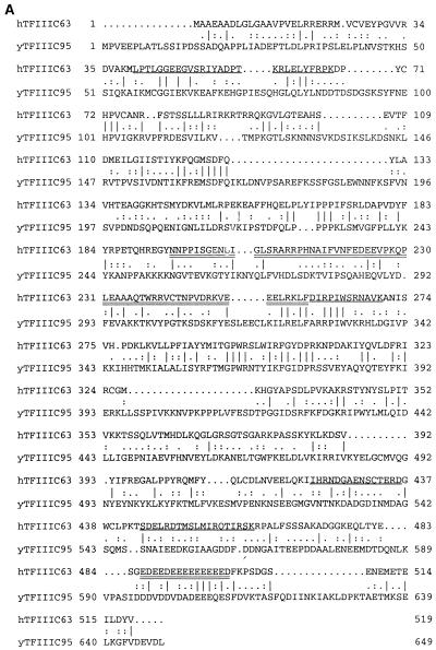
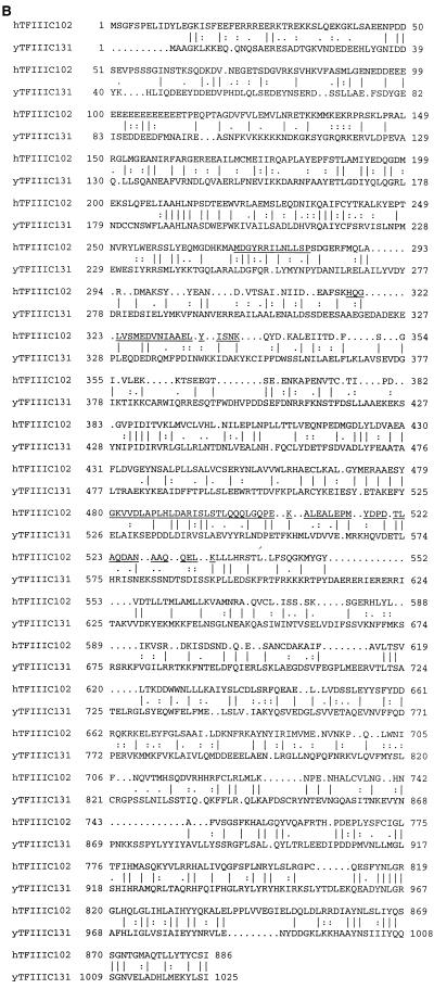
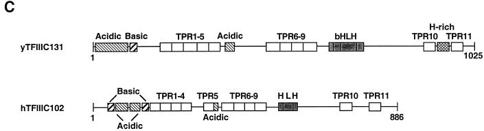
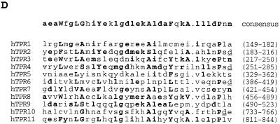
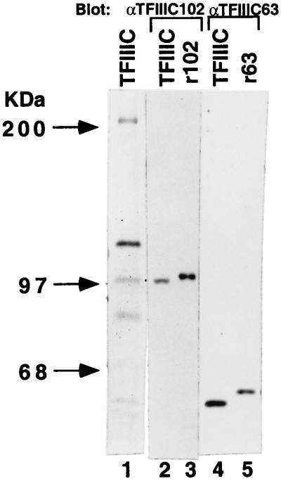
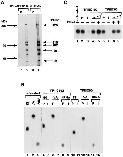
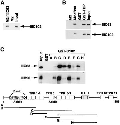
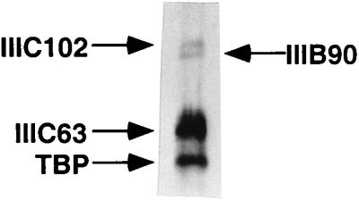
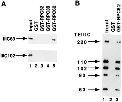
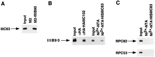
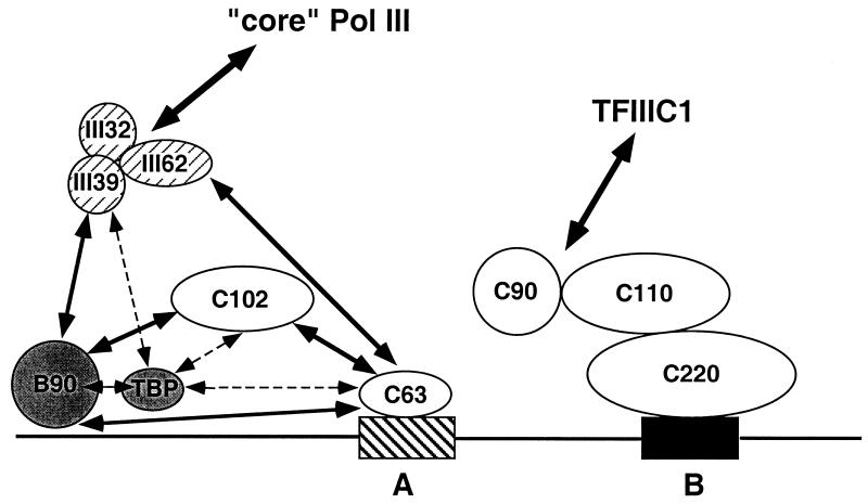
Human transcription factor IIIC (hTFIIIC) is a multisubunit complex that mediates transcription of class III genes through direct recognition of promoters (for tRNA and virus-associated RNA genes) or promoter-TFIIIA complexes (for the 5S RNA gene) and subsequent recruitment of TFIIIB and RNA polymerase III. We describe the cognate cDNA cloning and characterization of two subunits (hTFIIIC63 and hTFIIIC102) that are present within a DNA-binding subcomplex (TFIIIC2) of TFIIIC and are related in structure and function to two yeast TFIIIC subunits (yTFIIIC95 and yTFIIIC131) previously shown to interact, respectively, with the promoter (A box) and with a subunit of yeast TFIIIB. hTFIIIC63 and hTFIIIC102 show parallel in vitro interactions with the homologous human TFIIIB and RNA polymerase III components, as well as additional interactions that may facilitate both TFIIIB and RNA polymerase III recruitment. These include novel interactions of hTFIIIC63 with hTFIIIC102, with hTFIIIB90, and with hRPC62, in addition to the hTFIIIC102-hTFIIIB90 and hTFIIIB90-hRPC39 interactions that parallel the previously described interactions in yeast. As reported for yTFIIIC131, hTFIIIC102 contains acidic and basic regions, tetratricopeptide repeats (TPRs), and a helix-loop-helix domain, and mutagenesis studies have implicated the TPRs in interactions both with hTFIIIC63 and with hTFIIIB90. These observations further document conservation from yeast to human of the structure and function of the RNA polymerase III transcription machinery, but in addition, they provide new insights into the function of hTFIIIC and suggest direct involvement in recruitment of both TFIIIB and RNA polymerase III.


Figures

FIG. 1
FIG. 1
Predicted amino acid sequences and sequence alignments with yeast counterparts of the hTFIIIC102 and hTFIIIC63 subunits of the TFIIIC2 complex. The peptide sequences obtained by microsequence analyses are underlined. (A) Predicted amino acid sequence of hTFIIIC63 and alignment with yTFIIIC95 (31, 36). A highly acidic region at the C terminus and a central helix-turn-helix motif are double underlined. (B) Predicted amino acid sequence of hTFIIIC102 and alignment with yTFIIIC131 (28). (C) Schematic representation of hTFIIIC102 and yTFIIIC131 (28). Two basic regions are found at amino acid positions 23 to 34 and 130 to 147, and three acidic regions are found at amino acid positions 45 to 52, 92 to 114, and 363 to 374. The HLH motif is found at amino acid positions 552 to 600. (D) Consensus sequence of the TPR unit as defined by Sikorski et al. (33) and sequences of the 11 TPR units of hTFIIIC102. The four conserved residues that form “helix A” (residues 4, 7, 8, and 11), the three residues that form “helix B” (residues 20, 24, and 27), and the unique proline residue often found at position 32 (10, 14) are shown in boldfaced capital letters. Other residues that fit the TPR consensus sequence are shown in boldfaced lowercase letters. The following equivalences, based on the work of Marck et al. (28), are used: A, G, S, and V; E and D; K and R; I, L, M, and V; and L, F, Y, H, and W. Aspartate (D) residues, often found at positions 33 and 34 of TPR units, are underlined at those positions.
FIG. 1
FIG. 1
Predicted amino acid sequences and sequence alignments with yeast counterparts of the hTFIIIC102 and hTFIIIC63 subunits of the TFIIIC2 complex. The peptide sequences obtained by microsequence analyses are underlined. (A) Predicted amino acid sequence of hTFIIIC63 and alignment with yTFIIIC95 (31, 36). A highly acidic region at the C terminus and a central helix-turn-helix motif are double underlined. (B) Predicted amino acid sequence of hTFIIIC102 and alignment with yTFIIIC131 (28). (C) Schematic representation of hTFIIIC102 and yTFIIIC131 (28). Two basic regions are found at amino acid positions 23 to 34 and 130 to 147, and three acidic regions are found at amino acid positions 45 to 52, 92 to 114, and 363 to 374. The HLH motif is found at amino acid positions 552 to 600. (D) Consensus sequence of the TPR unit as defined by Sikorski et al. (33) and sequences of the 11 TPR units of hTFIIIC102. The four conserved residues that form “helix A” (residues 4, 7, 8, and 11), the three residues that form “helix B” (residues 20, 24, and 27), and the unique proline residue often found at position 32 (10, 14) are shown in boldfaced capital letters. Other residues that fit the TPR consensus sequence are shown in boldfaced lowercase letters. The following equivalences, based on the work of Marck et al. (28), are used: A, G, S, and V; E and D; K and R; I, L, M, and V; and L, F, Y, H, and W. Aspartate (D) residues, often found at positions 33 and 34 of TPR units, are underlined at those positions.
FIG. 2
FIG. 2
Identification of the cDNA-encoded proteins as the 102- and 63-kDa hTFIIIC2 subunits. The polypeptide components of a highly purified TFIIIC2 fraction were analyzed in a silver-stained gel (lane 1). Anti-hTFIIIC102 and anti-hTFIIIC63 antibodies were used to detect hTFIIIC102 and hTFIIIC63 in a highly purified TFIIIC2 fraction (lanes 2 and 4) and in purified reticulocyte lysate-expressed FLAG-hTFIIIC102 and baculovirus-expressed His10–hTFIIIC63 (lanes 3 and 5).
FIG. 3
FIG. 3
Immunoprecipitation of the TFIIIC2 complex and immunodepletion of TFIIIC transcription activity by anti-hTFIIIC102 and anti-hTFIIIC63 antibodies. (A) Immunoprecipitates from HeLa nuclear extracts treated with preimmune (P) and immune (I) anti-hTFIIIC102 (lanes 1 and 2) and anti-hTFIIIC63 (lanes 3 and 4) antibodies in BC500–0.1% NP-40 were subjected to Western blot analysis. The polypeptides were detected by a mixture of antibodies against hTFIIIC220, -110, -102, -90, and -63. The amounts of individual antibodies were adjusted so that the intensities of the hTFIIIC220, -110, -102, -90, and -63 immunoreactive bands were similar. The extra band between the 63- and 90-kDa polypeptides in lanes 2 and 4 reflects cross-reactivity with the anti-hTFIIIC110 antibodies and appears to represent a proteolytic breakdown product of hTFIIIC110 that was not consistently observed. (B) Nuclear extracts treated with preimmune (P) and immune (I) anti-hTFIIIC102 (lanes 4 to 9) and anti-hTFIIIC63 (lanes 10 to 15) antibodies were used for in vitro transcription assays with 5S RNA (lanes 1, 4, 5, 10, and 11), VAI RNA (lanes 2, 6, 7, 12, and 13), and tRNA (lanes 1, 8, 9, 14, and 15) templates. (C) Either 1 μl (lanes 4 and 8) or 2 μl (lanes 5 and 9) of an immunopurified TFIIIC was added to nuclear extracts depleted with immune anti-hTFIIIC102 (lanes 4 and 5) or with anti-hTFIIIC63 (lanes 8 and 9) and tested for transcription with the VAI RNA template.
FIG. 4
FIG. 4
Interactions of hTFIIIC102 with hTFIIIC63, hTFIIIB90, and TBP, and interactions of hTFIIIC63 with hTFIIIB90 and TBP. Input samples contained 10% of the amounts used for the interactions. (A) Purified baculovirus-expressed HA-hTFIIIC102 was incubated with M2 agarose or M2 agarose containing bound FLAG-hTFIIIC63 (M2-fIIIC63), and samples were washed and eluted as described in Materials and Methods. HA-hTFIIIC102 in input and eluted fractions was detected by immunoblotting with antisera against hTFIIIC102. (B) Purified baculovirus-expressed His10–hTFIIIC63 (top panel) or HA-hTFIIIC102 (bottom panel) was incubated with M2 agarose or M2 agarose-immobilized FLAG-hTFIIIB90 (M2-fB90) and with glutathione-Sepharose-immobilized GST or GST-TBP, and samples were washed and eluted as described in Materials and Methods. His10-hTFIIIC63 and HA-hTFIIIC102 were detected in input and eluted fractions by immunoblotting with anti-hTFIIIC63 and anti-hTFIIIC102 antibodies, respectively. (C) Sf9 cell extracts containing expressed His10–hTFIIIC63 (upper panel) or FLAG-hTFIIIB90 (lower panel) were incubated with glutathione-Sepharose-immobilized GST or with GST–truncated-hTFIIIC102 proteins (A, amino acids 1 to 148; B, amino acids 1 to 214; C, amino acids 207 to 507; D, amino acids 204 to 325; E, amino acids 419 to 711; F, amino acids 326 to 420; G, amino acids 419 to 531; and H, amino acids 527 to 724) in BC400–0.1% NP-40. After extensive washing of the beads with the same buffer, bound proteins were eluted by boiling in SDS sample buffer and analyzed by immunoblotting with antisera against hTFIIIC63 and hTFIIIB90. The amounts of GST–truncated-hTFIIIC102 proteins A through H and GST proteins were normalized by SDS-PAGE with Coomassie blue staining.
FIG. 5
FIG. 5
hTFIIIC102, hTFIIIC63, hTFIIIB90, and TBP form a subcomplex in vitro. The subcomplex formed from the baculovirus-expressed His10–hTFIIIC63, HA-hTFIIIC102, and FLAG-hTFIIIB90 and bacterially expressed GST-TBP was purified by successive affinity chromatography on Ni2+–NTA–agarose, HA antibodies linked to protein G-Sepharose, glutathione-Sepharose, and then M2 agarose. The preparative subcomplex was analyzed by immunoblotting with a mixture of antibodies against hTFIIIC102, hTFIIIC63, hTFIIIB90, and TBP. The relative amounts of the individual antibodies were different from the amounts used in Fig. 3A and 6B, such that the different intensities of the hTFIIIC102 and hTFIIIC63 immunoreactive bands do not necessarily reflect different stoichiometric amounts of these components.
FIG. 6
FIG. 6
Interactions of hTFIIIC102 and hTFIIIC63 with RNA polymerase III subunits hRPC32, hRPC39, and hRPC62. Input samples contained 10% of the amounts used for the interactions. (A) Purified baculovirus-expressed His10–hTFIIIC63 (top panel) or HA-hTFIIIC102 (bottom panel) was incubated with glutathione-Sepharose-immobilized GST-hRPC32 (lane 3), GST-hRPC39 (lane 4), GST-hRPC62 (lane 5), and GST (lane 2) proteins in BC150–0.1% NP-40. After extensive washes with the same buffer, the beads were boiled in SDS sample buffer. His10–hTFIIIC63 and HA-hTFIIIC102 in input and eluted fractions were detected on immunoblots by antisera against hTFIIIC63 and hTFIIIC102, respectively. The amounts of GST-hRPC32, GST-hRPC39, GST-hRPC62, and GST proteins in the inputs were normalized by SDS-PAGE with Coomassie blue staining. (B) Beads containing glutathione-Sepharose-immobilized GST-hRPC62 or GST were incubated with an immunopurified TFIIIC in BC150–0.1% NP-40. After extensive washes with the same buffer, the beads were boiled in SDS sample buffer. hTFIIIC220, -110, -102, -90, and -63 in the SDS eluates were detected by immunoblotting with a mixture of antisera against hTFIIIC220, -110, -102, -90, and -63. The amounts of individual antibodies were adjusted so that the intensities of the hTFIIIC220, -110, -102, -90, and -63 immunoreactive bands were similar.
FIG. 7
FIG. 7
Interactions between TFIIIB, TFIIIC, and RNA polymerase III. Input samples contained 10% of the amounts used for the interactions. (A) M2 agarose or M2 agarose-immobilized FLAG-hTFIIIB90 (M2-fIIIB90) was incubated with an immunopurified TFIIIC and washed with BC400–0.1% NP-40. Bound proteins were eluted with SDS buffer and analyzed by immunoblotting with antisera against hTFIIIC63. (B) Ni2+–NTA–agarose or Ni2+–NTA–agarose-immobilized His10–hTFIIIC63 and anti-HA agarose or anti-HA agarose-immobilized hTFIIIC102 were incubated with a purified core FLAG–hTFIIIB90–GST–TBP subcomplex. The latter subcomplex was isolated by successive affinity chromatography of a mixture of baculovirus-expressed FLAG-hTFIIIB90 and bacterially expressed GST-TBP on glutathione-Sepharose and M2 agarose, with BC400–0.1% NP-40 washes prior to elution (38). Bound proteins were eluted in the SDS buffer and analyzed by immunoblotting with antisera against hTFIIIB90. (C) Ni2+–NTA–agarose or Ni2+–NTA–agarose-immobilized His10–hTFIIIC63 was incubated with an immunopurified RNA polymerase III and washed with BC150–0.1% NP-40. Bound proteins were eluted in the SDS buffer and analyzed by immunoblotting with antisera against hRPC82 and hRPC53.
FIG. 8
FIG. 8
An integrated view of the interactions detected between subunits of human TFIIIB, TFIIIC, and RNA polymerase III. The observed protein-protein interactions are indicated by double-headed arrows. The A box and B box of the VAI/tRNA gene promoter are shown as hatched and solid rectangles, respectively. Hatched symbols, subcomplex of initiation-specific RNA polymerase III subunits; shaded symbols, TFIIIB subunits; open symbols, TFIIIC2 subunits. Individual subunits involved in interactions between TFIIIC2 and TFIIIC1 or between core RNA polymerase III and the three-subunit subcomplex are unknown. The positioning of the hTFIIIC220 subunit is based on cross-linking studies (21), whereas the positioning of the hTFIIIC63 and hTFIIIC102 subunits, as well as that of the TFIIIB subunits, is based on studies of the cognate yeast components.

Similar articles

Cited by

References

    1. Arrebola R, Manaud N, Rozenfeld S, Marsolier M C, Lefebvre O, Carles C, Thuriaux P, Conesa C, Sentenac A. τ91, an essential subunit of yeast transcription factor IIIC, cooperates with τ138 in DNA binding. Mol Cell Biol. 1998;18:1–9. - PMC - PubMed
    1. Bartholomew B, Kassavetis G A, Braun B R, Geiduschek E P. The subunit structure of Saccharomyces cerevisiae transcription factor IIIC probed with a novel photocrosslinking regent. EMBO J. 1990;9:2197–2205. - PMC - PubMed
    1. Bartholomew B, Kassavetis G A, Geiduschek E P. Two components of Saccharomyces cerevisiae TFIIIB are stereospecifically located upstream of a tRNA gene and interact with the second largest subunit of TFIIIC. Mol Cell Biol. 1991;11:5181–5189. - PMC - PubMed
    1. Bartholomew B, Durkovich D, Kassavetis G A, Geiduschek E P. Orientation and topography of RNA polymerase III in transcription complexes. Mol Cell Biol. 1993;13:942–952. - PMC - PubMed
    1. Bieker J J, Martin P L, Roeder R G. Formation of a rate-limiting intermediate in 5S RNA gene transcription. Cell. 1985;40:119–127. - PubMed

Publication types

Associated data