. 1999 Jul;19(7):4980-8.
doi: 10.1128/MCB.19.7.4980.

SOCS-3 is tyrosine phosphorylated in response to interleukin-2 and suppresses STAT5 phosphorylation and lymphocyte proliferation

Affiliations

SOCS-3 is tyrosine phosphorylated in response to interleukin-2 and suppresses STAT5 phosphorylation and lymphocyte proliferation

S J Cohney et al. Mol Cell Biol. 1999 Jul.

Abstract

Members of the recently discovered SOCS/CIS/SSI family have been proposed as regulators of cytokine signaling, and while targets and mechanisms have been suggested for some family members, the precise role of these proteins remains to be defined. To date no SOCS proteins have been specifically implicated in interleukin-2 (IL-2) signaling in T cells. Here we report SOCS-3 expression in response to IL-2 in both T-cell lines and human peripheral blood lymphocytes. SOCS-3 protein was detectable as early as 30 min following IL-2 stimulation, while CIS was seen only at low levels after 2 h. Unlike CIS, SOCS-3 was rapidly tyrosine phosphorylated in response to IL-2. Tyrosine phosphorylation of SOCS-3 was observed upon coexpression with Jak1 and Jak2 but only weakly with Jak3. In these experiments, SOCS-3 associated with Jak1 and inhibited Jak1 phosphorylation, and this inhibition was markedly enhanced by the presence of IL-2 receptor beta chain (IL-2Rbeta). Moreover, following IL-2 stimulation of T cells, SOCS-3 was able to interact with the IL-2 receptor complex, and in particular tyrosine phosphorylated Jak1 and IL-2Rbeta. Additionally, in lymphocytes expressing SOCS-3 but not CIS, IL-2-induced tyrosine phosphorylation of STAT5b was markedly reduced, while there was only a weak effect on IL-3-mediated STAT5b tyrosine phosphorylation. Finally, proliferation induced by both IL-2- and IL-3 was significantly inhibited in the presence of SOCS-3. The findings suggest that when SOCS-3 is rapidly induced by IL-2 in T cells, it acts to inhibit IL-2 responses in a classical negative feedback loop.


Figures
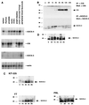
FIG. 1
FIG. 1
SOCS-3 is expressed in lymphocytes and regulated by IL-2 and IL-3. (A) Tissue mRNA blots were probed with cDNAs to CIS, SOCS-3, and SOCS-4, with β-actin as a control. (B) Kit-225 cells were treated as described above, and lysates were immunoprecipitated (IP) with antiserum to either SOCS-3 (αSOCS3) or CIS (αCIS) prior to SDS-PAGE. Filters were blotted with the corresponding antibody. (C) Kit-225 cells, YT cells, or PHA blasts were rested overnight in 2% FBS, stimulated with IL-2 for between 30 min and 8 h, lysed, immunoprecipitated, and immunoblotted with the antiserum to SOCS-3. (D) BaF3 cells were placed in cytokine-free medium overnight and treated with IL-3 for the indicated times, and the lysates were immunoprecipitated and immunoblotted with antiserum to SOCS-3. Sizes are indicated in kilodaltons.
FIG. 2
FIG. 2
Tyrosine phosphorylation of SOCS-3 in lymphocytes treated with IL-2 and by Jak1 in 293 T cells. (A) Kit-225 cells were treated as described in the text, and lysates were immunoprecipitated (IP) with antiserum to SOCS-3 (αSOCS-3). Immunoblotting was performed with the same antibody or antiphosphotyrosine antibody (αPTyr). (B) 293 T cells were transfected with Jak1 or Jak3 in combination with SOCS-3 and IL-2Rβ as indicated. After 48 h, cells were harvested and a portion of the lysate was used for immunoprecipitation with antiserum to SOCS-3. Immunoprecipitates and whole-cell lysates were immunoblotted with antiphosphotyrosine antibody or antiserum to SOCS-3, Jak1, or Jak3. Expression levels of Jak kinases and IL-2Rβ were tested on whole-cell lysates. (C) 293 T cells were transfected with various combinations of plasmids containing DNA encoding Jak1, IL-2Rβ, SOCS-1, SOCS-2, and SOCS-3 as indicated. After 48 h, cells were harvested and whole-cell lysates were subjected to SDS-PAGE and immunoblotting with antiphosphotyrosine antibody as described above. Blots were reprobed to check expression levels. (D) 293 T cells were transfected with plasmids encoding Jak3, IL-2Rβ, SOCS-1, SOCS-2, and SOCS-3 as indicated. After 48 h, cells were harvested and analyzed as described above. (E) 293 T cells were transfected with a plasmid encoding Jak1 (all lanes), IL-2Rβ (lanes 1 and 4), SOCS-2 (lane 2), or SOCS-3 (lanes 3 and 4); the lysates were immunoprecipitated with MAb M2, and in vitro kinase (IVK) assays performed on FLAG-tagged Jak1 and SOCS proteins. Sizes are indicated in kilodaltons.
FIG. 3
FIG. 3
SOCS-3 associates with the IL-2R complex following IL-2 stimulation of lymphocytes. Kit-225 cells were prepared as described in the text and treated with IL-2 for 15 min. Cells lysates were incubated with a GST–SOCS-3 fusion protein, and the immunoprecipitates (IP) were subjected to SDS-PAGE and membrane transfer. Filters were immunoblotted with antiphosphotyrosine (αPTyr) (A) or anti-Jak1 (αJak1) (B). (C) BaF3 cells expressing both IL-2Rβ and SOCS-3 were treated with MG132 (20 mM) for 30 min and were stimulated with IL-2 for 15 or 30 min; lysates were immunoprecipitated (IP) with antiserum to SOCS3 and blotted for Jak1 (upper panel) or SOCS-3 (lower panel). Sizes are indicated in kilodaltons.
FIG. 4
FIG. 4
SOCS-3 but not CIS inhibits IL-2-induced tyrosine phosphorylation of STAT5b and Jak1. (A) Two BaF3 clones (S3 CL2 and S3 CL3) expressing IL-2Rβ, in which SOCS-3 was absent due to the presence of tetracycline (tet) (+) or in which expression of SOCS-3 was induced by removal from tetracycline (−), were treated with IL-2 or IL-3 for 15 min, and lysates were subject to immunoblotting with antiphosphotyrosine (αPTyr) and immunoprecipitation (IP) with anti-STAT5b. SOCS-3 levels were checked by immunoblotting (lower panel). (B) BaF3 clones in which CIS or SOCS-3 expression was controlled by removal from tetracycline were treated as described above, and lysates were subjected to immunoblotting with antiphosphotyrosine and anti-STAT5b. CIS and SOCS-3 levels were checked by immunoblotting (lower panel). (C and D) SOCS-3-expressing cells were treated as described above with IL-2, immunoprecipitated anti-IL-2Rβ, and blotted for phosphotyrosine (C, top), Jak1 (C, middle), or anti-IL-2Rβ (C, bottom) or with IL-3, immunoprecipitated with anti-IL-3Rβc, and blotted for antiphosphotyrosine (D). Sizes are indicated in kilodaltons.
FIG. 5
FIG. 5
SOCS-3 inhibits cytokine-induced lymphocyte proliferation. Lymphocyte proliferation was assessed by [3H]thymidine incorporation as described in Materials and Methods. The experiments were performed on BaF3 clones (expressing CIS [A] or SOCS-3 [B]), in which SOCS-3 was absent due to the presence of tetracycline (Tet) (+) or in which expression of SOCS-3 was induced by removal from tetracycline (−). SOCS-3 expression in the absence of tetracycline was confirmed by immunoblotting (not shown). Med, cytokine-free medium.

Similar articles

Cited by

References

    1. Adams T E, Hansen J A, Starr R, Nicola N A, Hilton D J, Billestrup N. Growth hormone preferentially induces the rapid, transient expression of SOCS-3, a novel inhibitor of cytokine receptor signaling. J Biol Chem. 1998;273:1285–1287. - PubMed
    1. Auernhammer C J, Chesnokova V, Bousquet C, Melmed S. Pituitary corticotroph SOCS-3: novel intracellular regulation of leukemia-inhibitory factor-mediated proopiomelanocortin gene expression and adrenocorticotropin secretion. Mol Endocrinol. 1998;12:954–961. - PubMed
    1. Bjorbaek C, Elmquist J K, Frantz J D, Shoelson S E, Flier J S. Identification of SOCS-3 as a potential mediator of central leptin resistance. Mol Cell. 1998;1:619–625. - PubMed
    1. Cacalano N, Migone T, Bazan F, Hanson E, Chen M, Candotti F, O’Shea J J, Johnston J A. Autosomal SCID caused by a point mutation in the N-terminus of Jak3: mapping of the Jak3-receptor interaction domain. EMBO J. 1999;18:1549–1558. - PMC - PubMed
    1. Callus B A, Mathey-Prevot B. Interleukin-3-induced activation of the JAK/STAT pathway is prolonged by proteasome inhibitors. Blood. 1998;91:3182–3192. - PubMed

Publication types

MeSH terms

Substances