. 1999 Jul;19(7):5036-49.
doi: 10.1128/MCB.19.7.5036.

Distinct glucocorticoid receptor transcriptional regulatory surfaces mediate the cytotoxic and cytostatic effects of glucocorticoids

Affiliations

Distinct glucocorticoid receptor transcriptional regulatory surfaces mediate the cytotoxic and cytostatic effects of glucocorticoids

I Rogatsky et al. Mol Cell Biol. 1999 Jul.

Abstract

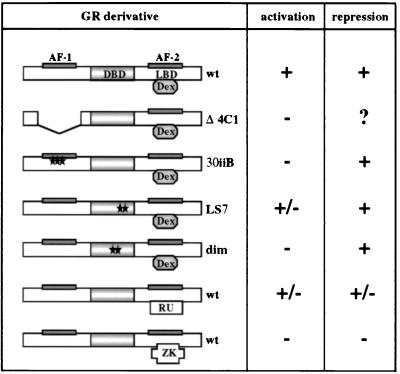
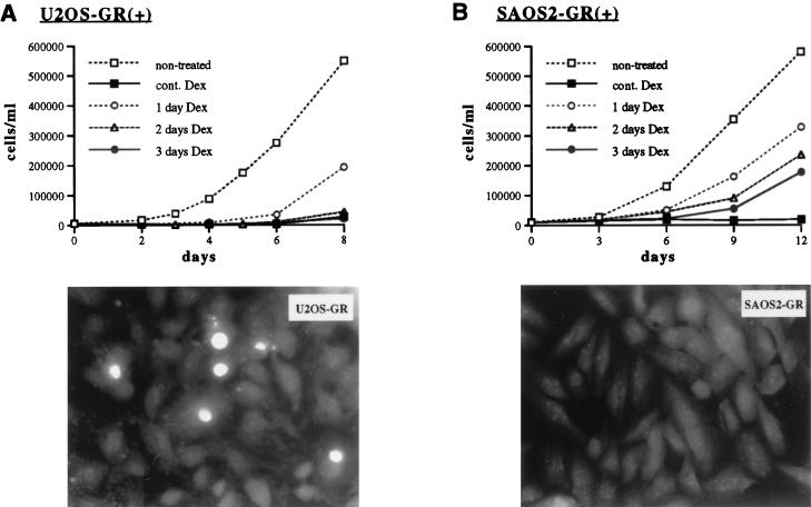
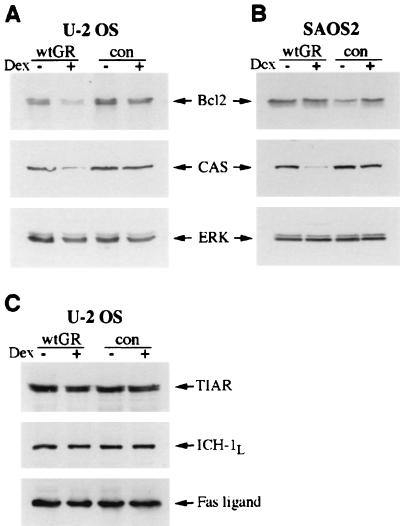
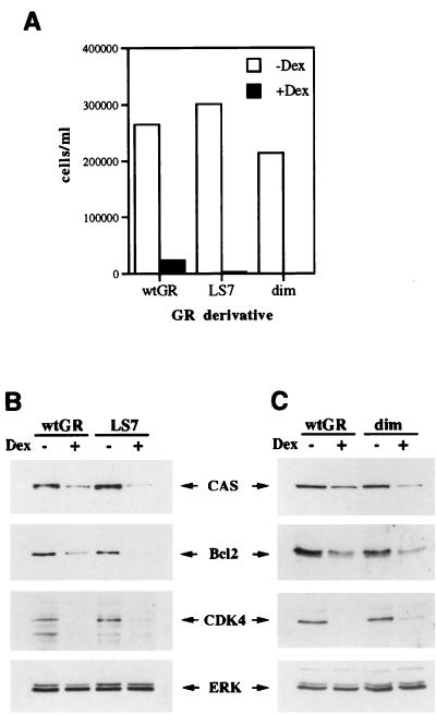
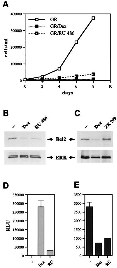
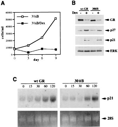
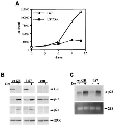
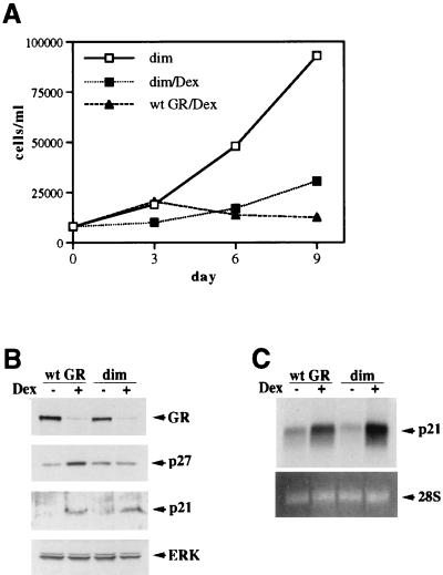
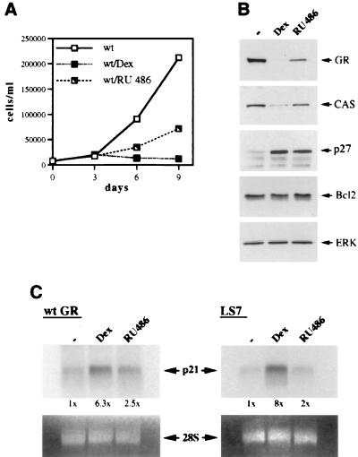
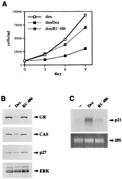
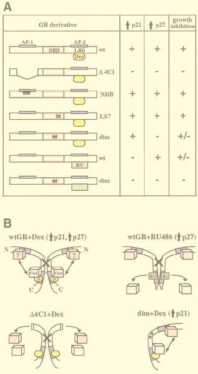
Glucocorticoids act through the glucocorticoid receptor (GR), which can function as a transcriptional activator or repressor, to elicit cytostatic and cytotoxic effects in a variety of cells. The molecular mechanisms regulating these events and the target genes affected by the activated receptor remain largely undefined. Using cultured human osteosarcoma cells as a model for the GR antiproliferative effect, we demonstrate that in U20S cells, GR activation leads to irreversible growth inhibition, apoptosis, and repression of Bcl2. This cytotoxic effect is mediated by GR's transcriptional repression function, since transactivation-deficient mutants and ligands still bring about apoptosis and Bcl2 down-regulation. In contrast, the antiproliferative effect of GR in SAOS2 cells is reversible, does not result in apoptosis or repression of Bcl2, and is a function of the receptor's ability to stimulate transcription. Thus, the cytotoxic versus cytostatic outcome of glucocorticoid treatment is cell context dependent. Interestingly, the cytostatic effect of glucocorticoids in SAOS2 cells involves multiple GR activation surfaces. GR mutants and ligands that disrupt individual transcriptional activation functions (activation function 1 [AF-1] and AF-2) or receptor dimerization fail to fully inhibit cellular proliferation and, remarkably, discriminate between the targets of GR's cytostatic action, the cyclin-dependent kinase inhibitors p21(Cip1) and p27(Kip1). Induction of p21(Cip1) is agonist dependent and requires AF-2 but not AF-1 or GR dimerization. In contrast, induction of p27(Kip1) is agonist independent, does not require AF-2 or AF-1, but depends on GR dimerization. Our findings indicate that multiple GR transcriptional regulatory mechanisms that employ distinct receptor surfaces are used to evoke either the cytostatic or cytotoxic response to glucocorticoids.


Figures

FIG. 1
FIG. 1
Transcriptional regulatory responses of GR derivatives and ligands. Rat GR derivatives ectopically expressed in U2OS and SAOS2 cells are shown. The GR DBD, LBD, and N-terminal and C-terminal transcriptional activation functions (AF-1 and AF-2, respectively) are indicated. The GR Δ4C1 mutant lacks amino acids 70 to 300. The 30iiB mutant contains three amino acid substitutions in AF-1 (E219K, F220L, and W234R). The LS7 mutant contains two point mutations, P493R and A494S, in the second zinc finger of the GR DBD. The dimer (dim) mutant contains two mutations, R479D and D481R, which disrupt the GR dimerization interface. The locations of the point mutations are shown with stars. The ability of each derivative to activate and repress transcription is summarized based on previously published studies (see text for references); +, −, and +/− represent a transcriptionally competent, a transcriptionally inactive, or a context-dependent phenotype, respectively. A question mark indicates that the phenotype has not been characterized. The transcriptional regulatory responses of the wt GR bound by the full agonist Dex, the partial agonist RU 486 (RU), and the antagonist ZK 299 (ZK) are also shown.
FIG. 2
FIG. 2
GR activation induces apoptosis in U2OS but not SAOS2 cells. (Top panels) U2OS-GR(+) (A) and SAOS2-GR(+) (B) cells were seeded on day 0 into six-well plates (15,000 and 25,000 cells/well, respectively) in the presence of 100 nM Dex where indicated. Cells were refed at 24 (1 day Dex), 48 (2 days Dex), or 72 (3 days Dex) h with hormone-free medium. Nontreated and continuously (cont.) Dex-treated cells were cultured in the absence or presence of Dex, respectively, throughout the experiment. On the indicated days cells were trypsinized, stained with trypan blue, and counted with a hemocytometer. (Bottom panels) GR-expressing U2OS (A) and SAOS2 (B) cells were cultured in the presence of 100 nM Dex for 24, 48, or 72 h and subjected to the TUNEL assay as described in Materials and Methods. Fluorescence microscopy was performed with a standard fluorescein filter set at a wavelength of 520 nm to view fluorescein-12-dUTP incorporated into DNA nicks. Note multiple apoptotic nuclei in U2OS-GR(+) cells treated with Dex for 24 h. No apoptotic nuclear morphology was detected in SAOS2-GR(+) cells treated with Dex for 24, 48 (shown), or 72 h.
FIG. 3
FIG. 3
Expression of apoptosis-related proteins in U2OS-GR(+) and SAOS2-GR(+) cells. U2OS (A and C) and SAOS2 (B) cells expressing wt GR or receptor-deficient control (con) GR-negative cells were cultured in the absence (−) or presence (+) of 100 nM Dex for 2 days, and whole-cell lysates were prepared (see Materials and Methods). Equal amounts of total protein were resolved on a Tris-glycine–4 to 20% gradient polyacrylamide gel, transferred to Immobilon paper, and probed with antibodies against Bcl2, CAS, ERK, TIAR, ICH-1L, and Fas ligand. Equal loading in each lane is demonstrated by probing with an anti-ERK antibody. Each blot is representative of two or more independent experiments.
FIG. 4
FIG. 4
The GR LS7 and dimer mutants function like the wt GR in U2OS cells. U2OS-LS7 and U2OS-dim clones were generated as described in Materials and Methods. (A) The GR LS7 and dimerization mutants induce cell cycle arrest in U2OS cells. U2OS-GR(+) (wt GR), U2OS-LS7 (LS7), and U2OS-dim (dim) clones were seeded on day 0 into six-well plates in duplicate and cultured in the absence or presence of 100 nM Dex as indicated. The total numbers of viable cells were determined on days 2, 4, and 6 by the trypan blue exclusion method. The graph represents cell counts on day 6. (B and C) The GR LS7 (B) and dim (C) mutants repress the expression of CAS, Bcl2, and CDK4. U2OS cells expressing the wt GR or the LS7 or dim mutant were cultured in the absence or presence of 100 nM Dex for 40 h and harvested, and the expression of CAS, Bcl2, CDK4, and ERK was assessed by immunoblotting as described in Materials and Methods. Similar results were obtained with at least three independent clones expressing each GR mutant.
FIG. 5
FIG. 5
RU 486 functions as a GR agonist in the context of U2OS-GR(+) cells. (A) RU 486 inhibits proliferation of U2OS-GR(+) cells. U2OS-GR(+) cells were seeded on day 0 into six-well plates (15,000 cells/well) in duplicate and cultured in the presence of an ethanol vehicle, 100 nM Dex, or 100 nM RU 486. The total number of viable cells was determined on the indicated days. (B) Expression of Bcl2 is repressed in both Dex-treated and RU 486-treated U2OS-GR(+) cells. U2OS-GR(+) cells were cultured in the presence of an ethanol vehicle, 100 nM Dex, or 100 nM RU 486 for 40 h, and expression of Bcl2 and ERK in whole-cell extracts was examined by immunoblotting as described in Materials and Methods. (C) ZK 299 is a pure GR antagonist in U2OS cells. U2OS-GR(+) cells were cultured in the presence of an ethanol vehicle, 100 nM Dex, or 100 nM ZK 299 for 40 h, and expression of Bcl2 and ERK in whole-cell extracts was examined by immunoblotting. (D) RU 486 is a weak agonist with respect to GR transcriptional activation in U2OS-GR(+) cells. U2OS-GR(+) cells were seeded in 6-cm-diameter dishes (120,000/dish) and transfected the following day via the calcium phosphate precipitation method with an XG46TL reporter plasmid (4 μg/dish) and a pCMV-LacZ plasmid (0.75 μg/dish) as an internal control for transfection efficiency. Transfected cells were treated with an ethanol vehicle (−), 100 nM Dex, or 100 nM RU 486 (RU) for 12 h, and GR transcriptional activation was assessed via luciferase assay, normalized to β-Gal activity, and expressed as relative luminescence units (RLU). (E) RU 486 is a potent agonist with respect to GR-mediated transcriptional repression in U2OS-GR(+) cells. Cells were transfected with an XAP1TL reporter plasmid and a pCMV-LacZ plasmid, and GR transcriptional repression was assessed as described for panel D.
FIG. 6
FIG. 6
Transcriptional activation by the GR AF-1 is dispensable for cell cycle arrest in SAOS2 cells. (A) The GR 30iiB mutant induces complete cell cycle arrest in SAOS2 cells. SAOS2-30iiB stable transformants were generated as described in Materials and Methods. SAOS2-30iiB clones were seeded on day 0 into six-well plates (20,000 cells/well) and cultured in the absence or presence of 100 nM Dex. Total numbers of viable cells were determined on the indicated days by using the trypan blue exclusion method. (B) Induction of p21 and p27 by the GR 30iiB mutant. SAOS2-GR(+) (wt GR) and SAOS2-30iiB (30iiB) cells were cultured in the presence of 100 nM Dex or ethanol vehicle for 3 days, and whole-cell extracts were prepared and subjected to immunoblotting for GR, p27, p21, and ERK. Note the induction of p21 and p27 by both the wt GR and the 30iiB mutant. (C) Time course of p21 mRNA induction. SAOS2-GR(+) (wt GR) and SAOS2-30iiB (30iiB) cells were treated with 100 nM Dex for 0, 15, 30, 60, and 120 min, and total RNA was isolated and subjected to Northern blot analysis with a 32P-labeled p21 cDNA probe (top panels). Equal loading in each lane is demonstrated by ethidium bromide staining of the 28S rRNA (bottom panels).
FIG. 7
FIG. 7
The GR LS7 mutant functions like the wt GR when stably expressed in SAOS2 cells. (A) Inhibition of SAOS2 cell proliferation by the GR LS7 mutant. Multiple SAOS2-LS7 clones were generated as described in Materials and Methods. SAOS2-LS7 cells were seeded on day 0 into six-well plates (20,000 cells/well) and cultured in the absence or presence of 100 nM Dex. Total numbers of viable cells were determined on the indicated days by using the trypan blue exclusion method. Similar cell growth kinetics were observed in three independent LS7-expressing clones. (B) The GR LS7 mutant induces the expression of p21 and p27 proteins. SAOS2-GR(+) (wt GR), SAOS2-LS7 (LS7), and GR-negative SAOS2 (con) cells were cultured in the presence of 100 nM Dex or ethanol vehicle for 3 days, and whole-cell extracts were prepared and subjected to immunoblotting for GR, p27, p21, and ERK. Note the increase in p21 and p27 protein in the wt GR- and LS7-expressing cells but not in the GR-deficient control cells. (C) Induction of the steady-state p21 mRNA level by the GR LS7 mutant. SAOS2-GR(+) (wt GR) and SAOS2-LS7 (LS7) cells were treated with 100 nM Dex for 2 h 30 min where indicated, and total RNA was isolated and hybridized to a 32P-labeled p21 cDNA probe (top panel). Equal loading in each lane is demonstrated by ethidium bromide staining of the 28S rRNA (bottom panel).
FIG. 8
FIG. 8
Dissociation of p21 induction from p27 induction in SAOS2 cells through the GR dimerization mutant. (A) Inhibition of cell proliferation by the GR dimerization-deficient mutant. SAOS2 clones stably expressing the GR dim mutant were generated as described in Materials and Methods. SAOS2-GR(+) and SAOS2-dim cells were seeded on day 0 into six-well plates (20,000 cells/well) and cultured in the absence or presence of 100 nM Dex. Total numbers of viable cells were determined on the indicated days. Note incomplete inhibition of cell proliferation by the dim mutant compared to the wt GR-expressing clone. Quantitation of cell counts reveals 76% growth inhibition for the dim mutant and 96% for the wt GR. Identical growth kinetics were demonstrated for three independent clones expressing the GR dim mutant. (B) Induction of p21 but not p27 expression by the dim mutant. SAOS2 cells expressing the wt GR or the dimerization-deficient mutant were cultured in the presence or absence of 100 nM Dex for 3 days, and whole-cell extracts were prepared and subjected to immunoblotting for GR, p27, p21, and ERK. Note the increase in p21 but not p27 protein in the dim-expressing cells. (C) Induction of the steady-state p21 mRNA level by the dim mutant. Hormone treatment, RNA isolation, and Northern hybridization were performed exactly as described for Fig. 7C. Potent Dex-dependent induction of p21 mRNA is observed in both SAOS2-GR(+) (wt GR) and SAOS2-dim (dim) cells.
FIG. 9
FIG. 9
Induction of CDI expression by the wt GR in SAOS2 cells is uncoupled by RU 486. (A) Differential effects of GR ligands on the proliferation of SAOS2-GR(+) cells. SAOS2-GR(+) cells were seeded on day 0 into six-well plates (20,000 cells/well) in duplicate and cultured in the presence of an ethanol vehicle, 100 nM Dex, or 100 nM RU 486. Total numbers of viable cells were determined on the indicated days. Note 69% inhibition of cell proliferation in the presence of RU 486, compared to 98% observed in the presence of Dex. (B) Alterations in protein expression induced by RU 486 in SAOS2-GR(+) cells. SAOS2-GR(+) cells were cultured in the presence of an ethanol vehicle, 100 nM Dex, or 100 nM RU 486 for 3 days, and expression of GR, CAS, p27, Bcl2, and ERK in whole-cell extracts was examined by immunoblotting. Note that comparatively mild repression of GR and CAS is observed in the presence of RU 486, whereas p27 induction is retained whether GR is activated with Dex or RU 486. Similar results were obtained with RU 486 concentrations of 500 nM and 1 μM (data not shown). (C) RU 486-activated wt GR or LS7 mutant fails to efficiently induce p21 mRNA expression in SAOS2 cells. SAOS2-GR(+) (wt GR) and SAOS2-LS7 (LS7) cells were treated with an ethanol vehicle, 100 nM Dex, or 100 nM RU 486 for 2 h 30 min, and total RNA was isolated and subjected to Northern hybridization with a 32P-labeled p21 cDNA probe (top panels). Equal loading in each lane is demonstrated by ethidium bromide staining of 28S rRNA (bottom panels). The results of autoradiography were quantitated by spot densitometry. The densitometric value obtained for mock-treated SAOS2-GR(+) or SAOS2-LS7 cells was arbitrarily set as 1×.
FIG. 10
FIG. 10
RU 486 loses partial agonist activity in the context of the GR dimerization-deficient mutant. (A) The GR dim mutant does not affect cell proliferation in the presence of RU 486. SAOS2-dim cells were seeded on day 0 into six-well plates (20,000 cells/well) in duplicate and cultured in the presence of an ethanol vehicle, 100 nM Dex, or 100 nM RU 486. Total numbers of viable cells were determined on the indicated days. (B) The RU 486-activated GR dimerization-deficient mutant fails to alter the expression of GR, CAS, or p27. SAOS2-dim cells were cultured in the presence of an ethanol vehicle, 100 nM Dex, or 100 nM RU 486 for 3 days, and expression of GR, CAS, p27, and ERK in the whole-cell extracts was examined by immunoblotting. (C) The RU 486-activated dimer mutant does not induce the steady-state level of p21 mRNA. SAOS2-dim cells were treated with an ethanol vehicle, 100 nM Dex, or 100 nM RU 486 for 2 h 30 min, and total RNA was isolated and hybridized to a 32P-labeled p21 cDNA probe (top panel). Equal loading into each lane is demonstrated by ethidium bromide staining of 28S rRNA (bottom panel).
FIG. 11
FIG. 11
Regulation of p21 and p27 in SAOS2 cells by different GR derivatives. (A) Effects of GR derivatives on p21 and p27 expression. The GR domains and receptor derivatives stably introduced into SAOS2 cells are described in Fig. 1. The ability of GR derivatives to induce p21 and p27 expression and to effect growth inhibition in SAOS2 cells was assessed in the presence of Dex (yellow ovals) or RU 486 (green rectangles). (B) A model for the differential regulation of CDIs by the wt and mutant GRs in SAOS2 cells. Schematically shown are two wt GR molecules (wtGR+Dex), which in the presence of the full agonist Dex (D, yellow ovals) dimerize through the DBD (indicated by a cross) and recruit a coactivator protein (CoA, blue cubes) to associate with the C-terminal AF-2 (shown in dark blue). An additional putative interacting protein (?, pink cubes) may associate with the GR N terminus and AF-1. Possible communication between the C-terminal coactivator and an unknown N terminus-interacting factor (indicated with an arrow) induces expression of p21 and p27. Deletion of the GR N terminus (Δ4C1+Dex) results in a loss of the unknown interactor, disrupting a putative interaction between the N and the C termini, such that the induction of both p21 and p27 is abolished. wt GR in the presence of the partial agonist RU 486 (wtGR+RU 486) is unable to recruit a C-terminal coactivator due to a conformational change in the LBD. The induction of p21 is abolished, suggesting a requirement for a functional AF-2 for this effect, whereas enhanced expression of p27 occurs in an AF-2-independent manner, possibly through the unknown N-terminal interactor and as-yet-unidentified transcriptional regulatory function at GR N terminus. Disruption of the GR dimerization interface (dim+Dex) eliminates p27 but not p21 induction, indicating that stimulated p27 expression depends on GR dimerization and GRE binding, whereas p21 induction is accomplished by GR in its monomeric form.

Similar articles

Cited by

References

    1. Adams J M, Cory S. The Bcl-2 protein family: arbiters of cell survival. Science. 1998;281:1322–1326. - PubMed
    1. Alam J, Cook J L. Reporter genes: application to the study of mammalian gene transcription. Anal Biochem. 1990;188:245–254. - PubMed
    1. Alessandrini A, Chiaur D S, Pagano M. Regulation of the cyclin-dependent kinase inhibitor p27 by degradation and phosphorylation. Leukemia. 1997;11:342–345. - PubMed
    1. Almlof T, Gustafsson J A, Wright A P. Role of hydrophobic amino acid clusters in the transactivation activity of the human glucocorticoid receptor. Mol Cell Biol. 1997;17:934–945. - PMC - PubMed
    1. Alnemri E S, Fernandes T F, Haldar S, Croce C M, Litwack G. Involvement of Bcl-2 in glucocorticoid-induced apoptosis of human pre-B-leukemias. Cancer Res. 1992;52:491–495. - PubMed

Publication types

MeSH terms