. 1999 Jul;19(7):5061-72.
doi: 10.1128/MCB.19.7.5061.

Domain swapping used to investigate the mechanism of protein kinase B regulation by 3-phosphoinositide-dependent protein kinase 1 and Ser473 kinase

Affiliations

Domain swapping used to investigate the mechanism of protein kinase B regulation by 3-phosphoinositide-dependent protein kinase 1 and Ser473 kinase

M Andjelković et al. Mol Cell Biol. 1999 Jul.

Abstract

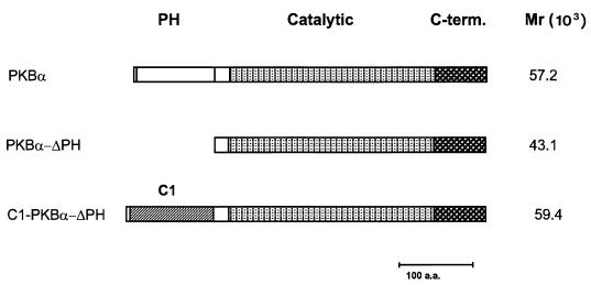
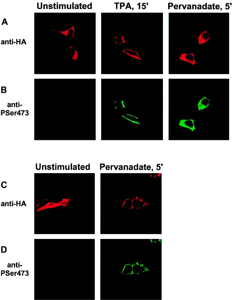
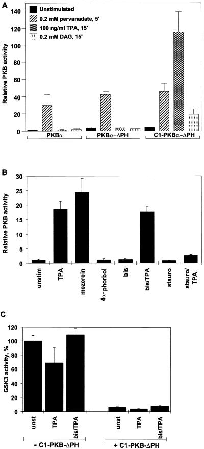
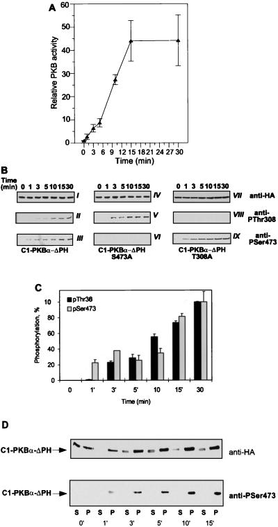
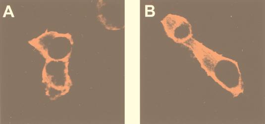
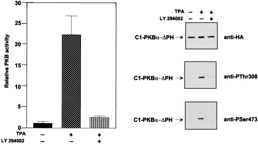
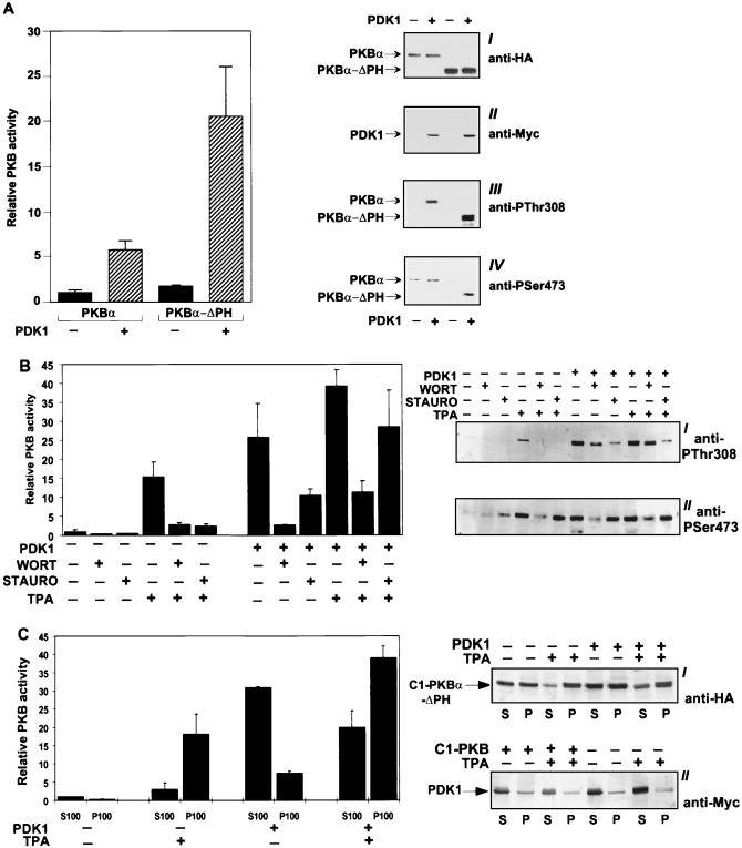
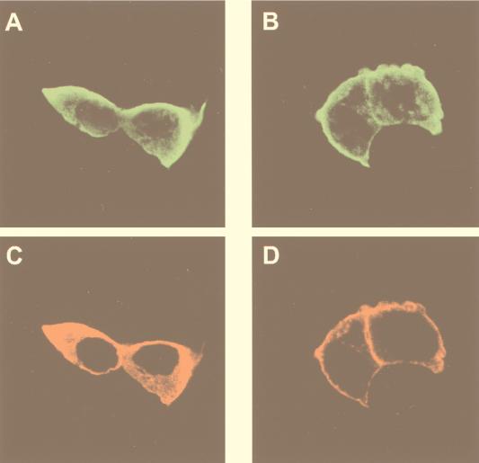
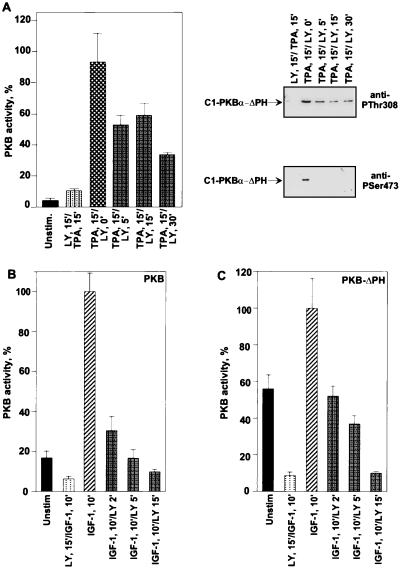
Protein kinase B (PKB or Akt), a downstream effector of phosphoinositide 3-kinase (PI 3-kinase), has been implicated in insulin signaling and cell survival. PKB is regulated by phosphorylation on Thr308 by 3-phosphoinositide-dependent protein kinase 1 (PDK1) and on Ser473 by an unidentified kinase. We have used chimeric molecules of PKB to define different steps in the activation mechanism. A chimera which allows inducible membrane translocation by lipid second messengers that activate in vivo protein kinase C and not PKB was created. Following membrane attachment, the PKB fusion protein was rapidly activated and phosphorylated at the two key regulatory sites, Ser473 and Thr308, in the absence of further cell stimulation. This finding indicated that both PDK1 and the Ser473 kinase may be localized at the membrane of unstimulated cells, which was confirmed for PDK1 by immunofluorescence studies. Significantly, PI 3-kinase inhibitors prevent the phosphorylation of both regulatory sites of the membrane-targeted PKB chimera. Furthermore, we show that PKB activated at the membrane was rapidly dephosphorylated following inhibition of PI 3-kinase, with Ser473 being a better substrate for protein phosphatase. Overall, the results demonstrate that PKB is stringently regulated by signaling pathways that control both phosphorylation/activation and dephosphorylation/inactivation of this pivotal protein kinase.


Figures

FIG. 1
FIG. 1
Schematic representation of PKBα constructs used to study kinase regulation by inducible membrane translocation.
FIG. 2
FIG. 2
Activation of C1-PKB-ΔPH by TPA is due to membrane translocation. HEK 293 cells plated on coverslips were transfected with C1-PKB-ΔPH (A and B) or wild-type PKB (C and D) and serum starved for 16 h prior to stimulation with TPA or pervanadate. Fixed and permeabilized cells were incubated with the anti-HA epitope monoclonal antibody 12CA5 (A and C) and a rabbit polyclonal antibody raised against the phosphoSer473 phosphopeptide (B and D), followed by a biotinylated anti-mouse antibody/streptavidin-conjugated Texas red and FITC-conjugated anti-rabbit antibody, and analyzed by confocal microscopy.
FIG. 3
FIG. 3
Activation of C1-PKB-ΔPH and effects on GSK-3β activity. (A) Activation of wild-type PKB, PKB-ΔPH, and C1-PKB-ΔPH by pervanadate, TPA, or DAG. The PKB constructs were expressed in HEK 293 cells. Transfected cells were starved 24 h prior to stimulation with pervanadate, TPA, or DAG, as indicated. (B) Effects of PKC inhibitors on C1-PKB-ΔPH activation. Transfected cells were serum starved for 24 h before 15 min of stimulation with TPA, mezerein, or 4α-phorbol. Pretreatment with bisindolylmaleimide I (bis) and staurosporine (stauro) was done for 30 min before the addition of vehicle or TPA. PKB activity is the average (±standard deviation) of two experiments with duplicate immunoprecipitates and was corrected for different expression levels for each construct. The activity of wild-type PKB (A) and C1-PKB-ΔPH (B) from unstimulated cells was taken as 1. (C) Inactivation of GSK-3β by C1-PKB-ΔPH. Myc epitope-tagged GSK-3β was expressed in HEK 293 cells either alone or together with C1-PKB-ΔPH. Transfected cells were serum starved for 24 h before 15 min of stimulation with TPA. Pretreatment with bisindolylmaleimide I was as described for panel B. GSK-3β activity is the average (±standard deviation) of two experiments with duplicate immunoprecipitates and was corrected for different expression levels in coexpression experiments. Kinase activity determined from unstimulated cells expressing GSK-3β was taken as 100%.
FIG. 4
FIG. 4
Time course of C1-PKB-ΔPH activation, phosphorylation, and translocation to the particulate fraction. (A) C1-PKB-ΔPH was expressed in HEK 293 cells that were serum starved 24 h prior to stimulation with TPA (100 ng/ml) for the indicated time periods. C1-PKB-ΔPH activity was determined following immunoprecipitation with the anti-HA epitope antibody. PKB activity is the average (±standard deviation) of two experiments with duplicate immunoprecipitates. The activity of C1-PKB-ΔPH from unstimulated cells was taken as 1. (B) Phosphorylation state of C1-PKB-ΔPH, C1-PKB-ΔPH-S473A, and C1-PKB-ΔPH-T308A was determined by immunoblot analysis using the antibody specific for phosphoThr308 phosphopeptide (gels II, V, and VIII) and phosphoSer473 phosphopeptide (gels III, VI, and IX). Expression levels for each C1-PKB-ΔPH construct were monitored by the anti-HA epitope antibody (gels I, IV, and VII). (C) Quantification of phosphorylation depicts the average (±standard deviation) of two representative experiments out of four. Phosphorylation levels of Thr308 and Ser473 at the 30-min time point were taken as 100%. (D) Transfected HEK 293 treated with TPA (100 ng/ml) for the indicated time periods were subjected to hypotonic lysis, and cytosolic (S100) and particulate (P100) fractions were prepared as described in Materials and Methods. Changes in the distribution of C1-PKB-ΔPH protein and phosphorylation were revealed by immunoblotting with the anti-HA epitope antibody (upper gel) and anti-phosphoSer473 antibody (lower gel), respectively.
FIG. 5
FIG. 5
Subcellular localization of PDK1. HEK 293 cells transfected on coverslips with PDK1 were either left unstimulated following 16 h of serum starvation (A) or stimulated with TPA (100 ng/ml) for 15 min (B). Cells were immunostained with the anti-Myc epitope monoclonal antibody 9E10, followed by biotinylated anti-mouse antibody/streptavidin-conjugated Texas red, and analyzed by confocal microscopy.
FIG. 6
FIG. 6
Activation of C1-PKB-ΔPH by TPA is sensitive to the PI 3-kinase inhibitor. HEK 293 cells expressing C1-PKB-ΔPH were subjected to a 24-h serum starvation before stimulation with TPA (100 ng/ml), with or without a 15-min pretreatment with LY 294402. C1-PKB-ΔPH was immunoprecipitated with the anti-HA epitope antibody. Kinase activity is the average (±standard deviation) of two experiments with duplicate immunoprecipitates. The activity of C1-PKB-ΔPH from unstimulated cells was taken as 1. Phosphorylation state was determined by immunoblot analysis using the antibody specific for the HA epitope (top gel), phosphoThr308 phosphopeptide (middle gel), or phosphoSer473 phosphopeptide (bottom gel).
FIG. 7
FIG. 7
Expression of PDK1 activates PKB, PKB-ΔPH, and C1-PKB-ΔPH in vivo. (A) HEK 293 cells were transfected with PKB and PKB-ΔPH alone or with an equal amount of Myc epitope-tagged PDK1. PKB was immunoprecipitated from cells serum starved for 24 h with the anti-HA epitope antibody. Kinase activity is the average (±standard deviation) of two experiments with duplicate immunoprecipitates and was corrected for different expression levels of the constructs. The activity of wild-type PKB from transfected cells was taken as 1. PKB and PDK1 expression was confirmed by immunoblot analysis using the anti-HA epitope antibody (gel I) and anti-Myc epitope antibody (gel II), respectively. Phosphorylation state was determined by immunoblot analysis using the antibody specific for phosphoThr308 phosphopeptide (gel III) and phosphoSer473 phosphopeptide (gel IV). (B) C1-PKB-ΔPH was transfected into 293 cells alone or together with an equal amount of Myc epitope-tagged PDK1. Cells were either treated with vehicle following 24 h of serum starvation or subjected to one of the following treatments before lysis: 15-min TPA stimulation (100 ng/ml), 30-min incubation with wortmannin (WORT; 200 nM), 30-min incubation with staurosporine (STAURO; 200 ng/ml), 30-min wortmannin treatment before TPA addition, or 30-min staurosporine treatment prior to TPA stimulation. C1-PKB-ΔPH was immunoprecipitated with the anti-HA epitope antibody. PKB activity is the average (±standard deviation) of two experiments with duplicate immunoprecipitates. The activity of C1-PKB-ΔPH from transfected, unstimulated cells was taken as 1. PKB phosphorylation state was determined by immunoblot analysis using the antibody specific for phosphoThr308 phosphopeptide (gel I) and phosphoSer473 phosphopeptide (gel II). (C) Subcellular distribution of C1-PKB-ΔPH activity and protein in the presence of PDK1 and TPA. HEK 293 cells cotransfected and serum starved as described for panel B were treated with either vehicle or TPA for 15 min. The cytosolic (S100) and particulate (P100) fractions were prepared as described in Materials and Methods. C1-PKB-ΔPH was immunoprecipitated from S100 and P100 with the anti-HA epitope antibody. PKB activity is the average (±standard deviation) of two experiments with duplicate immunoprecipitates. The activity of C1-PKB-ΔPH from the S100 fraction of transfected, unstimulated cells was taken as 1. Changes in C1-PKB-ΔPH and PDK1 distribution were detected by immunoblotting with the anti-HA epitope antibody (gel I) and anti-Myc antibody (gel II), respectively.
FIG. 8
FIG. 8
Activation of C1-PKB-ΔPH with TPA induces translocation of PDK1. HEK 293 cells cotransfected on coverslips with PDK1 and C1-PKB-ΔPH were left unstimulated following 16 h of serum starvation (A and C) or stimulated with TPA (100 ng/ml) for 15 min (B and D). Cells were immunostained with a polyclonal anti-PKB antibody (Abα469/480) (A and B) and the anti-Myc epitope 9E10 monoclonal antibody (C and D), followed by an FITC-conjugated anti-rabbit antibody and biotinylated anti-mouse antibody/streptavidin-conjugated Texas red, and analyzed by confocal microscopy.
FIG. 9
FIG. 9
Effects of LY 294003 posttreatment on TPA-induced activity of C1-PKB-ΔPH and IGF-1-induced activity of PKB and PKB-ΔPH. HEK 293 cells expressing C1-PKB-ΔPH (A), wild-type PKB (B), or PKB-ΔPH (C) were serum starved for 24 h before 15 min of stimulation with TPA (100 ng/ml) (A) or IGF-1 (100 ng/ml) (B and C), followed by treatment with 50 μM LY 294002 (LY) or vehicle for the indicated time periods, or pretreated with the same concentration of LY 294002 for 15 min before addition of agonist. Protein was precipitated with the anti-HA epitope antibody. PKB activity is the average (±standard deviation) of two experiments with duplicate immunoprecipitates. Kinase activity from stimulated cells was taken as 100%. PKB phosphorylation state in panel A was determined by immunoblot analysis using the antibodies specific for phosphoThr308 (top gel) and phosphoSer473 (bottom gel).

References

    1. Alessi D R, Andjelković M, Caudwell B, Cron P, Morrice N, Cohen P, Hemmings B A. Mechanism of activation of protein kinase B by insulin and IGF-1. EMBO J. 1996;15:6541–6551. - PMC - PubMed
    1. Alessi D R, James S R, Downes C P, Holmes A B, Gaffney P R J, Reese C, Cohen P. Characterization of a 3-phosphoinositide-dependent protein kinase which phosphorylates and activates protein kinase Bα. Curr Biol. 1997;7:261–269. - PubMed
    1. Alessi D R, Deak M, Casamayor A, Caudwell F B, Morrice N, Norman D G, Gaffney P, Reese C B, MacDoucall C N, Harbison D, Ashwarth A, Bownes M. 3-Phosphoinositide-dependent protein kinase-1 (PDK1): structural and functional homology with the Drosophila DSTPK61 kinase. Curr Biol. 1997;7:776–789. - PubMed
    1. Andersson S, Davie D N, Dahlbäck H, Jörnvall H, Russell D W. Cloning, structure, and expression of the mitochondrial cytochrome P-450 sterol 26-hydroxylase, a bile-acid biosynthetic enzyme. J Biol Chem. 1989;264:8222–8229. - PubMed
    1. Andjelković M, Jakubowicz T, Cron P, Ming X-F, Han J H, Hemmings B A. Activation and phosphorylation of a pleckstrin homology domain-containing protein-kinase (RAC-PK/PKB) promoted by serum and protein-phosphatase inhibitors. Proc Natl Acad Sci USA. 1996;93:5699–5704. - PMC - PubMed

Publication types

MeSH terms