. 1999 Jul;19(7):5073-82.
doi: 10.1128/MCB.19.7.5073.

Cell cycle withdrawal promotes myogenic induction of Akt, a positive modulator of myocyte survival

Affiliations

Cell cycle withdrawal promotes myogenic induction of Akt, a positive modulator of myocyte survival

Y Fujio et al. Mol Cell Biol. 1999 Jul.

Abstract

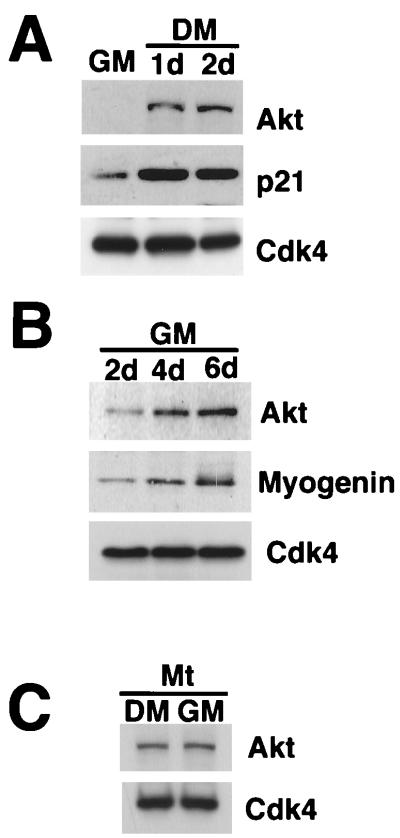
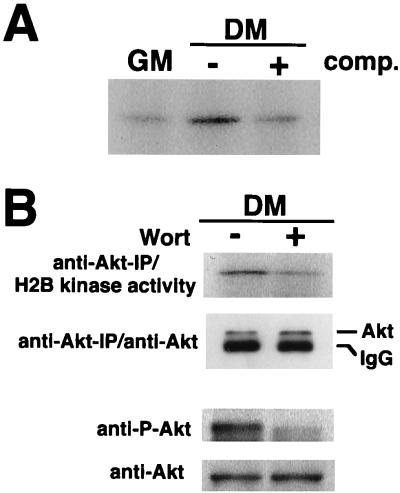
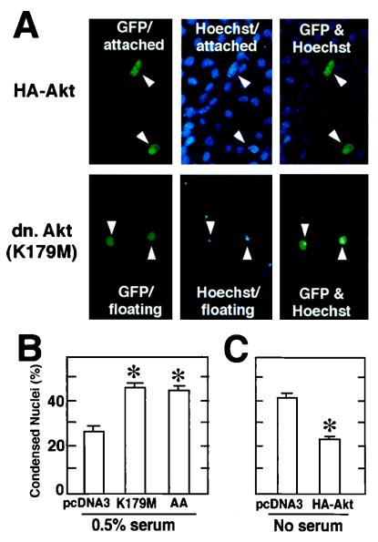
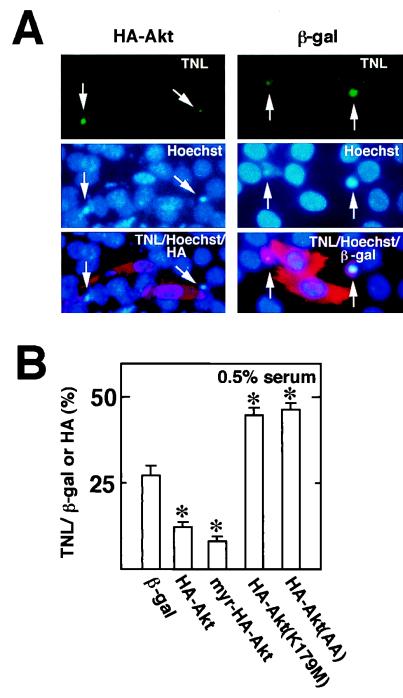
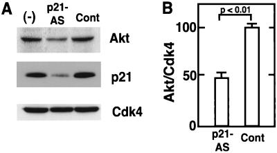
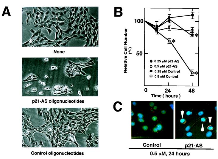
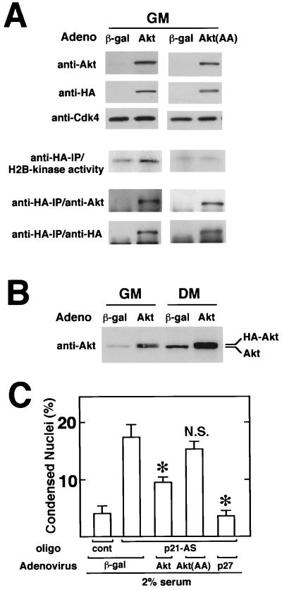
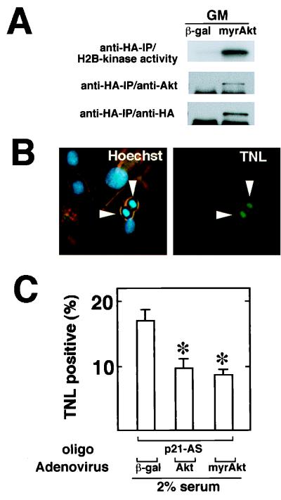
During myogenesis, proliferating myoblasts withdraw from the cell cycle, acquire an apoptosis-resistant phenotype, and differentiate into myotubes. Previous studies indicate that myogenic induction of the cyclin-dependent kinase inhibitor p21 results in an inhibition of apoptotic cell death in addition to its role as a negative cell cycle regulator. Here we demonstrate that the protein encoded by the Akt proto-oncogene is induced in C2C12 cells during myogenic differentiation with a corresponding increase in kinase activity. In differentiating cultures, expression of dominant-negative forms of Akt increase the frequency of cell death whereas expression of wild-type Akt protects against death, indicating that Akt is a positive modulator of myocyte survival. Antisense oligonucleotides against p21 block cell cycle withdrawal, inhibit Akt induction, and enhance cell death in differentiating myocyte cultures. Adenovirus-mediated transfer of wild-type or constitutively active Akt constructs confer partial resistance to cell death under conditions where cell cycle exit is blocked by the antisense oligonucleotides. Collectively, these data indicate that cell cycle withdrawal facilitates the induction of Akt during myogenesis, promoting myocyte survival.


Figures

FIG. 1
FIG. 1
Structures of replication-defective adenoviral vectors that express wild-type and mutated Akt. CMV, cytomegalovirus promoter/enhancer; SV40 pA, simian virus 40 polyadenylation site.
FIG. 2
FIG. 2
Akt is induced upon myocyte differentiation. (A) Akt protein upregulation during myogenesis induced by mitogen deprivation. C2C12 cells were cultured in growth medium (GM) or in differentiation medium (DM) for 24 h (1d) or for 48 h (2d). Cell lysates containing 20 μg protein were subjected to Western immunoblot analysis using anti-Akt, p21, and Cdk4 antibodies. (B) Akt protein upregulation induced by prolonged cultivation of confluent cultures in high-mitogen medium. C2C12 cells were cultured in growth medium (GM) for 2, 4, and 6 days. Cell lysates were prepared, and 20 μg of extract protein was immunoblotted with anti-Akt, myogenin, and Cdk4. (C) Akt protein expression is sustained in terminally differentiated cells following serum restimulation. C2C12 cells were induced to differentiate into myotubes by incubating cultures for 4 days in differentiation medium. These cultures were then cultured in fresh differentiation medium (DM) or growth medium (GM) for 24 h. Cell lysates (20 μg) were immunoblotted with anti-Akt and Cdk4 antibodies.
FIG. 3
FIG. 3
Akt kinase activity in differentiating myocyte cultures is sensitive to inhibition by wortmannin. (A) Cell lysates were prepared from C2C12 cells cultured in growth medium (GM) or differentiation medium (DM) for 48 h. Kinase activity was determined in anti-Akt immunoprecipitates, using histone H2B as a substrate. Specificity of the kinase reaction was analyzed by inclusion of 8 μg of Akt peptide (comp.) in the immunoprecipitation reaction with the extract prepared from differentiating cells. (B) Akt activity in differentiating myocytes is inhibited by wortmannin. C2C12 cells were cultured in differentiation medium (DM) for 48 h. Wortmannin (Wort) was added to the medium at final concentration of 200 nM, and cells were harvested after 1 h of incubation. Dimethyl sulfoxide was used as a vehicle at final concentration 0.1%. In the upper panel, Akt kinase activity immunoprecipitated (IP) with anti-Akt antibody was analyzed with histone H2B as a substrate. Immunoblot analysis with anti-Akt was also performed on anti-Akt-immunoprecipitated materials. In the lower panel, cell lysates were immunoblotted with anti-Akt and anti-phospho-Akt (anti-P-Akt) antibodies. IgG, immunoglobulin G.
FIG. 4
FIG. 4
Myogenic induction of Akt occurs through protein stabilization. (A) Akt mRNA is not upregulated during myogenesis. Total RNAs were prepared from the C2C12 cells in growth medium (GM) or in differentiation medium (DM) for 24 h (1d) or for 48 h (2d). Northern blot analysis was performed with a cDNA probe to the PH domain of Akt. The 28S RNA band is shown to indicate equal loading of the gel. (B) Akt protein stability increases upon myogenic differentiation. Cycloheximide was added to cultures exposed to growth medium (GM) or differentiation medium (DM) (2 days), and Western immunoblot analysis was performed on Akt and Cdc2 at the indicated time points (t). Cdc2 levels do not change significantly under the conditions of this assay (50), and it is used to control for differences in sample loading. The ratio of Akt to Cdc2 signal in the absence of cycloheximide is assigned a value of 1.0. (C) Proteasome inhibitor ALLN promotes Akt protein expression. C2C12 cells were cultured with the indicated concentrations of ALLN in growth medium for 12 h. Dimethyl sulfoxide was used as a vehicle at a final concentration of 0.1%. Cell lysates were immunoblotted with anti-Akt antibody.
FIG. 5
FIG. 5
Effects of expression of wild-type or mutant Akt on the frequency of myocytes displaying nuclear condensation during differentiation. Differentiating C2C12 cells were cotransfected with plasmids encoding dominant-negative (dn.) or wild-type Akt along with a plasmid encoding GFP as a transfection marker at a ratio of 9:1. (A) Representative photomicrographs showing adherent (attached) and floating cells in myocyte cultures transfected with the GFP expression plasmid and plasmids encoding wild-type Akt or the K179M Akt mutant. Note the condensed Hoechst 33342-stained nuclei that correspond to the floating GFP-positive cells in cultures transfected with K179M Akt. In contrast, cultures cotransfected with an expression plasmid encoding wild-type Akt revealed a high frequency of adherent GFP-positive cells with normal-appearing nuclei. (B) Dominant-negative Akt expression plasmids promote myocyte apoptosis during differentiation. Cells were cotransfected with empty vector (pcDNA3) or plasmids expressing the dominant-negative forms of Akt, using the LipofectAmine method. After transfection, cells were incubated in growth medium for 18 h and then in DMEM containing 0.5% horse serum. Low concentrations of serum were used for these assays to increase the frequency of apoptosis. After 24 h of incubation, cells were fixed and stained with Hoechst 33342 as described in Materials and Methods. The adherent and floating transfected GFP-positive cells were scored for normal or pyknotic nuclei. Data are shown as mean ± SEM (∗, P < 0.01). (C) Wild-type Akt promotes myocyte survival. Cells were cotransfected with the indicated expression plasmids and the GFP expression plasmid. After transfection and incubation in growth medium for 18 h, the medium was changed to DMEM without serum. Serum-free medium was used to maximize the number of cells undergoing apoptosis, such that the protective effects of wild-type Akt could be determined more accurately. After 24 h of incubation, the transfectants were scored as described for panel B. Data are shown as mean ± SEM (∗, P < 0.01).
FIG. 6
FIG. 6
Effects of expression of wild-type or mutant Akt on the frequency of TUNEL-positive cells during myogenic differentiation. Differentiating myocytes were transfected with plasmids encoding wild-type, constitutively active, or dominant-negative Akt tagged with HA. The plasmid encoding β-Gal (β-gal) was used as a control. After transfection, cells were incubated in growth medium for 18 h and then in DMEM containing 0.5% horse serum for 24 h. Transfected cells were identified by immunostaining with anti-HA or β-Gal antibodies. Apoptotic cells were detected by TUNEL (TNL). (A) Representative photomicrographs showing TUNEL-positive cells (arrows). Cells were treated as described above and stained by TUNEL (TNL; green), Hoechst 33342 (Hoechst; blue), and anti-HA or β-Gal antibody (HA or β-gal; red). (B) Percentage of cells that stained positive for plasmid-encoded transgene expression. For each condition, four separate cultures were transfected with the indicated plasmid, and at least 50 HA- or β-Gal-positive cells were counted on each plate. Data are shown as mean ± SEM.
FIG. 7
FIG. 7
Antisense oligonucleotides against p21 inhibit the induction of p21 and Akt during myogenic differentiation. (A) C2C12 cells were cultured in standard differentiation medium (−) or in differentiation medium containing Lipofectin with 0.5 μM p21-antisense (p21-AS) or control (Cont) oligonucleotides for 24 h. After 24 h of incubation, cells were harvested and cell lysates (20 μg protein) were immunoblotted with anti-Akt, anti-p21, and anti-Cdk4 antibodies. Representative data from one of five assays are shown. (B) Quantitative analysis of reduced Akt expression. The intensities of the Akt and Cdk4 bands in immunoblots from five independent experiments were quantified by gel densitometry. The ratios of Akt band intensity relative to Cdk4 band intensity were calculated and normalized to the ratios of band intensities in parallel cultures that were incubated in differentiation medium with no oligonucleotide or Lipofectin. Data are shown as mean ± SEM.
FIG. 8
FIG. 8
Antisense oligonucleotides against p21 promote cell death during differentiation. The cells were plated and transfected with the oligonucleotides. C2C12 cells were cultured in standard differentiation medium or in differentiation medium containing Lipofectin with the indicated oligonucleotides for 24 or 48 h. (A) Representative light photomicrographs of cultures exposed to differentiation medium in the absence (none) or presence of 0.5 μM p21-antisense (p21-AS) or control oligonucleotides for 48 h were obtained by a Nikon Diaphot light microscope at a magnification of ×100. (B) Cultures were washed with PBS and harvested by trypsinization. Cell number was determined with a hemacytometer. Data are shown as mean ± SEM of four parallel cultures that were treated according to the indicated conditions (∗, P < 0.01). (C) Representative photomicrographs showing Hoechst 33342-staining patterns of differentiating myocyte cultures exposed to 0.5 μM control or p21-antisense oligonucleotides for 24 h in medium containing 2% horse serum. Note the condensation of chromatin in cells exposed to p21-antisense oligonucleotides (arrowheads).
FIG. 9
FIG. 9
Adenovirus-mediated Akt gene transfer partially inhibits apoptosis in differentiating myocytes following p21 ablation. (A) Adeno-Akt expresses functional Akt, but the Adeno-Akt(AA)-encoded protein is kinase deficient. C2C12 cells were infected in growth medium (GM) with the adenoviral vectors expressing β-Gal or HA-tagged Akt or Akt(AA) at an MOI of 250 for 24 h prior to harvest. Cell lysates (10 μg of protein) were subjected to Western immunoblot analysis using anti-Akt, anti-HA, or anti-Cdk4 antibody. Histone H2B kinase activity in anti-HA immunoprecipitates (IP) was determined as described in Materials and Methods. Immunoblot analysis, with anti-Akt or anti-HA antibody was performed on the anti-HA-immunoprecipitated material to confirm the presence of transgene-encoded protein. (B) Akt overexpression following infection with Adeno-Akt. C2C12 cultures in growth medium were infected in parallel with Adeno-Akt or Adeno-βgal at an MOI of 250. After 15 h, the media was replaced with fresh growth medium (GM) or differentiation medium (DM) containing 2% horse serum for 24 h. Cell lysates (20 μg) were then analyzed by Western immunoblotting using anti-Akt antibody. (C) Akt expression partially inhibits p21-antisense-induced cell death. C2C12 cells were cultured in growth medium and infected with Adeno-Akt, Adeno-Akt(AA), Adeno-p27, or Adeno-βgal at an MOI of 250 for 15 h. Cultures were then transferred to 2% horse serum differentiation medium in the presence of 0.5 μM p21-antisense (p21-AS) or control (cont) oligonucleotide for 24 h. Frequency of apoptosis was determined by staining with Hoechst 33342 and scoring for nuclei with normal or condensed chromatin. Data are shown as mean ± SEM (n = 4). (∗, P < 0.01; NS, nonsignificant compared to Adeno-βgal-treated cultures exposed to p21-antisense oligonucleotides).
FIG. 10
FIG. 10
Constitutively active Akt partially inhibits apoptosis in differentiating myocytes following p21 ablation. (A) Adeno-myrAkt expresses functional Akt. C2C12 cells were infected in growth medium (GM) with the adenoviral vectors expression β-Gal or HA-tagged myrAkt at an MOI of 250 for 24 h prior to harvest. Histone H2B kinase activity in anti-HA immunoprecipitates (IP) was determined as described in Materials and Methods, and immunoblot analysis with anti-Akt and anti-HA antibodies was performed on the anti-HA-immunoprecipitated material to confirm the presence of transgene-encoded protein. (B) Analysis of apoptosis by TUNEL staining (TNL) in differentiating cultures of C2C12 cells following infection with adenoviral vectors. Note that many TUNEL-positive cells also display chromatin condensation when stained with Hoechst 33342. (C) Akt expression partially inhibits p21-antisense-induced cell death. C2C12 cells were cultured in growth media and infected with Adeno-myrAkt, Adeno-Akt, or Adeno-βgal at an MOI of 250 for 15 h. Cultures were then transferred to 2% horse serum differentiation medium in the presence of 0.5 μM p21-antisense (p21-AS) or control oligonucleotide for 24 h. Frequency of apoptosis was determined by scoring for nuclei that were TUNEL positive. Data are shown as mean ± SEM (n = 4). (∗, P < 0.01 compared to Adeno-βgal-infected cultures exposed to p21-antisense oligonucleotides.)

Similar articles

Cited by

References

    1. Alessi D R, Andjelkovic M, Caudwell B, Cron P, Morrice N, Cohen P, Hemmings B A. Mechanism of activation of protein kinase B by insulin and IGF-1. EMBO J. 1996;15:6541–6551. - PMC - PubMed
    1. Altomare D A, Guo K, Cheng J Q, Sonoda G, Walsh K, Testa J R. Cloning, chromosomal localization and expression analysis of the mouse Akt2 oncogene. Oncogene. 1995;11:1055–1060. - PubMed
    1. Altomare D A, Lyons G E, Mitsuuchi Y, Cheng J Q, Testa J R. Akt2 mRNA is highly expressed in embryonic brown fat and AKT2 kinase is activated by insulin. Oncogene. 1998;16:2407–2411. - PubMed
    1. Andrés V, Walsh K. Myogenin expression, cell cycle withdrawal and phenotypic differentiation are temporally separable events that precede cell fusion upon myogenesis. J Cell Biol. 1996;132:657–666. - PMC - PubMed
    1. Bellacosa A, Franke T F, Gonzalez-Portal M E, Datta K, Taguchi T, Gardner J, Cheng J Q, Testa J R, Tsichlis P N. Structure, expression and chromosomal mapping of c-akt: relationship to v-akt and its implications. Oncogene. 1993;8:745–754. - PubMed

Publication types

MeSH terms

LinkOut - more resources