. 1999 Jul 1;518(Pt 1):257-69.
doi: 10.1111/j.1469-7793.1999.0257r.x.

Interstitial cells of cajal generate electrical slow waves in the murine stomach

Affiliations

Interstitial cells of cajal generate electrical slow waves in the murine stomach

T Ordög et al. J Physiol. .

Abstract

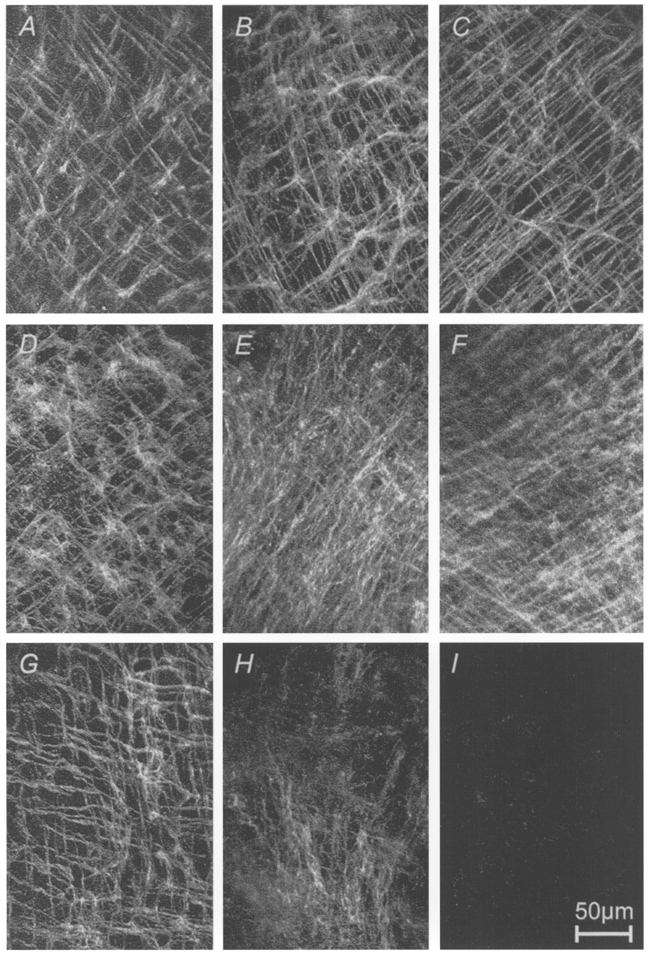
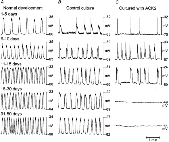
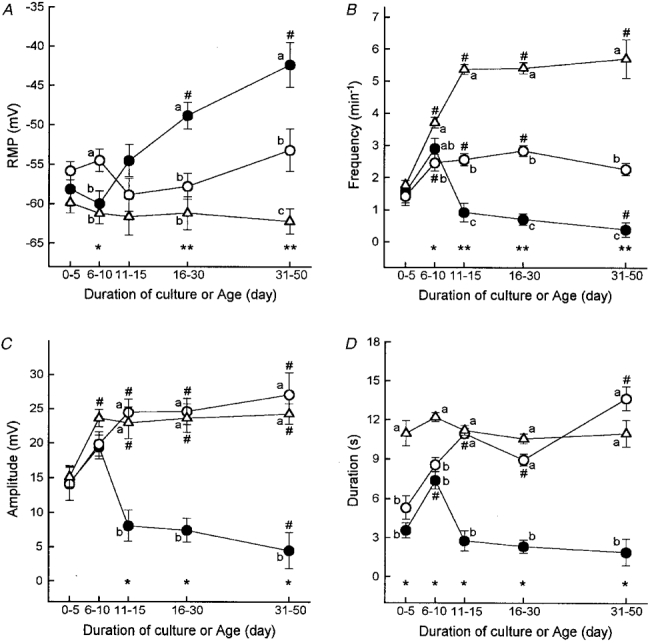
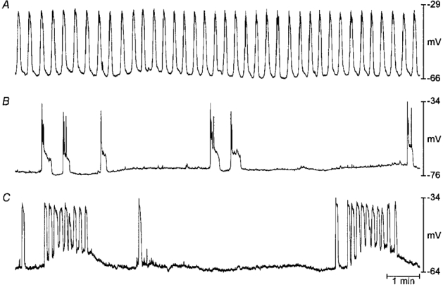
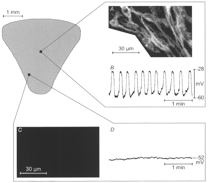
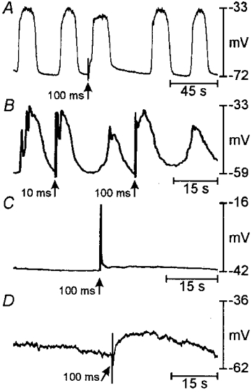
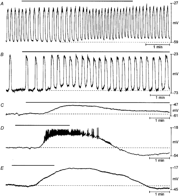
1. The gastric corpus and antrum contain interstitial cells of Cajal (ICC) within the tunica muscularis. We tested the hypothesis that ICC are involved in the generation and regeneration of electrical slow waves. 2. Normal, postnatal development of slow wave activity was characterized in tissues freshly removed from animals between birth and day 50 (D50). Slow wave amplitude and frequency increased during this period. Networks of myenteric ICC (IC-MY) were present in gastric muscles at birth and did not change significantly in appearance during the period of study as imaged by confocal immunofluorescence microscopy. 3. IC-MY networks were maintained and electrical rhythmicity developed in organ culture in a manner similar to normal postnatal development. Electrical activity was maintained for at least 48 days in culture. 4. Addition of a neutralizing antibody (ACK2) for the receptor tyrosine kinase, Kit, to the culture media caused progressive loss of Kit-immunoreactive cells. Loss of Kit-immunoreactive cells was associated with loss of slow wave activity. Most muscles became electrically quiescent after 3-4 weeks of exposure to ACK2. 5. In some muscles small clusters of Kit-immunoreactive IC-MY remained after culturing with ACK2. These muscles displayed slow wave activity but only in the immediate regions in which Kit-positive IC-MY remained. These data suggest that regions without Kit-immunoreactive cells cannot generate or regenerate slow waves. 6. After loss of Kit-immunoreactive cells, the muscles could not be paced by direct electrical stimulation. Stimulation with acetylcholine also failed to elicit slow waves. The data suggest that the generation of slow waves is an exclusive property of IC-MY; smooth muscle cells may not express the ionic apparatus necessary for generation of these events. 7. We conclude that IC-MY are an essential element in the spontaneous rhythmic electrical and contractile activity of gastric muscles. This class of ICC appears to generate slow wave activity and may provide a means for regeneration of slow waves.


Figures

Figure 1
Figure 1. Effects of neutralizing Kit antibodies on ICC networks of the murine gastric corpus and antrum
ICC networks in gastric muscle preparations were visualized by Kit immunohistochemistry and confocal microscopy. A-C, tissues freshly removed from normal mice on D1, D13 and D18. D-F, muscles removed from newborn mice (<= 1 day of postnatal age) and cultured for 0, 13 and 21 days under control conditions; G-I, muscles removed from newborn mice and cultured in the presence of neutralizing Kit antibodies for 0, 9 and 43 days. ICC networks were fully developed at birth in each animal (see A, D and G). Culturing did not appear to significantly alter the structure or distribution of ICC networks. Inclusion of neutralizing Kit antibody (ACK2) caused a time-dependent loss of ICC in the cultured muscles. Scale bar is 50 μm and applies to each panel.
Figure 2
Figure 2. Effects of neutralizing Kit antibody on gastric slow waves
Representative electrical activity recorded from muscles freshly removed from animals on various days after birth (as noted) and from muscles cultured for various periods in the absence and presence of ACK2. (Electrical recordings were made from cultured muscles at the same time periods as the records from freshly dissected muscles in A.) At birth regular slow wave activity was recorded in gastric muscles. The frequency increased with age. A similar trend was observed in cultured muscles (B); however, the maximum frequency did not reach the same level as in normal development (see text and Fig. 3 for details). In the presence of ACK2, slow waves began to develop, but in many muscles slow waves disappeared and the muscles became electrically quiescent (C). Loss of slow waves corresponded to the loss of ICC as shown in Fig. 1.
Figure 3
Figure 3. Summary of the effects of ACK2 on electrical and slow wave parameters
Resting membrane potential (RMP; A), and frequency (B), amplitude (C) and duration (D) of electrical slow waves recorded in freshly dissected gastric muscles during normal postnatal development (▵) and in preparations cultured from birth in the absence (○) or presence of Kit antibodies (•). Data were grouped into time bins, and data points in each panel are means ±s.e.m. from an average of 23 cells (range, 11-36) of at least 5 tissues from each time bin. # Significant difference from the first time bin (days 0-5) in each group of muscles (using one-way ANOVA followed by Bonferroni's t test or Kruskal-Wallis ANOVA on ranks followed by Dunn's test; ANOVA P values: A, ACK2-treated cultures, P < 0.001; B, normal development and ACK2-treated cultures, P < 0.001; control cultures, P < 0.003; C, all groups, P < 0.001; D, control and ACK2-treated cultures, P < 0.001; in multiple comparisons P < 0.05 was considered statistically significant). Asterisks indicate statistical significance between groups of muscles within the same time bins (one-way ANOVA followed by Student-Newman-Keuls test or Kruskal-Wallis ANOVA on ranks followed by Dunn's test). ANOVA P values were: A: *P < 0.005, **P < 0.001; in B: *P < 0.002, **P < 0.001; and in C and D: *P < 0.001. Data points within time bins designated by different lower case letters are significantly different (P < 0.05 by multiple comparisons; for example, a is different from b, and a and b are different from c).
Figure 4
Figure 4. Arrhythmic slow wave activity in gastric muscles cultured with neutralizing Kit antibodies
A shows typical slow wave activity recorded from a gastric muscle preparation cultured under control conditions for 15 days beginning at birth. B and C show patterns of arrhythmic slow wave activity recorded from muscles cultured with neutralizing Kit antibodies for 12 and 28 days, respectively.
Figure 5
Figure 5. Correlation between Kit-like immunoreactivity and electrical slow wave activity
Kit-LI (A and C) and intracellular electrical activity (B and D) from a gastric muscle preparation cultured with neutralizing Kit antibodies for 41 days beginning at birth. Slow waves were recorded from a small area in the mid-corpus and the co-ordinates were recorded as a percentage of length and width of the tissue with a micrometer eyepiece. The muscles were then fixed and processed for immunohistochemistry. The region from which slow waves were recorded also displayed Kit-LI. Other areas, including that shown in the antrum in C and D, were electrically quiescent and lacked Kit-LI.
Figure 6
Figure 6. Effects of ACK2 on responses to electrical field stimulation
Examples of responses to single square-wave electrical pulses (150 V, 10-100 ms). In A, slow waves were evoked prematurely by 100 ms pulses in a freshly dissected muscle obtained from a D10 mouse. B shows a record from a muscle cultured under control conditions from birth for 21 days. Premature slow waves were evoked by 100 ms pulses. C and D show records from muscles cultured from birth for 12 and 18 days, respectively, with ACK2. These muscles were electrically quiescent and 100 ms pulses (and longer pulses up to 500 ms) failed to evoke slow waves.
Figure 7
Figure 7. Effects of ACK2 on responses of gastric muscle to pharmacological stimulation
Examples of responses to 10−5 M acetylcholine (A-D) or to 100 mM KCl (E) in gastric muscle preparations. A shows the response of a freshly dissected muscle obtained from a 44-day-old mouse exposed to ACh (10−5 M; black bar). Resting membrane potential depolarized (note dotted line) and slow wave frequency was increased in response to ACh. B shows the response of a muscle cultured under control conditions beginning at birth for 13 days to ACh (10−5 M). Resting membrane potential was depolarized (dotted line) and the frequency of slow waves increased in response to ACh. C and D show responses of muscles cultured with ACK2 for 19 and 18 days, respectively. In these electrically quiescent muscles ACh (10−5 M) depolarized resting membrane potential but never elicited slow wave activity. In the example in D, brief, spike-like activity was superimposed upon the tonic depolarization. In E a muscle treated with ACK2 for 41 days was exposed to elevated external K+. This also caused tonic depolarization without stimulating oscillatory slow wave activity.

Similar articles

Cited by

References

    1. Berezin I, Huizinga JD, Daniel EE. Interstitial cells of Cajal in the canine colon: a special communication network at the inner border of the circular muscle. Journal of Comparative Neurology. 1988;273:42–51. - PubMed
    1. Burns AJ, Herbert TM, Ward SM, Sanders KM. Interstitial cells of Cajal in the guinea-pig gastrointestinal tract as revealed by c-Kit immunohistochemistry. Cell and Tissue Research. 1997;290:11–20. - PubMed
    1. Burns AJ, Lomax AEJ, Torihashi S, Sanders KM, Ward SM. Interstitial cells of Cajal mediate inhibitory neurotransmission in the stomach. Proceedings of the National Academy of Sciences of the USA. 1996;93:12008–12013. - PMC - PubMed
    1. Christensen J, Rick GA, Lowe LS. Distributions of interstitial cells of Cajal in stomach and colon of cat, dog, ferret, opossum, rat, guinea pig and rabbit. Journal of the Autonomic Nervous System. 1992;37:47–56. 10.1016/0165-1838(92)90144-6. - DOI - PubMed
    1. Connor JA, Connor C, Kreulen DL, Prosser CL, Weems WA. Pacemaker activity and propagation in intestinal smooth muscle. In: Worcel M, Vassort G, editors. Smooth Muscle Pharmacology and Physiology. Paris: INSERM; 1976. pp. 285–300.

Publication types

MeSH terms

Substances

LinkOut - more resources