. 1999 Jun 10;399(6736):597-601.
doi: 10.1038/21218.

Regulation of endothelium-derived nitric oxide production by the protein kinase Akt

Affiliations

Regulation of endothelium-derived nitric oxide production by the protein kinase Akt

D Fulton et al. Nature. .

Erratum in

  • Nature 1999 Aug 19;400(6746):792

Abstract

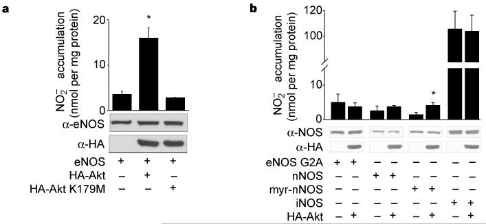
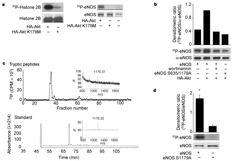
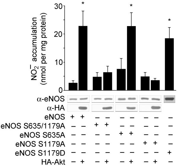
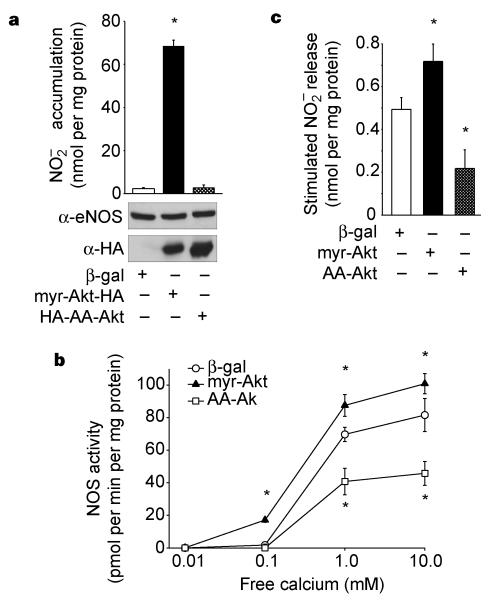
Endothelial nitric oxide synthase (eNOS) is the nitric oxide synthase isoform responsible for maintaining systemic blood pressure, vascular remodelling and angiogenesis. eNOS is phosphorylated in response to various forms of cellular stimulation, but the role of phosphorylation in the regulation of nitric oxide (NO) production and the kinase(s) responsible are not known. Here we show that the serine/threonine protein kinase Akt (protein kinase B) can directly phosphorylate eNOS on serine 1179 and activate the enzyme, leading to NO production, whereas mutant eNOS (S1179A) is resistant to phosphorylation and activation by Akt. Moreover, using adenovirus-mediated gene transfer, activated Akt increases basal NO release from endothelial cells, and activation-deficient Akt attenuates NO production stimulated by vascular endothelial growth factor. Thus, eNOS is a newly described Akt substrate linking signal transduction by Akt to the release of the gaseous second messenger NO.


Figures

Figure 1
Figure 1
Wild-type Akt, but not kinase-inactive Akt, increases NO release from cells expressing membrane-associated eNOS. a, COS cells were transfected with plasmids for eNOS, with or without Akt or kinase-inactive Akt (K179M). The production of NO (assayed as NO2) was determined by chemiluminescence. b, COS cells were transfected with various NOS plasmids as above. In both a and b, values for NO2 production were subtracted from levels obtained from cells transfected with the β-galactosidase cDNA only. The insets show the expression of proteins in total cell lysates. Data were mean ± s.e.m., n = 3-7 experiments; asterisk denotes P < 0.05.
Figure 2
Figure 2
Phosphorylation of eNOS by active Akt in vitro and in vivo. a, COS cells were transfected with HA-Akt or HA-Akt(K179M) and lysates were immunoprecipitated and placed into an in vitro kinase reaction with histone 2B (25 μg) or recombinant eNOS (3 μg) as substrates. Top panel: incorporation of 32P into the substrates; Bottom panel: amount of substrate by Coomassie staining of the gel. b, 32P-labelled wild-type or double-serine mutant of eNOS (eNOS S635/1179A) was affinity purified from transfected COS cells and subjected to autoradiography (upper panel) or western blotting (lower panel). The histogram shows the relative amount of labelled protein to the amount of immunoreactive eNOS in the gel. c, Labelled eNOS was digested with trypsin and peptides separated by RP-HPLC. The upper panel shows a predominant labelled tryptic peptide that comigrates with the unlabelled synthetic phosphopeptide standard (bottom panel). Insets: linear mode MS of labelled peptide (top) and phosphopeptide standard showing identical mass ions (bottom). d, Recombinant wild-type eNOS or eNOS S1179A were purified and equal amounts (2.4 μg) placed into an in vitro kinase reaction with recombinant Akt (see Methods). Top panel: the incorporation of 32P into eNOS; bottom panel; amount of substrate by Coomassie staining of the gel. Histogram (n = 3) reflects the relative amount of labelled eNOS to the mass of eNOS (Coomassie) in the in vitro kinase reaction.
Figure 3
Figure 3
Evidence that Ser 1179 is functionally important for Akt-stimulated NO release. COS cells were transfected with plasmids for wild-type eNOS or eNOS mutants, in the absence or presence of Akt, and the expression of the proteins and production of NO (assayed as NO2) were determined. Constructs with the S1179A mutation were not activated by Akt; the S1179D mutation resulted in a gain of function. Data are mean ± s.e.m. of 4-7 experiments; asterisks represent significant differences (P < 0.05).
Figure 4
Figure 4
Akt regulates the basal and stimulated production of NO in endothelial cells. a, BLMVEC were infected with adenoviral constructs (β-gal as control, myr-Akt and AA-Akt) and the amount of NO2 produced over 24 h was determined (n = 3). Inset: expression of eNOS and Akt. b, Lysates from adenovirus-infected BLMVEC were examined for NOS activity. Equal amounts of protein (50 μg) were incubated with various concentrations of free calcium, and the NOS activity was determined (n = 3 experiments). c, BLMVEC were infected with adenoviruses as above followed by stimulation with VEGF (40 ng per ml) for 30 min and NO2 release was quantified by chemiluminescence. Data are presented as VEGF-stimulated release of NO2 after subtraction of basal levels; mean ± s.e.m., n = 4; asterisks represent significant differences (P < 0.05).

References

    1. Shesely EG, et al. Elevated blood pressures in mice lacking endothelial nitric oxide synthase. Proc. Natl Acad. Sci. USA. 1996;93:13176–13181. - PMC - PubMed
    1. Huang PL, et al. Hypertension in mice lacking the gene for endothelial nitric oxide synthase. Nature. 1995;377:239–242. - PubMed
    1. Rudic RD, et al. Direct evidence for the importance of endothelium-derived nitric oxide in vascular remodeling. J. Clin. Invest. 1998;101:731–736. - PMC - PubMed
    1. Murohara T, et al. Nitric oxide synthase modulates angiogenesis in response to tissue ischemia. J. Clin. Invest. 1998;101:2567–2578. - PMC - PubMed
    1. Michel T, Li GK, Busconi L. Phosphorylation and subcellular translocation of endothelial nitric oxide synthase. Proc. Natl Acad. Sci. USA. 1993;90:6252–6256. - PMC - PubMed

Publication types