. 1999 Jul;67(7):3199-206.
doi: 10.1128/IAI.67.7.3199-3206.1999.

Survival of Mycobacterium avium and Mycobacterium tuberculosis in acidified vacuoles of murine macrophages

Affiliations

Survival of Mycobacterium avium and Mycobacterium tuberculosis in acidified vacuoles of murine macrophages

M S Gomes et al. Infect Immun. 1999 Jul.

Abstract

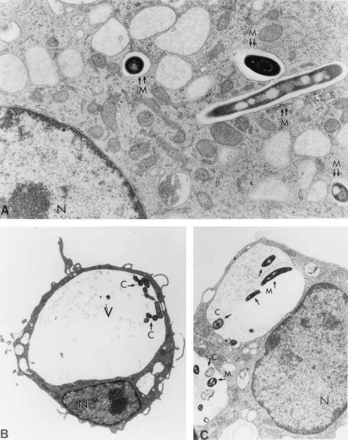
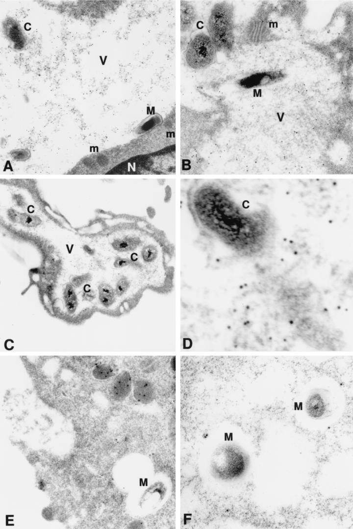
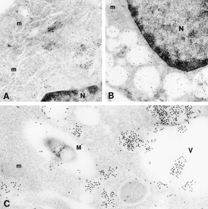
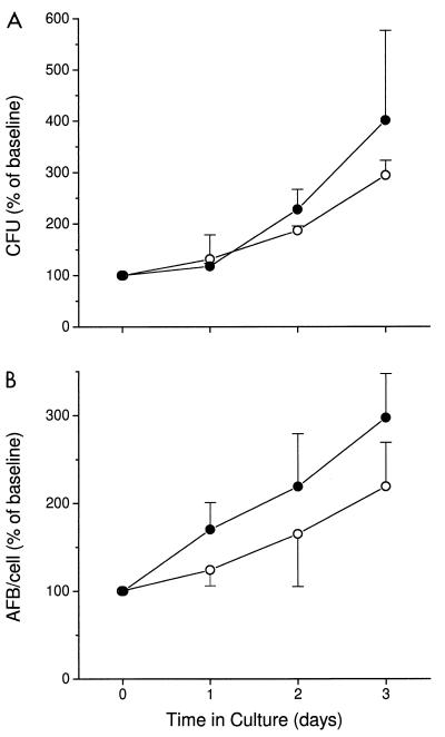
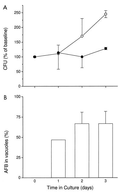
Despite the antimicrobial mechanisms of vertebrate phagocytes, mycobacteria can survive within the phagosomes of these cells. These organisms use various strategies to evade destruction, including inhibition of acidification of the phagosome and inhibition of phagosome-lysosome fusion. In contrast to mycobacteria, Coxiella burnetii, the etiologic agent of Q fever, inhabits a spacious acidified intracellular vacuole which is prone to fusion with other vacuoles of the host cell, including phagosomes containing mycobacteria. The Coxiella-infected cell thus provides a unique model for investigating the survival of mycobacteria in an acidified phagosome-like compartment. In the present study, murine bone marrow-derived macrophages were infected with either Mycobacterium avium or Mycobacterium tuberculosis and then coinfected with C. burnetii. We observed that the majority of phagocytosed mycobacteria colocalized to the C. burnetii-containing vacuole, which maintained its acidic properties. In coinfected macrophages, the growth of M. avium was not impaired following fusion with the acidified vacuole. In contrast, the growth rate of M. tuberculosis was reduced in acidified vacuoles. These results suggest that although both species of mycobacteria inhibit phagosome-lysosome fusion, they may be differentially susceptible to the toxic effects of the acidic environment in the mature phagolysosome.


Figures

FIG. 1
FIG. 1
(A) Electron micrograph of murine BMM infected with M. avium (M). Note the tight vacuole containing the bacterium (double arrows). Magnification, ×19,800. (B) Electron micrograph of murine BMM infected with C. burnetii (C). Note the large vacuole (V) occupying almost the entire cytoplasm of the cell and eccentric nucleus (N). Magnification, ×3,300. (C) Electron micrograph at 24 h after coinfection of murine BMM with M. avium and C. burnetii. Note colocalization of the two organisms in the same large vacuole (arrows point to the organisms). Magnification, ×8,300.
FIG. 2
FIG. 2
Electron micrograph of murine BMM infected with M. avium (M) and/or C. burnetii (C) and stained with DAMP and gold-conjugated antibody. (A and B) Coinfected cells with vacuoles containing M. avium and C. burnetii; (C and D) cells with vacuole containing C. burnetii only; (E and F) cells with vacuoles containing M. avium only. Note presence of gold particles, indicating the low-pH environment within the vacuoles (V) occupied by the two organisms and by C. burnetii only. Only a few gold particles are visible within the vacuoles containing M. avium only and in the nucleus (N) or mitochondria (m) of the cells. Magnifications, ×30,000 (A), ×30,000 (B), ×16,000 (C), ×86,000 (D), ×46,000 (E), and ×32,000 (F). Panel D is a higher magnification of panel C.
FIG. 3
FIG. 3
Electron micrograph of murine BMM stained with DAMP and gold-conjugated antibody. (A) Uninfected cell (24 h in culture) with small vacuoles (lysosomes) containing gold particles; (B) uninfected cell (5 days in culture) with larger vacuoles (secondary lysosomes) containing gold particles; (C) cell coinfected with C. burnetii (not seen in the figures) and M. avium (M). Vacuoles containing gold particles are seen fusing with the large vacuole (V) containing C. burnetii but are not seen fusing with the vacuole containing M. avium. Gold particles are not seen in the nucleus (N) or mitochondria (m) of the cells. Magnifications, ×43,000 (A), ×40,000 (B), ×48,000 (C).
FIG. 4
FIG. 4
Growth of M. avium in murine BMM (open circles) and in cells coinfected with C. burnetii (closed circles). BMM were infected with M. avium and then coinfected with C. burnetii on day 4 (designated in the graph as time zero). Growth of M. avium was evaluated by number of CFU (A) and by direct counting of AFB by light microscopy (B). Results are expressed as mean percentages of baseline values for three independent experiments. Error bars indicate standard deviations. Baseline values (measured at time zero) were (55 ± 11) × 104 and (56 ± 6) × 104 CFU per cell and 8.2 and 7.4 AFB per cell for M. avium alone and M. avium with C. burnetii, respectively. By an independent t test, P was >0.05 at all time points.
FIG. 5
FIG. 5
(A) Growth of M. tuberculosis in murine BMM (open circles) and in cells coinfected with C. burnetii (closed circles). BMM were infected with M. tuberculosis and then coinfected with C. burnetii on day 4 (designated in the graph as time zero). (B) Percent of M. tuberculosis colocalized within the C. burnetii-containing vacuole. Results are expressed as mean percentages of baseline values for three independent experiments. Error bars indicate standard deviations. Baseline values (measured at time zero) were (3.3 ± 0.8) × 104 and (4.6 ± 1.6) × 104 CFU for M. tuberculosis alone and for M. tuberculosis with C. burnetii, respectively. By an independent t test, P was 0.02 on day 3.

References

    1. Anderson R G W, Falck J R, Goldstein J L, Brown M S. Visualization of acidic organelles in intact cells by electron microscopy. Proc Natl Acad Sci USA. 1984;81:4838–4842. - PMC - PubMed
    1. Appelberg R, Sarmento A M. The role of macrophage activation and Bcg-encoded function(s) in the control of Mycobacterium avium infection in mice. Clin Exp Immunol. 1990;88:324–331. - PMC - PubMed
    1. Appelberg R, Orme I M. Effector mechanisms involved in cytokine-mediated bacteriostasis of Mycobacterium avium infections in murine macrophages. Immunology. 1993;80:352–359. - PMC - PubMed
    1. Armstrong J A, Hart P D. Response of cultured macrophages to mycobacterium tuberculosis with observation on fusion of lysosomes with phagosomes. J Exp Med. 1971;134:713–740. - PMC - PubMed
    1. Armstrong J A, Hart P D. Phagosome-lysosome interactions in cultured macrophages infected with virulent tubercle bacilli. Reversal of the usual nonfusion pattern and observations on bacterial survival. J Exp Med. 1975;142:1–16. - PMC - PubMed

Publication types

LinkOut - more resources