. 1999 Jul;67(7):3267-75.
doi: 10.1128/IAI.67.7.3267-3275.1999.

Innate antimicrobial activity of nasal secretions

Affiliations

Innate antimicrobial activity of nasal secretions

A M Cole et al. Infect Immun. 1999 Jul.

Abstract

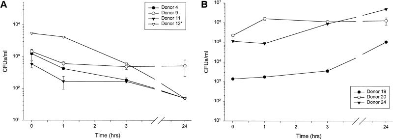
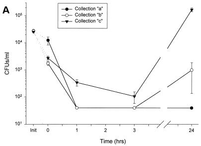
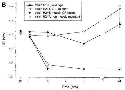
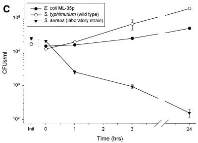
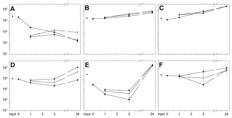
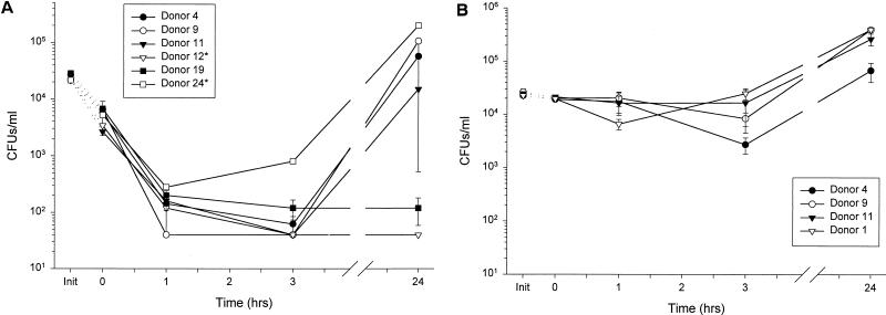
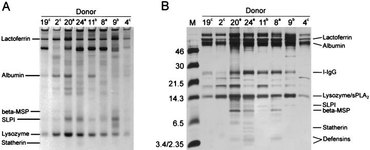
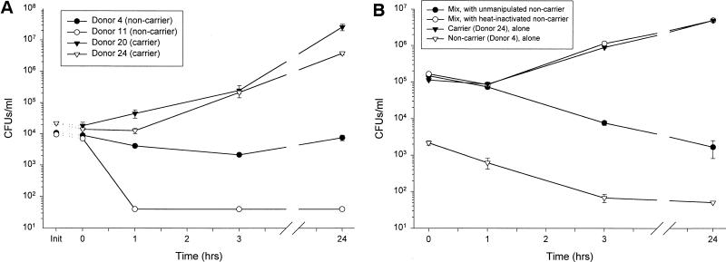
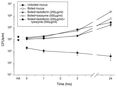
Minimally manipulated nasal secretions, an accessible form of airway surface fluid, were tested against indigenous and added bacteria by using CFU assays. Antimicrobial activity was found to vary between donors and with different target bacteria and was markedly diminished by dilution of the airway secretions. Donor-to-donor differences in electrophoresis patterns of nasal secretions in sodium dodecyl sulfate-polyacrylamide gel electrophoresis (PAGE) and acid urea-PAGE analyses were readily observed, suggesting that polymorphic genes encode the secreted proteins. Three donors (of twenty-four total), whose nasal fluid yielded similar protein band patterns and did not kill indigenous bacteria, were determined to be heavy nasal carriers of Staphylococcus aureus. Their fluid was deficient in microbicidal activity toward a colonizing strain of S. aureus but the defect was corrected in vitro by a 1:1 addition of nasal fluid from noncarriers. The microbicidal activity of normal fluid was inactivated by heating it for 10 min to 100 degrees C and could not be restored solely by the addition of two major nasal antimicrobial proteins, lysozyme and lactoferrin. Several other known antimicrobial proteins and peptides, including statherin, secretory phospholipase A2, and defensins, were identified in nasal secretions and likely contribute to their total antimicrobial properties. Nasal fluid may serve as a useful model for the analysis of lower-airway secretions and their role in host defense against airway colonization and pulmonary infections.


Figures

FIG. 1
FIG. 1
The fate of indigenous bacteria in minimally manipulated nasal secretions. (A) The majority of nasal fluid samples were inhibitory to indigenous microbes. (B) Nasal fluid samples from a minority of donors were permissive for indigenous microbes. Asterisk indicates n = 1, due to insufficient amounts of nasal secretions from the indicated donors.
FIG. 2
FIG. 2
Susceptibility of various strains of bacteria to a single donor’s nasal fluid. Nasal secretions from donor 4 were subjected to a modified CFU microassay of nasal fluid targeting four strains of P. aeruginosa, E. coli ML-35p, S. typhimurium wild type, and S. aureus clinical isolate. (A) Secretions tested at three different times showed similar activities against P. aeruginosa CF between 0 and 3 h but varied in the amount of regrowth at 24 h. (B) Fluid incubated with four different strains of P. aeruginosa showed strain-specific antimicrobial activity. (C) Nasal secretions permitted the slow growth of E. coli and S. typhimurium; however, S. aureus was effectively cleared. Init, inoculum (input) at time zero for each sample tested, without the addition of nasal fluid.
FIG. 2
FIG. 2
Susceptibility of various strains of bacteria to a single donor’s nasal fluid. Nasal secretions from donor 4 were subjected to a modified CFU microassay of nasal fluid targeting four strains of P. aeruginosa, E. coli ML-35p, S. typhimurium wild type, and S. aureus clinical isolate. (A) Secretions tested at three different times showed similar activities against P. aeruginosa CF between 0 and 3 h but varied in the amount of regrowth at 24 h. (B) Fluid incubated with four different strains of P. aeruginosa showed strain-specific antimicrobial activity. (C) Nasal secretions permitted the slow growth of E. coli and S. typhimurium; however, S. aureus was effectively cleared. Init, inoculum (input) at time zero for each sample tested, without the addition of nasal fluid.
FIG. 3
FIG. 3
Effect of fluid dilution on microbicidal activity. Six strains of bacteria were subjected to a CFU microassay of undiluted, 1:1-diluted, and 1:2-diluted minimally manipulated nasal fluid from donor 4. The CFU counts of S. aureus (clinical isolate) (A), E. coli (B), and S. typhimurium (C) were little affected. The antimicrobial effects of nasal secretions on S. aureus (isolate from nasal carrier donor 24) (D), P. aeruginosa CF (E), and P. aeruginosa H103 (F) were decreased with increasing dilution. The effects of undiluted, 1:1-diluted, and 1:2-diluted secretions are represented by closed circles, open triangles, and closed squares, respectively. The input inoculum (closed triangles) is shown for each graph.
FIG. 4
FIG. 4
Susceptibility of P. aeruginosa to multiple donors’ nasal fluids. Activities of multiple donors’ nasal secretions were tested individually against two strains of P. aeruginosa. Different donors’ fluids were tested in a CFU microassay against a cystic fibrosis clinical isolate (A) and the wild-type strain H103 (B). Init, inoculum (input) at time zero for each donor tested, without the addition of nasal fluid. Asterisks indicate n = 1, due to scant fluid from the indicated donors.
FIG. 5
FIG. 5
Donor-to-donor variability in the protein pattern of nasal secretions. (A) AU-PAGE of eight donor’s nasal secretions shows variations in lactoferrin, albumin, β-microseminoprotein (beta-MSP), SLPI, lysozyme, and statherin concentrations. (B) Reducing Tricine-SDS-PAGE of eight donors’ nasal secretions shows the variability in low-molecular-weight proteins. Superscripts indicate three donor groupings based on protein band similarities. The positions of identified components of nasal secretions are shown at the gel margins.
FIG. 6
FIG. 6
Nasal carriers of S. aureus are deficient in antimicrobial components that are restored by the addition of noncarrier secretions. (A) S. aureus, isolated from donor 24, was used as the target bacterium in a CFU microassay of nasal fluid from two noncarriers (donors 4 and 11) and two carriers (donors 20 and 24, gamma-irradiated fluid). While the noncarriers’ fluids were bacteriostatic or bactericidal, the carriers’ fluids did not inhibit bacterial growth. (B) The 1:1 mix of nasal secretions from an S. aureus carrier (donor 24) with fluid from a noncarrier (donor 4) is bactericidal to indigenous flora, while the mix of nasal secretions from an S. aureus carrier with heat-inactivated (boiling for 10 min) secretions from a noncarrier promoted the growth of indigenous bacteria.
FIG. 7
FIG. 7
Physiological concentrations of lysozyme and lactoferrin added to heat-inactivated nasal fluid (donor 4) did not restore microbicidal activity. While nonboiled fluid exhibited the expected bactericidal activity, heat-inactivated (boiling for 10 min) did not inhibit CFU growth. Physiological concentrations of lactoferrin and lysozyme, added separately and concurrently to heat-inactivated secretions, did not restore the antimicrobial activity.

Similar articles

Cited by

References

    1. Aho H J, Grenman R, Sipila J, Peuravuori H, Hartikainen J, Nevalainen T J. Group II phospholipase A2 in nasal fluid, mucosa and paranasal sinuses. Acta Otolaryngol. 1997;117:860–863. - PubMed
    1. Azen E A. Genetics of salivary protein polymorphisms. Crit Rev Oral Biol Med. 1993;4:479–485. - PubMed
    1. Biesbrock A R, Reddy M S, Levine M J. Interaction of a salivary mucin-secretory immunoglobulin A complex with mucosal pathogens. Infect Immun. 1991;59:3492–3497. - PMC - PubMed
    1. Brogden K A, Ackermann M, Huttner K M. Detection of anionic antimicrobial peptides in ovine bronchoalveolar lavage fluid and respiratory epithelium. Infect Immun. 1998;66:5948–5954. - PMC - PubMed
    1. Ellison R T, III, Giehl T J. Killing of gram-negative bacteria by lactoferrin and lysozyme. J Clin Invest. 1991;88:1080–1091. - PMC - PubMed

Publication types

MeSH terms

Substances