. 1999 Jul;67(7):3357-66.
doi: 10.1128/IAI.67.7.3357-3366.1999.

Role of hemolysin BL in the pathogenesis of extraintestinal Bacillus cereus infection assessed in an endophthalmitis model

Affiliations

Role of hemolysin BL in the pathogenesis of extraintestinal Bacillus cereus infection assessed in an endophthalmitis model

M C Callegan et al. Infect Immun. 1999 Jul.

Abstract

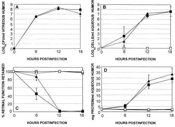
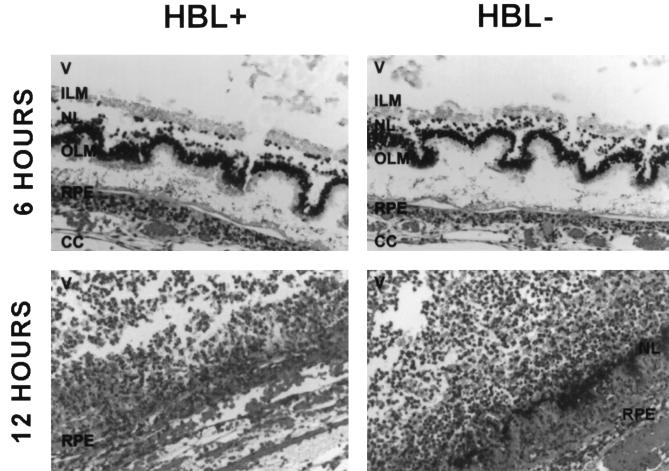
Bacillus cereus is a rare cause of serious human infection but, paradoxically, causes one of the most severe posttraumatic or endogenous infections of the eye, endophthalmitis, which frequently results in blindness. The virulence of B. cereus endophthalmitis historically has been attributed to toxin production. We therefore sought to examine the contribution of the dermonecrotic toxin, hemolysin BL, to the pathogenesis of B. cereus infection in an endophthalmitis system that is highly amenable to study. The pathogenesis of infection resulting from intravitreal injection of 10(2) CFU of either a clinical ocular isolate of B. cereus producing hemolysin BL (HBL+) or an isogenic mutant in this trait (HBL-) was assessed bacteriologically and by slit lamp biomicroscopy, electroretinography, histology, and inflammatory cell enumeration. Both HBL+ and HBL- strains evoked severe intraocular inflammatory responses as early as 12 h postinfection, with complete loss of retinal responsiveness by 12 h. The infections caused by both strains spread of the infection to adjacent tissues by 18 h. No significant differences in intraocular bacterial growth (P >/= 0.21) or inflammatory changes (P >/= 0.21) were observed in eyes infected with either HBL+ or HBL- strains during the course of infection. The level of retinal responsiveness was greater in HBL- infected eyes than in HBL+-infected eyes at 6 h only (P = 0.01). These results indicate that hemolysin BL makes no essential contribution to the severe and rapid course of infection in the endophthalmitis model.


Figures

FIG. 1
FIG. 1
Construction of pΔHBL for insertional mutagenesis of B. cereus hemolysin BL. Relevant restriction sites are shown. (A) A 3.3-kb PCR fragment of hemolysin BL was ligated into the multicloning region of pKRX, creating pKRX-hbl. The ermAM gene of pUC-ermAM was ligated to the 4.7-kb Eco47III/PstI fragment of pKRX-hbl to create pKRX-hbl::ermR. (B) The multicloning region of p3ERM was ligated to an inverse PCR fragment of pTV1OK containing the temperature-sensitive replicon from pWVO1 and a Kanr marker, to create pCASPER. (C) The EcoRI/PstI fragment of pKRX-hbl::ermR containing the disrupted hemolysin BL operon was ligated into the multicloning region of pCASPER to create pΔHBL.
FIG. 1
FIG. 1
Construction of pΔHBL for insertional mutagenesis of B. cereus hemolysin BL. Relevant restriction sites are shown. (A) A 3.3-kb PCR fragment of hemolysin BL was ligated into the multicloning region of pKRX, creating pKRX-hbl. The ermAM gene of pUC-ermAM was ligated to the 4.7-kb Eco47III/PstI fragment of pKRX-hbl to create pKRX-hbl::ermR. (B) The multicloning region of p3ERM was ligated to an inverse PCR fragment of pTV1OK containing the temperature-sensitive replicon from pWVO1 and a Kanr marker, to create pCASPER. (C) The EcoRI/PstI fragment of pKRX-hbl::ermR containing the disrupted hemolysin BL operon was ligated into the multicloning region of pCASPER to create pΔHBL.
FIG. 2
FIG. 2
(A) PCR analysis of wild-type and hemolysin BL-deficient strains. Samples of chromosomal DNA from strains MGBC145 (lane 1) and CJ145-1.1 (lane 2) were used to amplify hemolysin BL. PCR products were of the predicted sizes (indicated by lines on the left). (B) Chromosomal DNA from strains MGBC145 (lanes 1 and 3) and CJ145-1.1 (lanes 2 and 4) were digested with EcoRI, and fragments were separated on a 0.8% agarose gel. Lanes 1 and 2 were probed with 33P-labeled pKRX-hbl. Lanes 3 and 4 were probed with 33P-labeled pUC18-ermAM. MW, molecular weight markers.
FIG. 3
FIG. 3
Phenotypic analysis of wild-type and hemolysin BL-deficient strains. (A) Single colonies of MGBC145 (plate 1) and CJ145-1.1 (plate 2) were isolated on 2.5% sheep erythrocyte agar. Presence of the discontinuous zone of hemolysis (as seen in plate 1) indicated hemolysin BL activity. (B) Concentrated supernatants of MGBC145 (sample 1), uninoculated BHI (sample 2), or CJ145-1.1 (sample 3) were injected intradermally. Presence of a necrotic center surrounded by a blue (dark) zone (as seen in sample 1) indicated hemolysin BL activity.
FIG. 4
FIG. 4
Comparison of experimental B. cereus endophthalmitis initiated by wild type (HBL+) and isogenic HBL strains. Inocula of 102 CFU of either the HBL+ (■) or HBL (▴) strain were injected intravitreally, and infections were assessed at various times by bacterial enumeration (A), inflammatory cell enumeration in aqueous humor (B), ERG (C), and total protein concentration in aqueous humor (D). Also included were sham-injected (BHI [□] or BHI supplemented with 25 μg of erythromycin per ml [▵]) and uninjected (○) controls. All values represent the mean ± standard deviation for ≥4 eyes per group.
FIG. 5
FIG. 5
Retinal sections of eyes intravitreally infected with B. cereus MGBC145 (HBL+) or CJ145-1.1 (HBL) at 6 and 12 h postinfection. All sections were stained with hematoxylin and eosin. Photoreceptor layer folding and B. cereus residing between retinal layers were observed at 6 h. Retinal layers were virtually indistinguishable at 12 h. V, vitreous; ILM, inner limiting membrane; NL, nuclear layer; OLM, outer limiting membrane; RPE, retinal pigment epithelium; CC, choriocapillaris. Magnification of all panels, ×200.

References

    1. Affeldt J C, Flynn H W, Forster R K, Mandelbaum S, Clarkson J G, Jarus G D. Microbial endophthalmitis resulting from ocular trauma. Ophthalmology. 1987;94:407–413. - PubMed
    1. Akesson A, Hedstrom S A, Ripa T. Bacillus cereus: a significant pathogen in postoperative and post-traumatic wounds on orthopaedic wards. Scand J Infect Dis. 1991;23:71–77. - PubMed
    1. Altschul S F, Gish W, Miller W, Myers E W, Lipman D J. Basic local alignment search tool. J Mol Biol. 1990;215:403–410. - PubMed
    1. Beecher D J, Macmillan J D. Characterization of the components of hemolysin BL from Bacillus cereus. Infect Immun. 1991;59:1778–1784. - PMC - PubMed
    1. Beecher D J, Pulido J S, Barney N P, Wong A C L. Extracellular virulence factors in Bacillus cereus endophthalmitis: methods and implication of involvement of hemolysin BL. Infect Immun. 1995;63:632–639. - PMC - PubMed

Publication types