. 1999 Jul;67(7):3469-80.
doi: 10.1128/IAI.67.7.3469-3480.1999.

Neisseria gonorrhoeae coordinately uses Pili and Opa to activate HEC-1-B cell microvilli, which causes engulfment of the gonococci

Affiliations

Neisseria gonorrhoeae coordinately uses Pili and Opa to activate HEC-1-B cell microvilli, which causes engulfment of the gonococci

J M Griffiss et al. Infect Immun. 1999 Jul.

Abstract

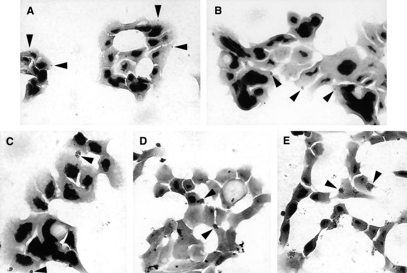
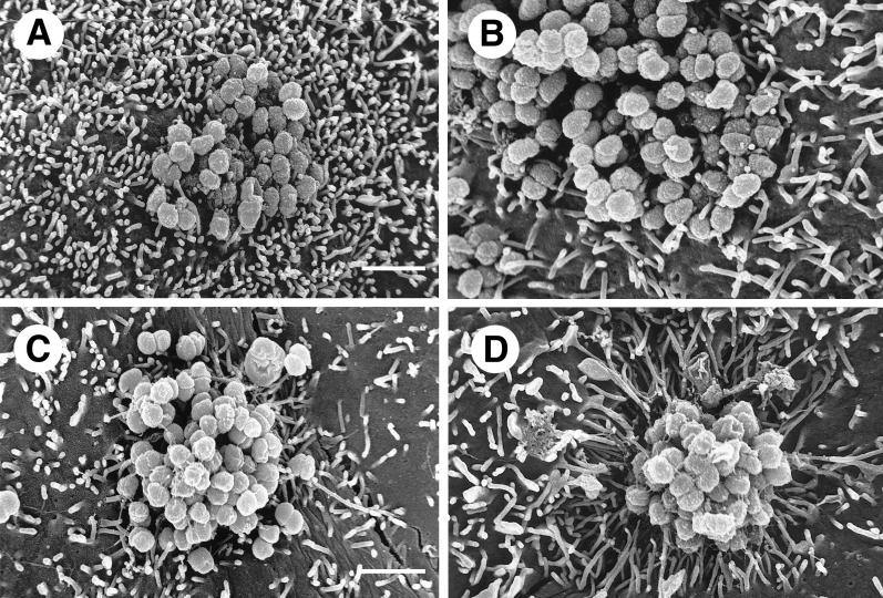
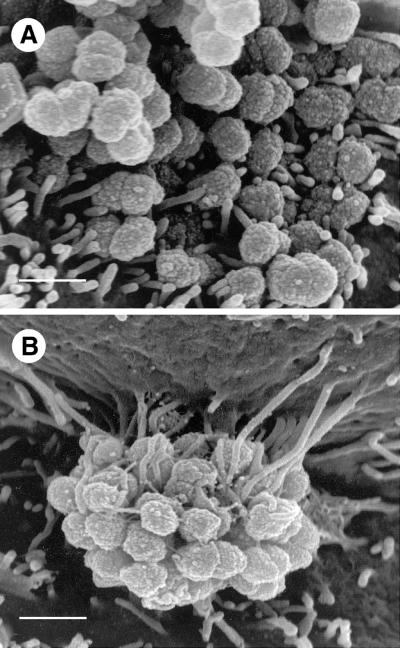
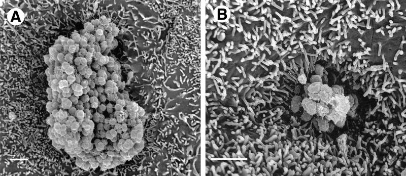
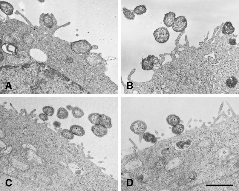
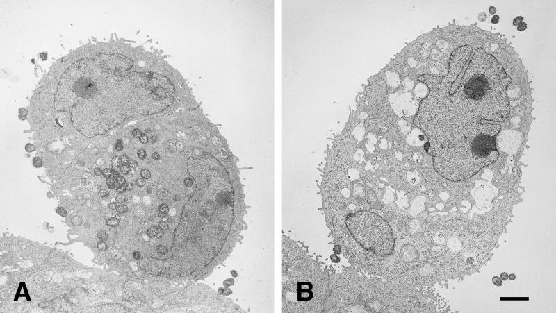
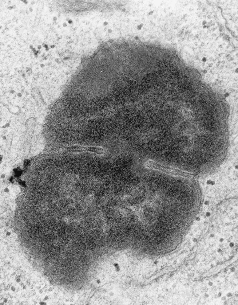
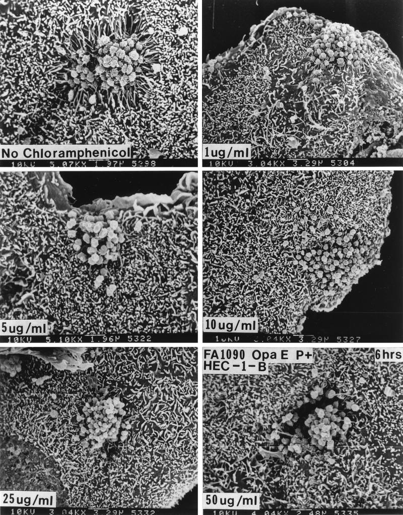
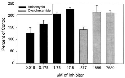
This study was undertaken to examine concomitant roles of pili and colony opacity-associated proteins (Opa) in promoting Neisseria gonorrhoeae adherence to and invasion of human endometrial HEC-1-B cells. Adherence of N. gonorrhoeae to cultured HEC-1-B cells was saturable, even though organisms adhered to <50% of the cells. During 4 to 6 h of incubation, adherent mono- and diplococci formed microcolonies on the surfaces of the cells. Microvilli of the HEC-1-B cells adhered by their distal ends to individual cocci within the microcolonies. When the microcolonies grew from isogenic pilus-negative (P-) Opa-, P- Opa+, or P+ Opa- gonococci, microvilli did not elongate, and the colonies were not engulfed. In contrast, the microvilli markedly elongated during exposure to P+ Opa+ gonococci. The microvilli adhered to the organisms along their full lengths and appeared to actively participate in the engulfment of the microcolonies. Internalized microcolonies, with P+ Opa+ gonococci, contained dividing cocci and appeared to be surrounded by cell membrane but were not clearly within vacuoles. In contrast, degenerate individual organisms were within vacuoles. Low doses of chloramphenicol, which inhibits protein synthesis by both prokaryotes and eukaryotes, prevented the microvillar response to and internalization of the P+ Opa+ gonococci; higher doses caused internalization without microvillus activation. Cycloheximide and anisomycin, which inhibit only eukaryotic protein synthesis, caused dose-dependent enhancement of uptake. Cytochalasins reduced engulfment; colchicine had no effect. These results show that gonococci must express both pili and Opa to be engulfed efficiently by HEC-1-B cells.


Figures

FIG. 1
FIG. 1
Gonococci form microcolonies on or in HEC-1-B cells. Representative micrographs of Gram-stained cell monolayers after 30 min (A), 60 min (B), 90 min (C), 2 h (D), and 4 h (E) of incubation are shown. Fewer than half of the cells appear to be colonized by gonococci. The gonococcal strain is FA1090 OpaE. Representative colonies are indicated by arrowheads; other colonies are also visible. At 30 min (A) adherent organisms appear as mono- and diplococci. By 60 min (B) tetracoccal and larger colonies are visible. Microcolonies are clearly visible along with new diplococcal and tetracoccal colonies. As colonies continue to grow (C), new microcolonies continue to be established (D). After 4 h of incubation, quite large colonies are visible adherent to or within the HEC-1-B cells (E).
FIG. 2
FIG. 2
Gonococci must express both Opa and pili (P) in order to signal HEC-1-B cells to engulf them. (A and B) SEM photomicrographs of typical microcolonies of MS11mk P organisms after 6 hours of growth on HEC-1-B cells. The bacteria in panel B also make Opa. Pili are not seen. (C and D) SEM photomicrographs of typical microcolonies of MS11mk P+ organisms. The bacteria in panel D also make Opa. Strands of pili are visible. (A) The P Opa microcolony is loose, with moderate bacterium-bacterium adhesion. Microvilli can be seen protruding from between individual gonococci, but there is little, if any, distortion of microvillus architecture other than that by simple contiguity. Some microvillus distal ends appear to have adhered to individual cocci. (B) The interaction between microvilli and the P Opa+ microcolony is not different from that seen with P Opa organisms (panel A). Opa expression appears to have little influence on the reaction of the epithelial cells to the organisms. (C) Pili appear to “lash” individual P+ Opa gonococci together to form a more compact colony. Some pili have bound microvilli together or pulled them toward the microcolony, modestly distorting microvillus architecture. Other strands of pili are visible radiating some distance from the microcolony along the surface of the epithelial cell. (D) The microvillus architecture has been altered dramatically as microvilli extend toward the microcolony of P+ Opa+ organisms. The microcolony consists of tightly adherent organisms that are bound together. More peripheral microvilli have extended themselves toward the microcolony to extraordinary lengths. As highly elongated peripheral microvilli adhere to the colony, the microvilli appear to pull the HEC-1-B cell around the colony and lift the HEC-1-B cell from the coverslip. Magnification, ×8,000; bar, 2 μm.
FIG. 3
FIG. 3
HEC-1-B cell microvilli adhere to P+ Opa+ gonococci along the full lengths of the microvilli. (A) At this SEM magnification it is clear that after 6 h of incubation only the distal ends of microvilli adhere to MS11mk P Opa+ organisms. Microvilli have not been elongated. Both true diplococci and adherent individual cocci are visible. (B) In this SEM photomicrograph microvilli can be seen to have wrapped themselves tightly around MS11mk P+ Opa+ cocci in the microcolony after 6 h of incubation on HEC-1-B cells. Microvilli adhere tightly to individual cocci up to the full lengths of the microvilli. Microvillus adherence is clearly separate from bacterium-pilus interactions. Microvilli at the top of the micrograph appear to have pulled the cell surface up along the side of the colony, creating the appearance of a microcolony hanging from a pedestal of microvilli. (The electron beam is focused vertically, not horizontally.) The extent of microvillus elongation is remarkable, and the contrast between microvillar responses to piliated and nonpiliated Opa+ bacteria is striking. Magnification, ×15,500; bar, 1 μm.
FIG. 4
FIG. 4
HEC-1-B cells engulf microcolonies with P+ Opa+ gonococci. After 6 h of incubation, the MS11mk P+ Opa+ microcolonies appear to sink into the HEC-1-B cell in these SEM photomicrographs as activated microvilli pull the underlying cell surfaces up around the colony. Magnification, ×8,000; bar, 2 μm.
FIG. 5
FIG. 5
TEM of P+ Opa+ organisms with activated microvilli adherent along their full lengths. (A and B) FA1090 P+ OpaE after 10 h of incubation on HEC-1-B cells. Microvilli have elongated and are adhering along their full lengths to gonococci, causing a perturbation of the HEC-1-B cell surface. Internalized cocci that are visible in both panels A and B appear to be free in the cell cytoplasm; vacuole membranes are not clearly visible (A). A bundle of pili is visible extending from the surface of a nonadherent coccus in panel B. (C and D) FA1090 P OpaE after 10 h of incubation on HEC-1-B cells. Microvilli have not elongated, and the HEC-1-B cell surface is not perturbed. Some of these nonpiliated organisms lie in contiguity with the cell surface, but they are not being internalized. Some vacuoles within the cell cytoplasm appear to contain degenerating organisms. Bacterium-bacterium adhesion is apparent in both panels; tip adherence by microvilli also can be seen in both panels. Bar, 1 μm.
FIG. 6
FIG. 6
Only P+ Opa+ organisms are found within HEC-1-B cells. TEM photomicrographs of HEC-1-B cells after 10 h of incubation with FA1090 P+ OpaE (A) and FA1090 P OpaE (B) organisms are shown. (A) Two cells are closely apposed, appearing as a single, ovoid cell. (Note the two nuclei, the tight junctions at the periphery, and microvilli in the space below the upper nucleus.) The lower (and larger) of the two cells contains what appears to be an internalized microcolony in which the individual cocci remain tightly adherent to each other; cocci may be in the process of being internalized into the upper cell. Vacuole membranes are not apparent around the ingested organisms. (B) Two cells are closely apposed, appearing as a single, ovoid cell. (Note the two nuceli, the tight junctions at the periphery, and the microvilli in the space between the nuclei.) The lower (and smaller) of the two cells contains a single internalized coccus, visible at the lower right; it appears to be within a vacuole. Other vacuoles within the cytoplasm are empty. (The “vacuoles” between the nuclei are intercellular spaces.) A second coccus is in close contact with the cell membrane. Bar, 1 μm.
FIG. 7
FIG. 7
High magnification (×50,000) of an intracellular FA1090 P+ OpaE organism that is in the process of dividing. Fragments of host cell membrane are visible in very close apposition to the bacterial outer membrane. This is most visible at the “waist” of the dividing coccus, but redundant fragments can be seen surrounding most of the organism. Note the absence of a clear vacuolar space between the host cell and bacterial cell membranes.
FIG. 8
FIG. 8
Chloramphenicol abolishes microvillus elongation and bacterial internalization at low doses but causes microvillus-independent HEC-1-B cell engulfment at higher doses. For these experiments FA1090 P+ OpaE variants were used, as these organisms stimulate microvillus elongation within 1 h. The HEC-1-B cells were incubated for 30 min with 0 to 50 μg of chloramphenicol per ml, the spent medium was aspirated, the HEC-1-B cells were inoculated with gonococci that had been diluted in medium containing the appropriate concentration of chloramphenicol, and the HEC-1-B cells were incubated for various times. As little as 5 μg of chloramphenicol per ml abolished microvillus elongation at 6 h; 10 μg of chloramphenicol per ml was needed to abolish microvillus elongation within the first hour. (A) FA1090 P+ OpaE after 6 h of incubation with HEC-1-B cells in the absence of chloramphenicol. (B to D) FA1090 P+ OpaE after 6 h of incubation with HEC-1-B cells in the presence of 1, 5, and 10 μg of chloramphenicol per ml. Note that microvillus-dependent engulfment appears to be lost at these concentrations of chloramphenicol. (E and F) FA1090 P+ OpaE after 6 h of incubation with HEC-1-B cells in the presence of 25 and 50 μg of chloramphenicol per ml. Note that the microcolonies are sinking into the HEC-1-B cells at these concentrations of chloramphenicol but that microvilli are not participating in their internalization. The magnification is given at the bottom of each panel.
FIG. 9
FIG. 9
Effect of eukaryotic cell protein synthesis inhibitors anisomycin and cyclohexamide on invasion of HEC-1-B cells by FA1090 P+ OpaE after 6 h of incubation. Means and standard errors for two or three experiments are shown.

Similar articles

Cited by

References

    1. Backman M, Källström H, Jonsson A-B. The phase-variable pilus-associated protein PilC is commonly expressed in clinical isolates of Neisseria gonorrhoeae, and shows sequence variability among strains. Microbiology. 1998;144:149–156. - PubMed
    1. Barritt D S, Schwalbe R S, Klapper D G, Cannon J G. Antigenic and structural differences among six proteins II expressed by a single strain of Neisseria gonorrhoeae. Infect Immun. 1987;55:2026–2031. - PMC - PubMed
    1. Bessen D, Gotschlich E C. Interactions of gonococci with HeLa cells: attachment, detachment, replication, penetration, and the role of protein II. Infect Immun. 1986;54:154–160. - PMC - PubMed
    1. Bhat K S, Gibbs C P, Barrera O, Morrison S G, Jähnig F, Stern A, Kupsch E M, Meyer T F, Swanson J. The opacity proteins of Neisseria gonorrhoeae strain MS11 are encoded by a family of 11 complete genes. Mol Microbiol. 1991;5:1889–1901. - PubMed
    1. Black W J, Schwalbe R S, Nachamkin I, Cannon J G. Characterization of Neisseria gonorrhoeae protein II phase variation by use of monoclonal antibodies. Infect Immun. 1984;45:453–457. - PMC - PubMed

Publication types

LinkOut - more resources