. 1999 Jul;67(7):3504-11.
doi: 10.1128/IAI.67.7.3504-3511.1999.

Differential roles of segmented filamentous bacteria and clostridia in development of the intestinal immune system

Affiliations

Differential roles of segmented filamentous bacteria and clostridia in development of the intestinal immune system

Y Umesaki et al. Infect Immun. 1999 Jul.

Abstract

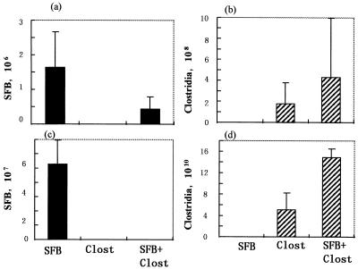
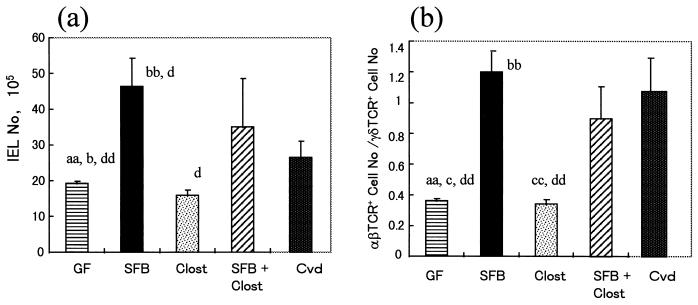
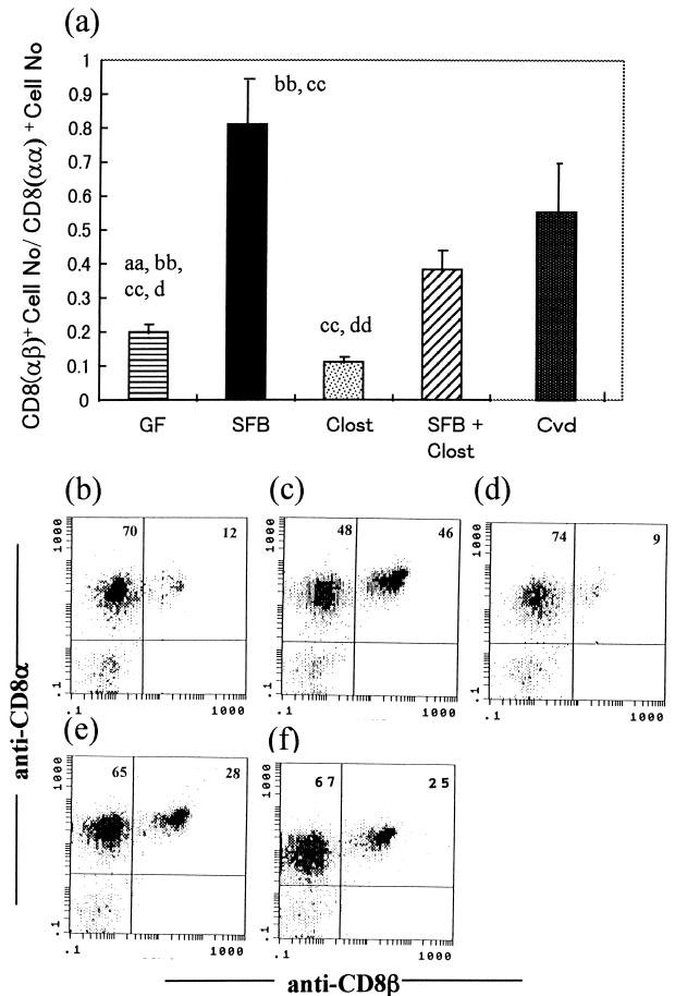
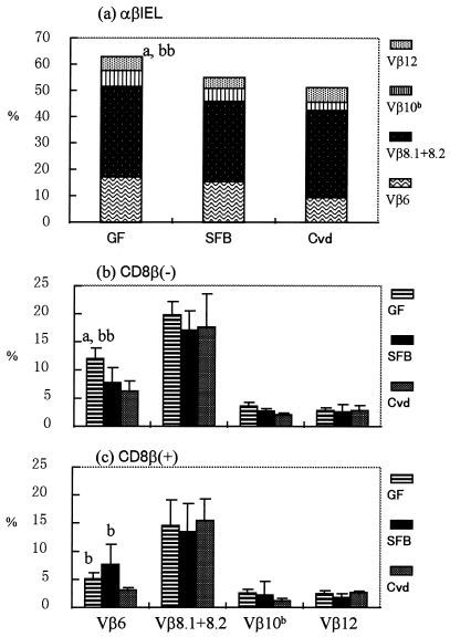
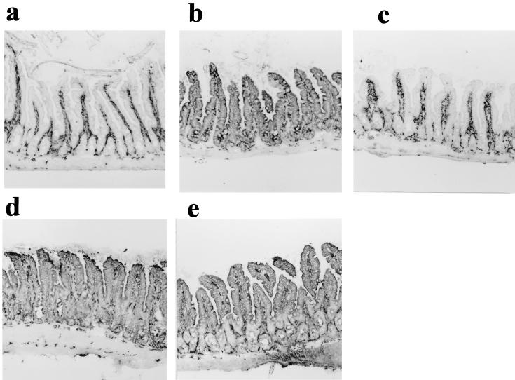
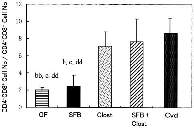
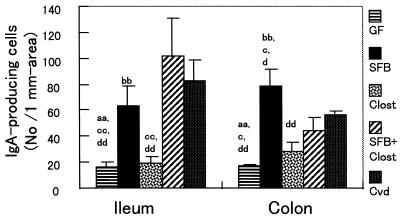
The presence of microflora in the digestive tract promotes the development of the intestinal immune system. In this study, to evaluate the roles of two types of indigenous microbe, segmented filamentous bacteria (SFB) and clostridia, whose habitats are the small and large intestines, respectively, in this immunological development, we analyzed three kinds of gnotobiotic mice contaminated with SFB, clostridia, and both SFB and clostridia, respectively, in comparison with germfree (GF) or conventionalized (Cvd) mice associated with specific-pathogen-free flora. In the small intestine, the number of alpha beta T-cell receptor-bearing intraepithelial lymphocytes (alpha betaIEL) increased in SFB-associated mice (SFB-mice) but not in clostridium-associated mice (Clost-mice). There was no great difference in Vbeta usage among GF mice, Cvd mice, and these gnotobiotic mice, although the association with SFB decreased the proportion of Vbeta6(+) cells in CD8beta- subsets to some extent, compared to that in GF mice. The expression of major histocompatibility complex class II molecules on the epithelial cells was observed in SFB-mice but not in Clost-mice. On the other hand, in the large intestine, the ratio of the number of CD4(-) CD8(+) cells to that of CD4(+) CD8(-) cells in alpha betaIEL increased in Clost-mice but not in SFB-mice. On association with both SFB and clostridia, the numbers and phenotypes of IEL in the small and large intestines changed to become similar to those in Cvd mice. In particular, the ratio of the number of CD8alpha beta+ cells to that of CD8alpha alpha+ cells in alpha betaIEL, unusually elevated in the small intestines of SFB-mice, decreased to the level in Cvd mice on contamination with both SFB and clostridia. The number of immunoglobulin A (IgA)-producing cells in the lamina propria was more elevated in SFB-mice than in Clost-mice, not only in the ileum but also in the colon. The number of IgA-producing cells in the colons of Clost-mice was a little increased compared to that in GF mice. Taken together, SFB and clostridia promoted the development of both IEL and IgA-producing cells in the small intestine and that of only IEL in the large intestine, respectively, suggesting the occurrence of compartmentalization of the immunological responses to the indigenous bacteria between the small and large intestines.


Figures

FIG. 1
FIG. 1
Microbiological status of the small intestines (ileum) and large intestines (feces) of SFB-mice, Clost-mice, and mice associated with both SFB and clostridia. The bacterial numbers were determined by microscopy using smear preparation of the ileal contents (a and b) and feces (c and d). The data are numbers of bacteria per gram of ileal contents or feces.
FIG. 2
FIG. 2
Total numbers of IEL (a) and ratios of the number of αβIEL to that of γδIEL (b) in the small intestines of GF mice, SFB-mice, Clost-mice, mice associated with both SFB and clostridia (SFB + Clost), and Cvd mice. The gnotobiotic mice were analyzed at 16 weeks of age in the second generation and compared with age-matched GF mice. The data represent means plus the standard deviations (n = 3 or 4) and were also reproducible in the experiment involving the first generation. The letters a, b, c, and d near the columns indicate statistically significant differences from the SFB, Clost, SFB + Clost, and Cvd groups, respectively. Single letter, P < 0.05; double letters, P < 0.01.
FIG. 3
FIG. 3
Ratios of the number of CD8αβ-bearing αβIEL to that of CD8αα-bearing αβIEL in GF mice, SFB-mice, Clost-mice, mice associated with both SFB and clostridia (SFB + Clost), and Cvd mice. The data in panel a are means plus standard deviations (n = 3 or 4) and were reproducible in the experiment involving the first generation. The letters a, b, c, and d near the columns indicate statistically significantly differences from the SFB, Clost, SFB + Clost, and Cvd groups, respectively. Single letter, P < 0.05; double letters, P < 0.01. (b to f) Representative data for the quadrants on three-color analysis gating of αβIEL in the GF (b), SFB (c), Clost (d), SFB + Clost (e), and Cvd (f) groups.
FIG. 4
FIG. 4
Vβ usage in GF mice, SFB-mice, and Cvd mice. (a) Data showing the sum of Vβ6+, Vβ8.1+8.2+, Vβ10b+, and Vβ12+ cells in each group. (b and c) Vβ6+, Vβ8.1+8.2+, Vβ10b+, and Vβ12+ cells in CD8β (b) and CD8β+ (c) subsets determined by three-color analysis of GF mice, SFB-mice, and Cvd mice. The data are means plus standard deviations (n = 4 to 7). The letters a and b near the columns indicate statistically significant differences from the SFB and Cvd groups, respectively. Single letter, P < 0.05; double letters, P < 0.01.
FIG. 5
FIG. 5
MHC class II molecule expression in ileal epithelial cells of GF mice (a), SFB-mice (b), Clost-mice (c), mice associated with both SFB and clostridia (d), and Cvd mice (e). Shown are longitudinal sections of the ileum stained with an admixture of biotinylated anti-I-Ad and anti-I-Ed antibodies, followed by peroxidase-labelled streptavidin. The data are representative of the mice in each group and were reproducible in the experiment involving the first generation. Magnification, ×85.
FIG. 6
FIG. 6
Ratio of the number of CD4 CD8 cells to that of CD4 CD8+ cells among αβBIEL in the large intestines of GF mice, SFB-mice, Clost-mice, mice associated with both SFB and clostridia (SFB + Clost), and Cvd mice. The data are means plus the standard deviations (n = 3 or 4) and were reproducible in the experiment involving the first generation. The letters b, c, and d indicate statistically significant differences from the Clost, SFB + Clost, and Cvd groups, respectively. Single letter, P < 0.05; double letters, P < 0.01.
FIG. 7
FIG. 7
Numbers of IgA-producing cells in the ileal lamina propria and colonic lamina propria of GF mice, SFB-mice, Clost-mice, mice associated with both SFB and clostridia (SFB + Clost), and Cvd mice. The data are means plus the standard deviations (n = 3 or 4) and were reproducible in the experiment involving the first generation. The letters a, b, c, and d indicate statistically significant differences from the SFB, Clost, SFB + Clost, and Cvd groups, respectively. Single letter, P < 0.05; double letters, P < 0.01.

References

    1. Badiner G, Goodman T G, Lefrancois L. Selection of intestinal intraepithelial lymphocyte T cell receptors: evidence for a dynamic tissue specific process. Int Immunol. 1993;5:223–226. - PubMed
    1. Balk S P, Ebert E C, Blumenthal R L, Mcdermott F V, Wucherpfenning K W, Landau S B, Blumberg R S. Oligoclonal expansion and CD1 recognition by human intestinal intraepithelial lymphocytes. Science. 1991;253:1411–1415. - PubMed
    1. Barnard J A, Warwick G J, Gold L I. Localization of transforming growth factor β isoforms in the normal murine small intestine and colon. Gastroenterology. 1993;105:67–73. - PubMed
    1. Beagley K W, Eldridge J H, Kiyono H, Everson M P, Koopman J P, Hirano Y, Kishimoto T, McGhee J R. Interleukins and IgA synthesis. Human and murine interleukin 6 induce high rate IgA secretion in IgA-committed B cells. J Exp Med. 1989;169:2133–2148. - PMC - PubMed
    1. Berg R D, Savage D C. Immune responses of specific pathogen-free and gnotobiotic mice to antigens of indigenous and nonindigenous microorganisms. Infect Immun. 1975;11:320–329. - PMC - PubMed

Substances