. 1999 Jul;67(7):3548-57.
doi: 10.1128/IAI.67.7.3548-3557.1999.

Role of the 85-kilobase plasmid and plasmid-encoded virulence-associated protein A in intracellular survival and virulence of Rhodococcus equi

Affiliations

Role of the 85-kilobase plasmid and plasmid-encoded virulence-associated protein A in intracellular survival and virulence of Rhodococcus equi

S Giguère et al. Infect Immun. 1999 Jul.

Abstract

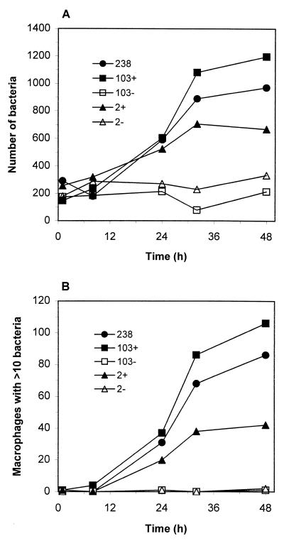
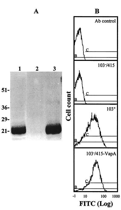
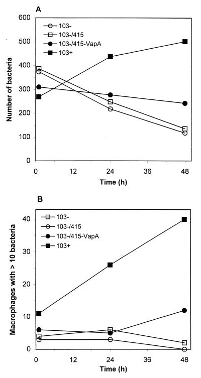
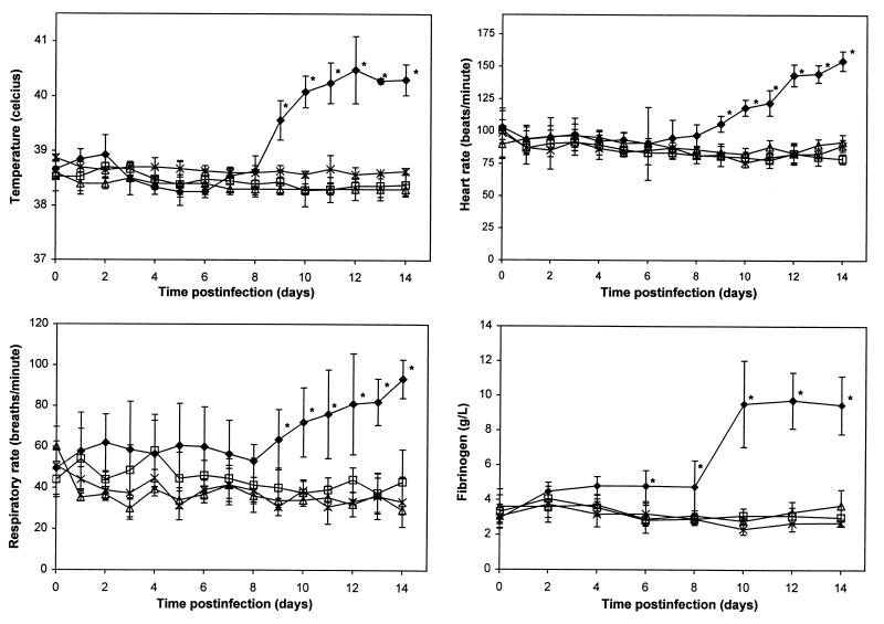
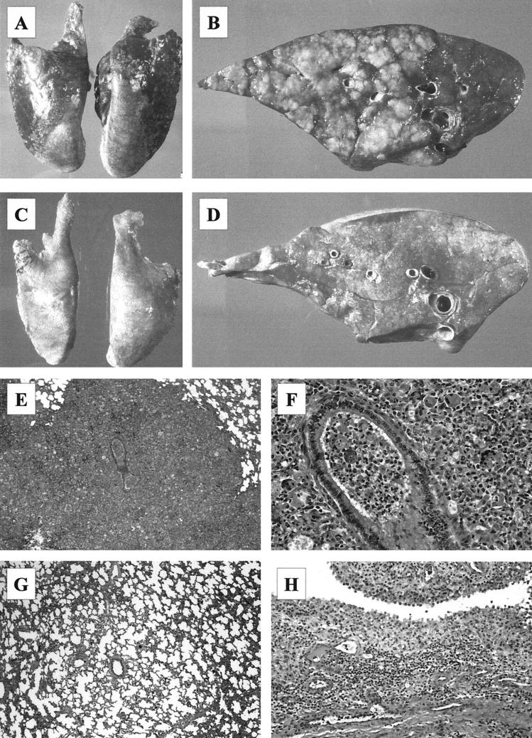
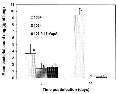
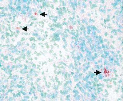
Rhodococcus equi is a facultative intracellular pathogen of macrophages and a cause of pneumonia in young horses (foals) and immunocompromised people. Isolates of R. equi from pneumonic foals typically contain large, 85- or 90-kb plasmids encoding a highly immunogenic virulence-associated protein (VapA). The objective of this study was to determine the role of the 85-kb plasmid and VapA in the intracellular survival and virulence of R. equi. Clinical isolates containing the plasmid and expressing VapA efficiently replicated within mouse macrophages in vitro, while plasmid-cured derivatives of these organisms did not multiply intracellularly. An isolate harboring the large plasmid also replicated in the tissues of experimentally infected mice, whereas its plasmid-cured derivative was rapidly cleared. All foals experimentally infected with a plasmid-containing clinical isolate developed severe bronchopneumonia, whereas the foals infected with its plasmid-cured derivative remained asymptomatic and free of visible lung lesions. By day 14 postinfection, lung bacterial burdens had increased considerably in foals challenged with the plasmid-containing clinical isolate. In contrast, bacteria could no longer be cultured from the lungs of foals challenged with the isogenic plasmid-cured derivative. A recombinant, plasmid-cured derivative expressing wild-type levels of VapA failed to replicate in macrophages and remained avirulent for both mice and foals. These results show that the 85-kb plasmid of R. equi is essential for intracellular replication within macrophages and for development of disease in the native host, the foal. However, expression of VapA alone is not sufficient to restore the virulence phenotype.


Figures

FIG. 1
FIG. 1
Infection of murine peritoneal macrophages with isogenic strains of R. equi. At various times postinfection, macrophage monolayers were fixed with methanol, and the bacteria were immunostained and visualized by fluorescence microscopy. (A) The numbers of bacteria (ordinate) associated with 200 macrophages were visually counted by fluorescence microscopy. (B) The numbers of macrophages with 10 or more bacteria were also recorded. Each strain was evaluated a minimum of three times with similar results.
FIG. 2
FIG. 2
Expression of VapA by strains of R. equi. (A) Immunoblot of R. equi with a MAb against VapA. Lanes: 1, recombinant strain 103/415-VapA; 2, strain 103; 3, strain 103+. Sizes of protein molecular mass markers are indicated to the left in kilodaltons. (B) Flow cytometry profile showing the expression of VapA on the surface of R. equi. Bacteria were grown in liquid broth, washed, and stained with a MAb to VapA. The relative mean fluorescence values of wild-type isolate 103+, pYUB415 vector-electroporated plasmid-cured derivative (103/415), and the recombinant pMH1-electroporated strain 103 (103/415-VapA) were compared. In addition, all analyses included a comparison to bacteria stained with an irrelevant MAb (Ab control).
FIG. 3
FIG. 3
Infection of macrophages with the VapA-expressing recombinant strain. Bacteria were added to J774A.1 cells, a murine monocyte-macrophage-like cell line. At 1, 24, and 48 h postinfection, parallel monolayers were fixed, and bacteria were immunostained and visualized by fluorescence microscopy. Bacterial growth is expressed as both the total number of bacteria per 200 macrophages (A) and the number of macrophages (of 200 counted) containing 10 or more bacteria (B). Each data point represents the average of data from two coverslips. This figure is representative of four independent experiments.
FIG. 4
FIG. 4
Temperatures, heart rates, respiratory rates, and fibrinogen concentrations of control foals (▵) and foals infected with R. equi 103+ (⧫), 103 (□), or 103/415-VapA (×). Values are shown as the means ± SD (error bars) within each group. An asterisk indicates that P is <0.05 compared to baseline values for the same group and the other three groups at the same time point.
FIG. 5
FIG. 5
Pathological findings in foals infected intrabronchially with R. equi 103+ or 103. (A) Severe bilateral consolidation of the lungs on day 14 postinfection in a foal infected with R. equi 103+. (B) Cross section of a cranioventral lung lobe in a foal infected with R. equi 103+ showing multiple well-defined nodular areas of pulmonary consolidation on day 14 postinfection. (C) Lungs of a foals infected with R. equi 103 showing no gross lesions. (D) Cross section of the lung shown in panel C. (E) Extensive bronchopneumonia in the lungs of a strain 103+-infected foal on day 14. Magnification, ×10. (F) Higher magnification of the lesion shown in panel A showing pyogranulomatous inflammation. Magnification, ×68. (G) Mild atelectasis and hypercellularity of the alveolar septi in a strain 103-infected foal on day 14. Magnification, ×12. (H) Severe inflammation of the synovial membrane in the hock of a strain 103+-infected foal on day 14. Magnification, ×38.
FIG. 6
FIG. 6
Bacterial numbers in the lungs of foals infected with R. equi 103+, 103, or 103/415-VapA. Numbers of R. equi in four dispersed and preselected areas of both lungs were determined by culturing serial dilutions of lung homogenates and counting the CFU. Letters differing between bacterial groups or time points indicate a statistically significant difference in bacterial numbers (P < 0.01).
FIG. 7
FIG. 7
Immunohistochemical staining of the lungs of a foal infected with strain 103/415-VapA showing VapA expression in vivo (arrows). Magnification, ×400.

References

    1. Arlotti M, Zoboli G, Moscatelli G L, Magnati G, Maserati R, Borghi V, Andreoni M, Libanore M, Bonazzi L, Piscina A, Ciammarughi R. Rhodococcus equi infection in HIV-positive subjects: a retrospective analysis of 24 cases. Scand J Infect Dis. 1996;28:463–467. - PubMed
    1. Byrne B A, Prescott J F, Palmer G H, Hines S A. Conference handbook of the 8th International Conference on Equine Infectious Diseases, Dubai, United Arab Emirates. 1998. Characterization of a virulence-associated gene family in Rhodococcus equi, abstr. P-63; p. 236.
    1. De la Pena-Moctezuma A, Prescott J F. Association with HeLa cells by Rhodococcus equi with and without the virulence plasmid. Vet Microbiol. 1995;46:383–392. - PubMed
    1. De la Pena-Moctezuma A, Prescott J F. Attempts to find phenotypic markers of the virulence plasmid of Rhodococcus equi. Can J Vet Res. 1996;60:29–33. - PMC - PubMed
    1. Donisi A, Suardi M G, Casari S, Longo M, Cadeo G P, Carosi G. Rhodococcus equi infection in HIV-infected patients. AIDS. 1996;10:359–362. - PubMed

Publication types

LinkOut - more resources