. 1999 Jul;67(7):3571-9.
doi: 10.1128/IAI.67.7.3571-3579.1999.

Fatal granuloma necrosis without exacerbated mycobacterial growth in tumor necrosis factor receptor p55 gene-deficient mice intravenously infected with Mycobacterium avium

Affiliations

Fatal granuloma necrosis without exacerbated mycobacterial growth in tumor necrosis factor receptor p55 gene-deficient mice intravenously infected with Mycobacterium avium

S Ehlers et al. Infect Immun. 1999 Jul.

Abstract

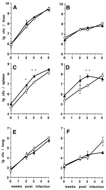
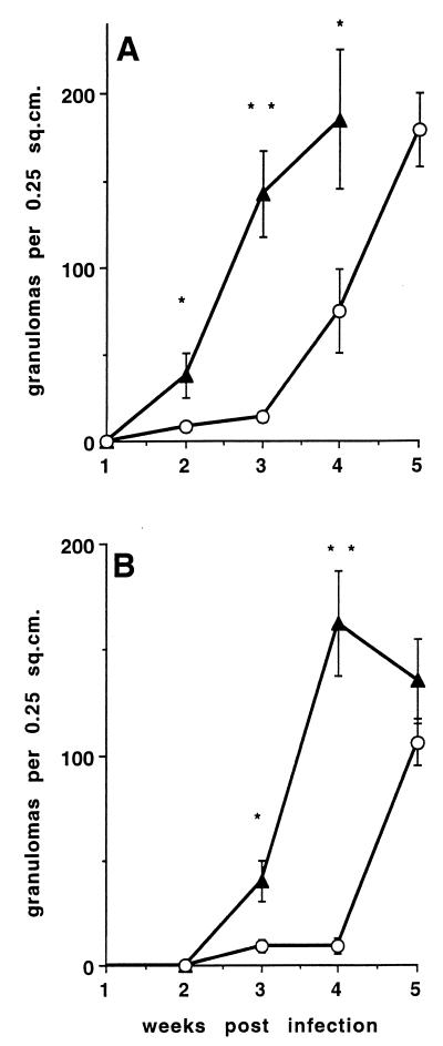
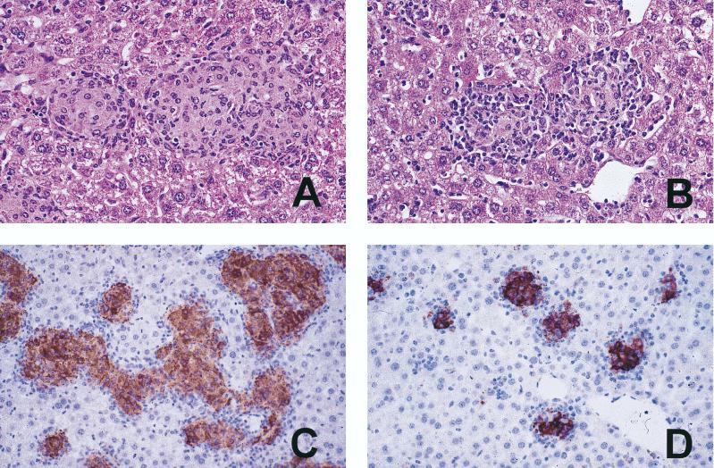
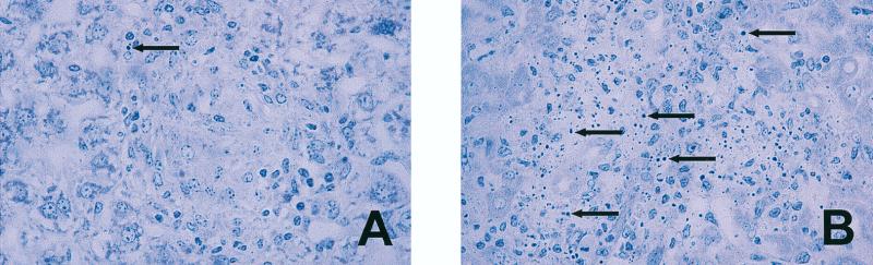
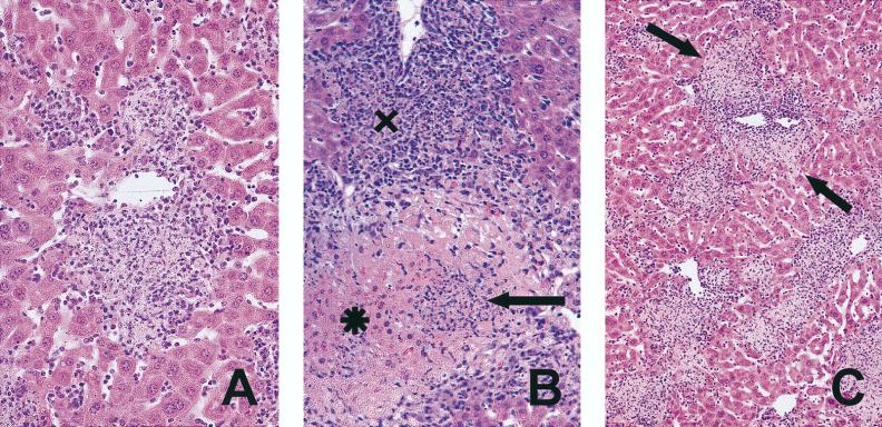
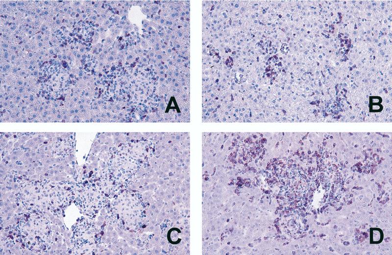
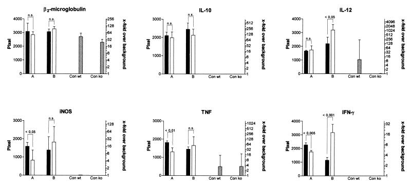
The pathogenesis of mycobacterial infections is associated with the formation of granulomas in which both antibacterial protection and tissue damage take place concomitantly. We used murine Mycobacterium avium infection to compare the development of granulomatous lesions in intravenously infected tumor necrosis factor receptor p55 (TNFRp55) gene-deficient (p55(-/-)) mice to the development of granulomatous lesions in M. avium-infected syngeneic C57BL/6 (p55(+/+)) mice. Up to 5 weeks after infection with either the highly virulent M. avium strain TMC724 or the intermediately virulent M. avium strain SE01, bacterial counts in the liver, spleen, and lung of p55(-/-) mice were identical to or lower than those in infected p55(+/+) mice. However, the formation of mononuclear cell foci in the liver was delayed by approximately 2 to 3 weeks in p55(-/-) mice compared to the results obtained for infected p55(+/+) mice. Despite comparable bacterial loads, granulomas in p55(-/-) mice underwent progressive necrosis, causing damage to the surrounding liver tissue. The appearance of necrotizing granulomas was associated with the death of all infected p55(-/-) mice, regardless of the virulence of the M. avium strain used for infection. Granulomatous lesions in the liver contained three times as many CD3(+) cells in p55(-/-) mice yet appeared more diffuse than in p55(+/+) mice. Semiquantitative reverse transcription-PCR studies revealed that prior to mouse death, interleukin-12 (IL-12) and gamma interferon mRNA levels were up regulated in the livers of infected p55(-/-) mice, while mRNA levels for tumor necrosis factor, the inducible isoform of nitric-oxide synthase (iNOS), and IL-10 were similar to those found in infected p55(+/+) mice. In response to persistent mycobacterial infection, the absence of TNFRp55 causes the disregulation of T-cell-macrophage interactions and results in fatal granuloma necrosis even when adequate antibacterial functions are maintained.


Figures

FIG. 1
FIG. 1
Course of M. avium infection in p55+/+ and p55−/− C57BL/6 mice. Mice were intravenously infected with either 1 × 10⁶ CFU of M. avium TMC724 (A, C, and E) or 8 × 10⁶ CFU of M. avium SE01 (B, D, and F), and bacterial counts were determined in the liver (A and B), spleen (C and D), and lungs (E and F) at indicated time points. Each point reflects the means and SD (error bars) of four mice per group. Triangles, p55+/+ mice; circles, p55−/− mice; ∗, P < 0.05; ∗∗, P < 0.005.
FIG. 2
FIG. 2
Granuloma formation in M. avium-infected p55+/+ and p55−/− C57BL/6 mice. Mice were intravenously infected with 105 CFU of either M. avium TMC724 (A) or M. avium SE01 (B). At indicated time points, the number of granulomas in an area of 0.25 cm2 were determined on nonsequential sections of HE-stained liver tissue. Each point reflects the means and SD (error bars) of 20 determinations (five sections per mouse, four mice per group). Triangles, p55+/+ mice; circles, p55−/− mice; ∗, P < 0.005; ∗∗, P < 0.0001.
FIG. 3
FIG. 3
Granuloma morphology in M. avium-infected p55+/+ and p55−/− mice. Mice were intravenously infected with 106 CFU of M. avium TMC724 and sacrificed 5 weeks postinfection. Liver sections were stained with HE (A and B) or with a rabbit anti-iNOS antiserum (immunoperoxidase stain; C and D). (A) Large epithelioid granulomas in a p55+/+ mouse (magnification, ×200). (B) Smaller, less well organized granulomas in a p55−/− mouse (×200). (C) Large, confluent, iNOS-positive granulomas in a p55+/+ mouse (×64). (D) Small, circumscript, iNOS-positive granulomas in a p55−/− mouse (×64).
FIG. 4
FIG. 4
Apoptotic cells within granulomas of M. avium-infected p55−/− mice. Mice were intravenously infected with 106 CFU of M. avium TMC724 and sacrificed 5 to 6 weeks postinfection for histological analysis of the liver. (A) Well-structured granuloma with epithelioid macrophages and a rare apoptotic cell in a p55+/+ mouse; (B) apoptotic cells and multiple pyknotic nuclei in early disintegrating granuloma of a p55−/− mouse (toluidine blue; magnification, ×128). Arrows, apoptotic cells.
FIG. 5
FIG. 5
Granuloma necrosis in M. avium-infected p55−/− mice. Mice were intravenously infected with 106 CFU of M. avium TMC724 and sacrificed 5 to 6 weeks postinfection for histological analysis of the liver. (A) Necrotizing granuloma (HE; magnification, ×200); (B) early necrotizing granuloma (X) and completely disintegrated granuloma (arrow) with leakage into adjacent liver tissue (✶) (HE; magnification, ×128); (C) low-power view of multiple foci of granuloma necrosis with incipient damage (arrows) to surrounding liver tissue (HE; magnification, ×64).
FIG. 6
FIG. 6
CD3+ cells in lesions of M. avium-infected p55+/+ and p55−/− mice. Mice were intravenously infected with 106 CFU of M. avium TMC724 and sacrificed 3 weeks (A and B) or 5 weeks (C and D) after infection. Immunohistology was performed on paraffin-embedded liver sections with an anti-CD3 monoclonal antibody and peroxidase-linked secondary antibodies. (A and C) p55+/+ mice; (B and D) p55−/− mice (×64).
FIG. 7
FIG. 7
Semiquantitative RT-PCR in the livers of p55+/+ and p55−/− mice. RT-PCR with specific cytokine primers and hybridization with labelled internal probes were performed on liver samples taken at 3 weeks (columns A) or 5 weeks (columns B) after infection with TMC724. Mean pixel values of four individual mice ± SDs per experimental group are shown. Significant differences in pixel values between p55+/+ mice (black columns) and p55−/− mice (white columns) are indicated. Crosshatched columns, uninfected controls; Con wt, p55+/+ control; Con ko, p55−/− control. Twofold serial dilutions of positive control cDNA were included in each reaction to ascertain linear amplification rates and provide a scale of comparison for samples from different experimental groups. Background X-ray film exposure was arbitrarily set at a pixel value of 1, and calculated x-fold increases over background pixel values are shown on the right. P values used in statistical comparisons are indicated above pixel columns. n.s., not significantly different.

References

    1. Appelberg R, Castro A G, Pedrosa J, Silva R A, Orme I M, Minóprio P. Role of gamma interferon and tumor necrosis factor alpha during T-cell-independent and -dependent phases of Mycobacterium avium infection. Infect Immun. 1994;62:3962–3971. - PMC - PubMed
    1. Appelberg R, Sarmento A, Castro A G. Tumour necrosis factor alpha in the host resistance to mycobacteria of distinct virulence. Clin Exp Immunol. 1995;101:308–313. - PMC - PubMed
    1. Benini, J., E. M. Ehlers, and S. Ehlers. Different types of pulmonary grnuloma necrosis in immunocompetent vs. TNFRp55-gene-deficient mice aerogenically infected with highly virulent Mycobacterium avium. J. Pathol., in press. - PubMed
    1. Blake J A, Eppig J T, Richardson J E, Davisson M T the Mouse Genome Informatics Group. The mouse genome database (MGD)—a community resource. Status and enhancement. Nucleic Acids Res. 1998;26:130–137. - PMC - PubMed
    1. Cattoretti G, Piteri S, Parraricini C, Becker M A G, Poggi S, Bifulco C, Key G, D’Amato L, Sabattini E, Fendale E, Geynolds F, Gerdes J, Rilke F. Antigen unmasking on formalin-fixed, paraffin-embedded tissue sections. J Pathol. 1993;171:83–98. - PubMed

Publication types

Substances