. 1999 Jun;103(12):1641-50.
doi: 10.1172/JCI6380.

Inhibition of TGF-beta 3 restores the invasive capability of extravillous trophoblasts in preeclamptic pregnancies

Affiliations

Inhibition of TGF-beta 3 restores the invasive capability of extravillous trophoblasts in preeclamptic pregnancies

I Caniggia et al. J Clin Invest. 1999 Jun.

Abstract

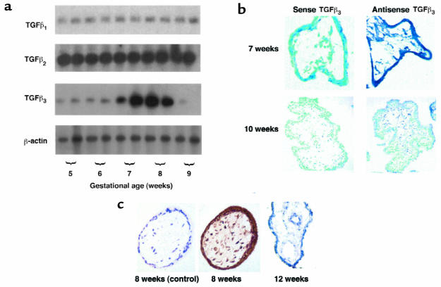
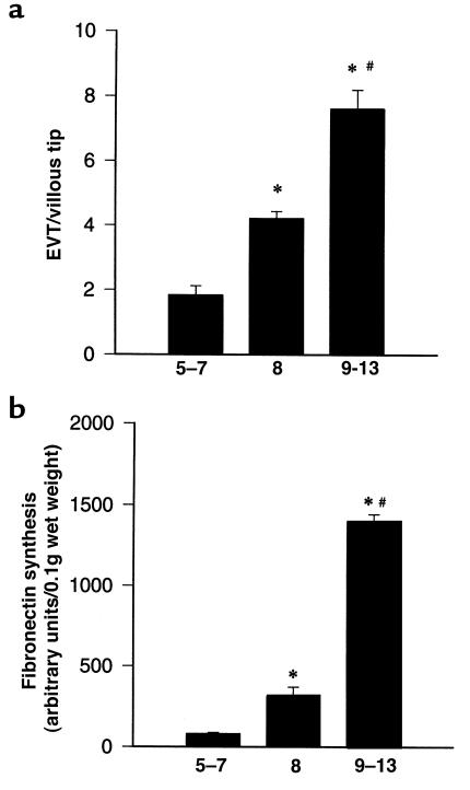
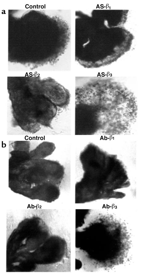
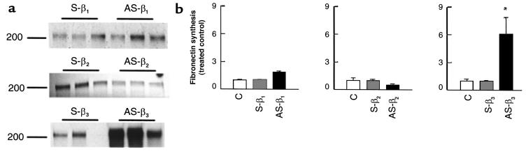
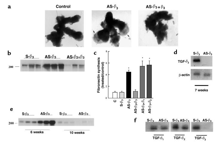
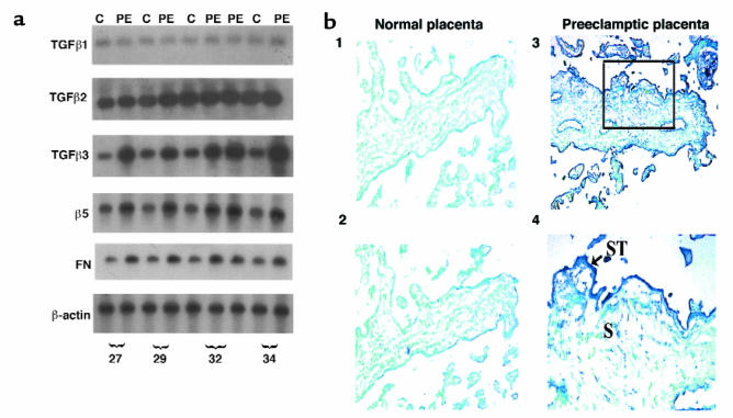
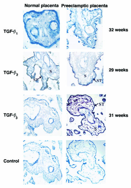
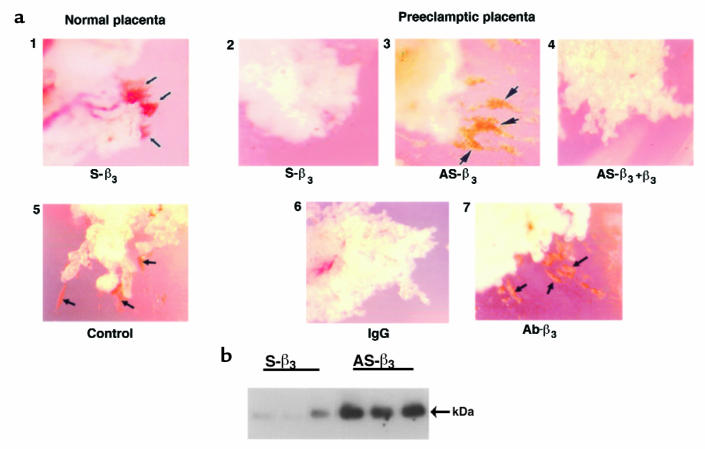
Preeclampsia, the major cause of maternal morbidity and mortality in developed countries, is associated with abnormalities of placenta function due to shallow invasion of the maternal decidua by trophoblasts. Data suggest that TGF-beta may play a role in inhibiting trophoblast outgrowth or invasion, or both. We report that placental TGF-beta 3 expression is high in early pregnancy but falls at around 9 weeks' gestation. This pattern is inversely correlated with trophoblast outgrowth and fibronectin synthesis, markers of early trophoblast differentiation toward an invasive phenotype. We demonstrate that TGF-beta 3 is overexpressed in preeclamptic placentae. In contrast to control placentae, explants from preeclamptic pregnancies fail to exhibit spontaneous invasion in vitro. Significantly, antisense-induced inhibition of TGF-beta 3 expression, and inhibition of TGF-beta 3 activity with antibodies, induces the formation of columns of trophoblast cells, which migrate out of the explant into the underlying Matrigel. To our knowledge, this is the first demonstration that the hypoinvasive placental phenotype characteristic of preeclampsia can be essentially normalized in vitro by biochemical manipulation. We speculate that a failure to downregulate expression of TGF-beta 3 at around 9 weeks' gestation results in shallow trophoblast invasion and predisposes the pregnancy to preeclampsia.


Figures

Figure 1
Figure 1
Expression of TGF-β isoforms in human placenta in the first trimester of gestation. (a) Message expression of TGF-β isoforms was assessed by low-cycle RT-PCR followed by Southern blot analysis using specific probes for TGF-β1, TGF-β2, TGF-β3, and the control housekeeping gene β-actin. Note that TGF-β3 expression increases at around 7–8 weeks’ gestation and declines thereafter. (b) Expression of TGF-β3 mRNA was also assessed by in situ hybridization to placental sections at 7 and 10 weeks’ gestation with digoxigenin-labeled sense and antisense TGF-β3 riboprobes. Endogenous alkaline phosphatases were blocked by the addition of levamisole. Sections were counterstained with methyl green. Note that TGF-β3 mRNA expression, viewed by blue staining, is high at 7 weeks in chorionic villi and decreases around 10 weeks. Control experiments were performed using sense TGF-β3 riboprobes. ×100. (c) Immunoperoxidase staining of TGF-β3 was performed in placental sections at 8 and 12 weeks’ gestation. Sections of placental tissue of 8 weeks’ gestation show strong positive immunoreactivity viewed as brown staining in the cytotrophoblast, syncytiotrophoblast, and stromal cells of the chorionic villi. Sections of placenta at 12 weeks’ gestation demonstrate low or absent TGF-β3 immunoreactivity in the villi. There is no immunoreactivity when antiserum was preincubated with an excess of TGF-β3 competing peptide (8 weeks, control). ×200.
Figure 2
Figure 2
Trophoblast differentiative capability of villous explants is gestational age dependent. Villous explants from first-trimester gestation (5–13 weeks) were cultured for 6 days. Differentiation was monitored by measuring morphological and biochemical markers of trophoblast differentiation. (a) EVT cell outgrowth and migration were expressed as the ratio of EVT outgrowths per villous tip, where the nominator, EVT outgrowths, represents the number of EVT columns sprouting from the villous tips plus the number of islands of EVT invading into the Matrigel. The denominator represents the total number of villous tips in a single explant culture. (b) At day 5, explants were metabolically labeled with [35S]methionine for 18 hours. Fibronectin was isolated from conditioned medium using gelatin-Sepharose beads on the protein extract separated by SDS PAGE. Radiolabeled bands were quantified with the use of a PhosphorImager. Data represent the mean ± SEM of 6–10 different experiments performed in triplicate. *P < 0.05, by ANOVA, compared with 5–7 weeks’ gestation. #P < 0.05, by ANOVA, compared with 8 weeks’ gestation.
Figure 3
Figure 3
Antisense oligonucleotides and antibody to TGF-β3, but not to TGF-β2 or TGF-β1, induce trophoblast outgrowth and migration in villous explant cultures. (a) Villous explants from 5–8 weeks’ gestation were maintained in culture for 5 days in the presence of 10 μM antisense oligonucleotides to TGF-β1 (AS-β1), TGF-β2 (AS-β2), and TGF-β3 (AS-β3). Control experiments were run in parallel using explants from the same placenta cultured in either medium alone or medium containing sense oligonucleotides. (b) Villous explants from 5–8 weeks’ gestation were maintained in culture for 5 days in the presence of 10 μg/mL of neutralizing antibody to TGF-β1 (Ab-β1), TGF-β2 (Ab-β2), and TGF-β3 (Ab-β3). Control experiments were run in parallel using explants from the same placenta cultured in either medium alone or in medium containing IgG control. Note that both antisense TGF-β3 (AS-β3) and antibody TGF-β3 (Ab-β3) treatments dramatically increase budding and outgrowth of EVT from the distal end of the villous tips when compared with control villous explants or explants exposed to TGF-β1 and TGF-β2 antisense oligonucleotides and antibodies. ×50.
Figure 4
Figure 4
Antisense oligonucleotides to TGF-β3, but not to TGF-β2 or TGF-β1, increase fibronectin synthesis in villous explant cultures. Explants were incubated for 4 days in medium alone, or in the presence of antisense oligonucleotides to TGF-β1 (AS-β1), TGF-β2 (AS-β2), TGF-β3 (AS-β3), and sense oligonucleotides (S-β1, S-β2, S-β3) (10 μM), and then metabolically labeled with [35S]methionine for 18 hours in the presence or absence of oligonucleotides. (a) Fibronectin was isolated from conditioned medium using gelatin-Sepharose beads and subjected to SDS-PAGE. The position of the marker with Mr = 200,000 is indicated. (b) Radiolabeled bands were quantified with the use of a PhosphorImager. Shown are the changes in fibronectin estimated after normalization to control cultures. Antisense TGF-β3 treatment (AS-β3; filled bar in third panel) significantly increased the amount of labeled fibronectin compared with both medium alone (C; open bars) or sense (S-β3; cross-hatched bar in third panel), whereas antisense TGF-β1 (AS-β1; filled bar in first panel) and TGF-β2 (AS-β2; filled bar in middle panel) did not. *P < 0.05 by ANOVA. All data are expressed as the mean ± SEM of 5 separate experiments carried out in triplicate.
Figure 5
Figure 5
The antisense TGF-β3 stimulatory effect on trophoblast migration and on fibronectin production is specific. Explants of 5–8 weeks’ gestation were treated for 5 days with 10 μM antisense oligonucleotides to TGF-β3 (AS-β3), plus 10 ng/mL recombinant TGF-β3 (AS-β33), TGF-β1 (AS-β31), or TGF-β2 (AS-β32). Control experiments were run in parallel using sense TGF-β3 (S-β3) or medium alone (C). (a) Shown is a representative experiment demonstrating that addition of recombinant TGF-β3 to antisense TGF-β3–treated explants (AS-β33) abolishes the antisense stimulatory effect on trophoblast budding and outgrowth (arrows). ×25. (b) Similar reversal of AS-β3 stimulation of fibronectin synthesis by exogenous TGF-β3. A representative analysis of triplicate samples from a single experiment is shown. The position of the marker with Mr = 200,000 is indicated. (c) Radiolabeled bands were quantified with the use of a PhosphorImager. Shown are the changes in fibronectin estimated after normalization to control cultures. Antisense TGF-β3 treatment (AS-β3) significantly increased the amount of labeled fibronectin compared with both medium alone (C) or sense oligonucleotide (S-β3). Addition of exogenous TGF-β3 (AS-β33), but not TGF-β1(AS-β31) or TGF-β2 (AS-β32), to the antisense-treated explants abolished the antisense stimulatory effect on fibronectin production. (d) mRNA expression of TGF-β3 in explants treated with antisense (AS-β3) or control sense (S-β3) oligonucleotides was measured by low-cycle RT-PCR followed by Southern blot analysis using specific probes for TGF-β3 and the control housekeeping gene β-actin. (e) The antisense TGF-β3 stimulatory effect on fibronectin production is lost by 10 weeks’ gestation. Explants of 6 and 10 weeks’ gestation were treated with 10 μM antisense (AS-β3) or control sense (S-β3) oligonucleotides to TGF-β3. Newly synthesized fibronectin was isolated from the medium as already described here. Representative analysis of triplicate samples from a single experiment is shown. (f) Antisense TGF-β3 treatment of villous explants (AS-β3) inhibits TGF-β3 mRNA but not TGF-β1 or TGF-β2 mRNA.
Figure 6
Figure 6
TGF-β3 is overexpressed in preeclamptic placentae. (a) Message expression of TGF-β isoforms, α5 integrin receptor, and fibronectin in preeclamptic (PE) and age-matched control placentae (C) was assessed by low-cycle RT-PCR followed by Southern blot analysis using appropriate probes. Note that expression of TGF-β3, α5, and fibronectin, but not TGF-β1 or TGF-β2, was higher in preeclamptic placentae than in controls. (b) Expression of TGF-β3 mRNA was also assessed by in situ hybridization to placental sections from normal and preeclamptic pregnancies using digoxigenin-labeled sense and antisense TGF-β3 riboprobes. Sections were counterstained with methyl green. Endogenous alkaline phosphatase were blocked by the addition of levamisole. Panel 2 shows a section of normal placenta at 29 weeks, with little or absent expression of TGF-β3. Panels 3 and 4 show sections of preeclamptic placental tissue of the same gestation, with high TGF-β3 expression viewed by blue staining in the syncytiotrophoblast (ST; arrow) and to a lesser extent in stromal cells (S) of the chorionic villi. Control experiments were performed using sense TGF-β3 riboprobes (panel 1). Panels 1–3: ×100; panel 4: ×200.
Figure 7
Figure 7
Immunoperoxidase staining of TGF-β1, TGF-β2, and TGF-β3 was performed in placental sections from normal pregnancies and pregnancies complicated by preeclampsia. Sections of both normal and preeclamptic placental tissue of 29 weeks’ gestation show low/absent TGF-β1 immunoreactivity in cells of the chorionic villi. Sections of both normal and preeclamptic placental tissue of 32 weeks’ gestation show positive TGF-β2 immunoreactivity in the syncytium of the chorionic villi (ST, arrow). Sections of normal placental tissue of 31 weeks’ gestation show low/absent TGF-β3 immunoreactivity in cells of the chorionic villi. Sections of preeclamptic placental tissue of the same gestation show strong TGF-β3 immunoreactivity viewed by brown staining in the syncytiotrophoblast (ST, arrow) and in stromal cells (S) of the chorionic villi. Control experiments were performed using antiserum preabsorbed with an excess of peptide. ×100. Immunostaining for TGF-β1, TGF-β2 , and TGF-β3 was repeated in 5 different preeclamptic and age-matched control placentae ranging from 27 to 34 weeks’ gestation.
Figure 8
Figure 8
Antisense oligonucleotides and antibody to TGF-β3 induce the formation of columns of trophoblast cells and gelatinase expression in preeclamptic villous explants. Villous explant cultures were prepared from preeclamptic and age-matched control placentae. (a) Explants were maintained in culture in the presence of either control sense or antisense oligonucleotide to TGF-β3 for 5 days. Shown is a representative experiment (n = 3 experiments carried out in triplicate). Panel 1 shows explants from a normal placenta (32 weeks) exposed to sense oligonucleotide (or medium alone; data not shown) spontaneously form columns of trophoblast cells that migrate and invade into the surrounding Matrigel (arrows), whereas explants from a preeclamptic placenta (32 weeks) do not (panel 2). Panel 3 shows antisense treatment of explants from a preeclamptic placenta (AS-β3) triggers the formation of invading trophoblast columns (arrows), an effect that is reversed by addition of recombinant TGF-β3 (panel 4). Similarly, explants from a normal placenta (29 weeks) exposed to control IgG (or medium alone; data not shown) spontaneously form columns of invasive trophoblast cells (arrows) (panel 5), whereas explants from a preeclamptic placenta (29 weeks) do not (panel 6). Panel 7 shows that antibody treatment (Ab-β3) triggers the formation of invading trophoblast columns (arrows) (n = 1 experiment carried out in triplicate). ×50. (b) Explants from preeclamptic placentae (32 weeks) were treated with antisense (AS-β3) or control sense (S-β3) oligonucleotides to TGF-β3 for 5 days. Samples of conditioned medium were collected at day 5 and subjected to Western blotting with MMP-9 antisera. Arrows indicate positions of MMP-9 (92 kDa).

Similar articles

Cited by

References

    1. Cross JC, Werb Z, Fisher SJ. Implantation and the placenta: key pieces of the development puzzle. Science. 1994;266:1508–1518. - PubMed
    1. Strickland S, Richards WG. Invasion of trophoblasts. Cell. 1992;71:355–357. - PubMed
    1. Damsky CH, Fitzgerald ML, Fisher SJ. Distribution patterns of extracellular matrix components and adhesion receptors are intricately modulated during first trimester cytotrophoblast differentiation along the invasive pathway in vivo. J Clin Invest. 1992;89:221–222. - PMC - PubMed
    1. Aplin JD. Implantation, trophoblast differentiation and haemochorial placentation: mechanistic evidence in vivo and in vitro. J Cell Sci. 1991;99:681–692. - PubMed
    1. Redman CWG. Current topic: pre-eclampsia and the placenta. Placenta. 1991;12:301–308. - PubMed

Publication types

MeSH terms

Substances