. 1999 Jun;103(12):1697-705.
doi: 10.1172/JCI6117.

Macrophage lipoprotein lipase promotes foam cell formation and atherosclerosis in vivo

Affiliations

Macrophage lipoprotein lipase promotes foam cell formation and atherosclerosis in vivo

V R Babaev et al. J Clin Invest. 1999 Jun.

Abstract

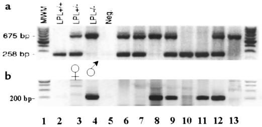
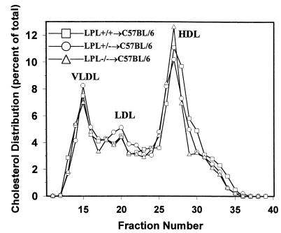
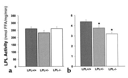
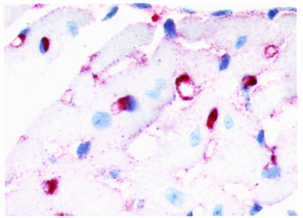
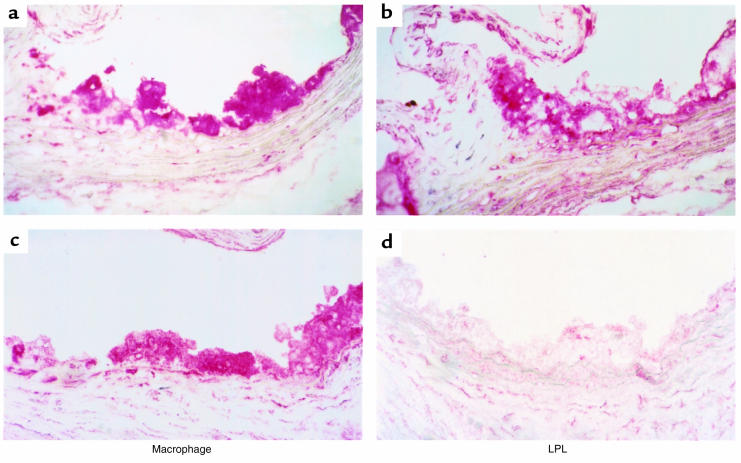
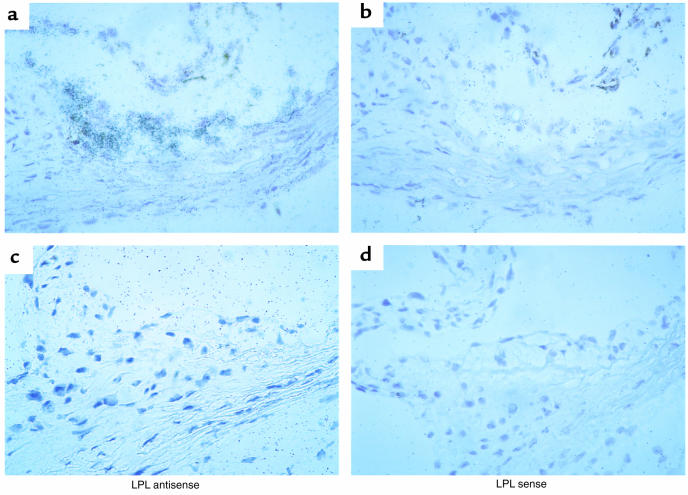
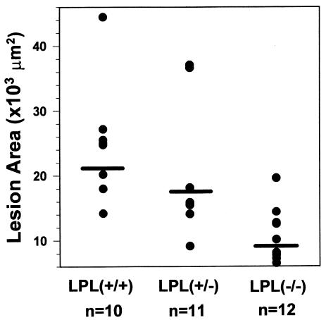
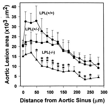
Expression of lipoprotein lipase (LPL) by the macrophage has been proposed to promote foam cell formation and atherosclerosis, primarily on the basis of in vitro studies. LPL-deficient mice might provide a model for testing the role of LPL secretion by the macrophage in an in vivo system. Unfortunately, homozygous deficiency of LPL in the mouse is lethal shortly after birth. Because the fetal liver is the major site of hematopoiesis in the developing fetus, transplantation of C57BL/6 mice with LPL-/- fetal liver cells (FLCs) was used to investigate the physiologic role of macrophage LPL expression in vivo. Thirty-four female C57BL/6 mice were lethally irradiated and reconstituted with FLCs from day 14 LPL+/+, LPL+/-, and LPL-/- donors. No significant differences were detected in plasma levels of post-heparin LPL activity or in serum cholesterol or triglyceride levels between the 3 groups on either a chow diet or an atherogenic diet. After 19 weeks on the atherogenic diet, aortae were collected for quantitative analysis of the extent of aortic atherosclerosis. LPL expression was detected by immunocytochemistry and in situ hybridization in macrophages of aortic atherosclerotic lesions of LPL+/+-->C57BL/6 and LPL+/--->C57BL/6 mice, but not in LPL-/--->C57BL/6 mice, whereas myocardial cells expressed LPL in all groups. The mean aortic lesion area was reduced by 55% in LPL-/--->C57BL/6 mice compared with LPL+/+-->C57BL/6 mice and by 45% compared with LPL+/--->C57BL/6 mice, respectively. These data demonstrate in vivo that LPL expression by macrophages in the artery wall promotes foam cell formation and atherosclerosis. off


Figures

Figure 1
Figure 1
Rapid PCR for identification of LPL genotype and sex of fetuses. Tail tissue was digested in proteinase K and used directly in PCR reaction. After PCR, the samples were separated on a 1.5% agarose gel containing ethidium bromide to view the DNA bands on ultraviolet illumination. (a) A combination of 3 oligonucleotide primers amplify a 258-bp band from exon 8 of the wild-type mouse LPL allele and a 675-bp band from the targeted LPL allele. Lanes 2–5 are control samples: LPL+/+, LPL+/–, LPL–/–, and no DNA, respectively. Lanes 6–13 are DNA samples from 8 fetuses from the same litter. (b) Amplification of a 200-bp band from the Zfy gene of the Y chromosome indicates male sex. Lanes 3 and 4 are female and male control samples, respectively. Lanes 2 and 5 contain no DNA. Lanes 6–13 are DNA samples from the same 8 fetuses as in a.
Figure 2
Figure 2
Lipoprotein distribution in C57BL/6 mice transplanted with LPL+/+, LPL+/– and LPL–/– FLCs after 8 weeks on the atherogenic diet. Mice were fasted for 4 hours. Lipoprotein distribution was determined by FPLC followed by cholesterol analysis of each fraction. Data are represented as an average (n = 3) percent distribution of total cholesterol. Fractions 14–17 contain VLDL; fractions 18–24 are IDL/LDL; and fractions 25–29 contain HDL. Fractions 30–40 are the non–lipoprotein-associated proteins.
Figure 3
Figure 3
LPL activity in post-heparin plasma and heart tissue of C57BL/6 mice transplanted with LPL+/+, LPL+/–, and LPL–/– FLCs. (a) Post-heparin plasma LPL activity. For analysis of plasma LPL activity, mice were fasted for 4 hours and then injected with 200 U heparin (Sigma Chemical Co.) in PBS; 30 minutes later, mice were bled, post-heparin plasma was collected, and LPL activity was measured as described previously (20). (b) Heart tissue LPL activity. Hearts were collected from mice after a 4-hour fast, and the apex of each was snap-frozen in liquid nitrogen. The heart tissue was homogenized later in assay buffer as described previously (20). Data are represented as an average of the 3 mice per group. *P < 0.05 vs. LPL+/+→C57BL/6 mice.
Figure 4
Figure 4
Immunocytochemical detection of macrophages in the myocardium of LPL+/+→C57BL/6 mice. Macrophages are stained with rat mAb MOMA-2 as the primary antibody, followed by biotinylated goat anti-rat IgG as the secondary antibody. The sections were incubated with avidin-biotin complex labeled with alkaline phosphatase, and the enzymatic activity was viewed with Fast Red TR/Naphthol AS-NX substrate. The sections were counterstained with hematoxylin. The primary antibody was omitted during the incubation of negative control sections, and no red staining was seen in these sections (data not shown). ×40.
Figure 5
Figure 5
Immunocytochemical detection of macrophages and LPL in proximal aorta of LPL+/+→C57BL/6 (a and b) and LPL–/–→C57BL/6 (c and d) mice. Macrophages are stained with rat mAb MOMA-2, and LPL is detected with a chicken antibody to recombinant human LPL. Note that LPL expression in control mice is colocalized with macrophages, whereas macrophages in LPL–/–→C57BL/6 mice do not stain for LPL. ×40.
Figure 6
Figure 6
LPL mRNA expression in aortic lesions of mice transplanted with LPL+/+ or LPL–/– FLCs. Expression of LPL mRNA is detected by hybridization of the antisense probe to mouse LPL in foam cell lesions of LPL+/+→C57BL/6 mice (a) but not of LPL–/–→C57BL/6 mice (c). Shown in b and d are the corresponding control serial sections after hybridization with the sense probe for LPL. ×40.
Figure 7
Figure 7
Quantification of atherosclerotic lesion area in mice transplanted with LPL+/+, LPL+/–, and LPL–/– FLCs. The extent of atherosclerotic lesions was quantified after 19 weeks of the atherogenic diet, using oil red O–stained sections from 300 μm of the proximal aorta. Fifteen alternate 10-μm sections from the proximal aorta were examined for each mouse, using a computer-assisted video imaging system. Data are represented as the average mean lesion area for each group. P < 0.001 vs. LPL+/+→C57BL/6 mice; P < 0.002 vs. LPL+/–→C57BL/6.
Figure 8
Figure 8
Comparison of the mean lesion area in progressive sections of the proximal aorta from mice transplanted with LPL+/+, LPL+/–, and LPL–/– FLCs. Mean lesion area per section for 15 alternate 10-μm sections spanning the proximal aorta starting from the aortic sinus, with a 10-μm interval between sections; error bars represent the SEM. *P < 0.05 vs. LPL+/+→C57BL/6 mice. **P < 0.05 vs. LPL+/+→C57BL/6 and LPL+/–→C57BL/6 mice.

References

    1. Brunzell, J.D. 1995. Familial lipoprotein lipase deficiency and other causes of the chylomicronemia syndrome. In The metabolic basis of inherited disease. Volume 2. C.R. Scriver, A.L. Beaudet, W.S. Sly, and D. Valle, editors. McGraw-Hill. New York, NY. 1913–1932.
    1. Saxena U, Klein MG, Goldberg IJ. Identification and characterization of the endothelial cell surface lipoprotein lipase receptor. J Biol Chem. 1991;266:17516–17521. - PubMed
    1. Yla-Herttuala S, et al. Macrophages and smooth muscle cells express lipoprotein lipase in human and rabbit atherosclerotic lesions. Proc Natl Acad Sci USA. 1991;88:10143–10147. - PMC - PubMed
    1. O’Brien KD, Gordon D, Deeb S, Ferguson M, Chait A. Lipoprotein lipase is synthesized by macrophage-derived foam cells in human coronary atherosclerotic plaques. J Clin Invest. 1992;89:1544–1550. - PMC - PubMed
    1. Zilversmit DB. Atherogenesis: a postprandial phenomenon. Circulation. 1979;60:473–485. - PubMed

Publication types

MeSH terms