. 1999 Jun;103(12):1729-35.
doi: 10.1172/JCI6871.

Mitogen-activated protein kinase inhibits 1,25-dihydroxyvitamin D3-dependent signal transduction by phosphorylating human retinoid X receptor alpha

Affiliations

Mitogen-activated protein kinase inhibits 1,25-dihydroxyvitamin D3-dependent signal transduction by phosphorylating human retinoid X receptor alpha

C Solomon et al. J Clin Invest. 1999 Jun.

Abstract

Human retinoid X receptor alpha (hRXR alpha) is a member of the nuclear receptor family of transcriptional regulators. It regulates transcription through its association with several heterodimeric partners, including the vitamin D3 receptor (VDR). Signaling through the VDR is essential for normal calcium homeostasis and has been shown to inhibit the proliferation of cancer cells derived from a number of tissues. Here we show that phosphorylation of hRXR alpha in ras-transformed human keratinocytes through the activated Ras-Raf-mitogen-activated protein kinase (Ras-Raf-MAP kinase) pathway results in attenuated transactivation by the VDR and resistance to the growth inhibitory action of 1,25 dihydroxyvitamin D3 [1,25(OH)2D3] and RXR-specific agonist LG1069 (4-[1-(5,6,7, 8-tetrahydro-3,5,5,8,8-pentamethyl-2-naphthalenyl) ethenyl]-benzoic acid). Phosphorylation of hRXR alpha occurs at serine 260, a consensus MAP kinase site. Inhibition of MAP kinase activity or point mutagenesis of serine 260 of hRXR alpha reverses the observed resistance to 1,25(OH)2D3 and LG1069. Thus, hRXR alpha is a downstream target of MAP kinase, and its phosphorylation may play an important role in malignant transformation.


Figures

Figure 1
Figure 1
Effect of 1,25(OH)2D3 and PD098059 on cell growth in HPK1A and HPK1Aras cells. Cells were transfected with the mOP3 reporter plasmid (20) and the growth hormone expression plasmid pLTR-GH, and then were treated with increasing concentrations of 1,25(OH)2D3 and 5% charcoal-stripped FBS in the absence or presence of 25 μM PD098059. (a) CAT activity was assayed after 24 hours and normalized for transfection efficiency by the corresponding hGH activity. (b) Cells were treated as above, and cell numbers were expressed as percent of vehicle-treated control cell numbers after 96 hours. Each value represents the mean ± SD of 3 determinations and is representative of 3 different experiments. Asterisks indicate a significant difference from vehicle-treated control values, whereas open circles indicate a significant difference between HPK1A and HPK1Aras cells in the absence of PD098059 at the 1,25(OH)2D3 concentration indicated.
Figure 2
Figure 2
Effect of 1,25(OH)2D3 and PD098059 on VDR/RXR complex formation in HPK1A and HPK1Aras cells. (a) A mOP VDRE was 32P labeled and incubated with nuclear extracts from HPK1A cells (lanes 2–4), PD098059-treated HPK1A cells (lanes 5–7), HPK1Aras cells (lanes 8–10), or PD098059-treated HPK1Aras cells (lanes 11–13), each in the presence or absence of either an anti-VDR antibody that recognizes the COOH-terminal domain of VDR (VDR Ab) or an anti-RXR antibody that recognizes the LBD of RXR (RXR Ab). The diamond indicates the presence of putative VDR/RXR complexes, the circle indicates supershifted complexes containing RXR, and the asterisk indicates supershifted complexes containing VDR. In lane 1, no nuclear extracts were incubated with the probe. (b) A mOP VDRE was 32P labeled and incubated with nuclear extracts from HPK1A cells (lanes 1 and 2) or HPK1Aras cells (lanes 3 and 4) in the absence or presence of an anti-RXR antibody that recognizes the NH2-terminal domain of hRXRα (RXR Ab) and inhibits VDR/RXR complex formation on the VDRE. The diamond indicates the presence of putative VDR/RXR complexes. (c) Nuclear extracts prepared from HPK1A and HPK1Aras cells were immunoprecipitated with an anti-RXRα antibody, followed by SDS-PAGE and Western blotting. The blots were treated with an anti-phosphoserine antibody, anti-phosphothreonine antibody, or an anti-RXRα antibody to confirm equal loading of the protein as indicated. The diamond indicates the presence of the hRXRα protein.
Figure 3
Figure 3
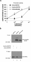
MAP kinase–dependent phosphorylation of hRXRα in HPK1A and HPK1Aras cells. (a) HPK1A cells were transfected with activated or inactivated MAPKK expression plasmids and were treated with increasing concentrations of 1,25(OH)2D3 and 5% charcoal-stripped FBS. CAT activity was assayed after 24 hours, as in Figure 1a. Asterisks indicate a significant difference from vehicle-treated control values, whereas open circles indicate a significant difference between cells overexpressing active and inactive MAPKK. (b) Nuclear extracts prepared from these cells were immunoprecipitated with an anti-RXRα antibody, followed by SDS-PAGE and Western blotting. The blots were treated with an anti-phosphoserine antibody or an anti-RXRα antibody to confirm equal loading of the protein as indicated. The diamond indicates the presence of the hRXRα protein.
Figure 4
Figure 4
Mutation of hRXRα at ser260 abrogates MAP kinase–dependent phosphorylation and eliminates partial resistance to 1,25(OH)2D3. (a) HPK1Aras cells were transfected with vectors expressing either wild-type hRXRα or hRXRα containing the ala260 mutation. Cells were treated with 5% charcoal-stripped FBS and increasing concentrations of 1,25(OH)2D3 as indicated. CAT activity was assayed after 24 hours as in Figure 1a. Asterisks indicate a significant difference from vehicle-treated control values, whereas open circles indicate a significant difference between cells overexpressing wild-type and mutant hRXRα. (b) HPK1A cells transfected with HA-tagged wild-type (lane 1) or mutant (lane 2) hRXRα and the constitutively active MAPKK expression plasmid, and HPK1Aras cells transfected with HA-tagged wild-type (lane 3) or mutant (lane 4) hRXRα, were extracted and immunoprecipitated with an anti-HA antibody. The immunoprecipitates were analyzed by Western blotting. The membrane was incubated with an anti-phosphoserine antibody or with an anti-RXRα antibody as a control for hRXRα levels. The diamond indicates the presence of the hRXRα protein. (c) A mOP VDRE was 32P labeled and incubated with COS-7 cell extracts from cells transfected with wild-type hRXRα and hVDR and treated (lanes 1–3) or not (lane 4) with recombinant MAP kinase, or extracts from cells transfected with the ala260 hRXRα mutant and hVDR, and treated with recombinant MAP kinase (lanes 5-7). The diamond indicates the presence of putative VDR/RXR complexes, and the circle indicates supershifted complexes containing RXR.
Figure 5
Figure 5
Effect of an RXR-specific ligand, LG1069, on HPK1A and HPK1Aras cells. (a) Cells were treated with increasing concentrations of LG1069 (10–10 to 10–6 M) in the absence or presence of 25 μM PD098059 and 5% charcoal-stripped FBS. Cell numbers were expressed as percent of DMSO vehicle-treated control cell numbers after 96 hours. Each value represents the mean ± SD of 3 determinations. Asterisks indicate a significant difference from vehicle-treated control values, whereas open circles indicate a significant difference between HPK1A and HPK1Aras cells at the LG1069 concentration indicated. (b) MTT-Microculture Tetrazolium Assay for Cell Growth (Promega Corp.). Cells were treated as in a, and formazan production was monitored by absorbance as described in Methods. Each value represents the mean ± SD of 4 determinations. Asterisks indicate a significant difference from vehicle-treated control values, whereas open circles indicate a significant difference between HPK1A and HPK1Aras cells at the LG1069 concentration indicated.

References

    1. McClay EF, Albright KD, Jones JA, Christen RD, Howell SB. Tamoxifen delays the development of resistance to cisplatin in human melanoma and ovarian cancer cell lines. Br J Cancer. 1994;70:449–452. - PMC - PubMed
    1. Zachos G, Spandidos GA. Expression of ras proto-oncogenes: regulation and implications in the development of human tumors. Crit Rev Oncol Hematol. 1997;26:65–75. - PubMed
    1. Davis RJ. The mitogen-activated protein kinase signal transduction pathway. J Biol Chem. 1993;268:14553–14556. - PubMed
    1. Weigel, N.L. 1994. Receptor phosphorylation in molecular biology intelligence unit. In Mechanism of steroid hormone regulation of gene transcription. M-J. Tsai, editor. R.G. Landes Co. Austin, Texas. 93–110.
    1. Haussler MR, et al. The nuclear vitamin D receptor: biological and molecular regulatory properties revealed. J Bone Miner Res. 1998;13:325–349. - PubMed

Publication types

MeSH terms