Comparative Study
. 1999 Jun 21;189(12):1907-21.
doi: 10.1084/jem.189.12.1907.

An invariant T cell receptor alpha chain defines a novel TAP-independent major histocompatibility complex class Ib-restricted alpha/beta T cell subpopulation in mammals

Affiliations
Comparative Study

An invariant T cell receptor alpha chain defines a novel TAP-independent major histocompatibility complex class Ib-restricted alpha/beta T cell subpopulation in mammals

F Tilloy et al. J Exp Med. .

Abstract

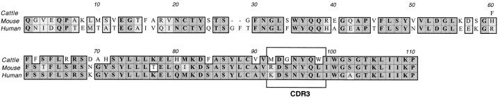
We describe here a new subset of T cells, found in humans, mice, and cattle. These cells bear a canonical T cell receptor (TCR) alpha chain containing hAV7S2 and AJ33 in humans and the homologous AV19-AJ33 in mice and cattle with a CDR3 of constant length. These T cells are CD4(-)CD8(-) double-negative (DN) T cells in the three species and also CD8alphaalpha in humans. In humans, their frequency was approximately 1/10 in DN, 1/50 in CD8alpha+, and 1/6,000 in CD4(+) lymphocytes, and they display an activated/memory phenotype (CD45RAloCD45RO+). They preferentially use hBV2S1 and hBV13 segments and have an oligoclonal Vbeta repertoire suggesting peripheral expansions. These cells were present in major histocompatibility complex (MHC) class II- and transporter associated with antigen processing (TAP)-deficient humans and mice and also in classical MHC class I- and CD1-deficient mice but were absent from beta2-microglobulin-deficient mice, indicating their probable selection by a nonclassical MHC class Ib molecule distinct from CD1. The conservation between mammalian species, the abundance, and the unique selection pattern suggest an important role for cells using this novel canonical TCR alpha chain.


Figures

Figure 1
Figure 1
High levels of hAV7S2/AV19-AJ33 TCR α chain are present in DN T cells from three mammal species: the indicated cell subpopulations were purified by FACS® sorting from human (A and B) or bovine (C and D) PBLs and from mouse lymph nodes (E and F). Duplicate RNA preparations were obtained and, after reverse transcription, PCR was carried out for the indicated gene segments. The amount of amplicon was quantified by ELISA at the indicated cycle. Averaged values of duplicates are shown for C–F. CD8α+ fraction contains both CD8αα+ and CD8αβ+ TCR α/β+ T cells. In cattle, no amplification could be obtained with boAV19-AJ33 primers in the CD8α+ fraction, and thus the corresponding curves are not displayed.
Figure 2
Figure 2
(A) The CDR3 of hAV7S2-AJ33 TCR α chain is of constant length in DN T cells and in human CD8αα+ T cells. PCR amplifications from hAV7S2 to Cα (V-C) or to AJ33 (V-J) were carried out on cDNA from the T cell subpopulations indicated, and polyclonal sequencing was carried out using the same AV7S2/AV19 primer as used for amplification. Panel B displays polyclonal sequencing of FACS®-sorted CD8αβ+ or CD8αα+ TCR α/β+ human PBLs.
Figure 3
Figure 3
High homology of human, cattle, and mouse hAV7S2/AV19-AJ33 α chains. Homologies were scored using the default PAM250 of ClustalW as implemented in MacVector software (Oxford Technology). Identities are bold and shaded in gray; similarities are light gray. The CDR3 region is boxed.
Figure 4
Figure 4
Direct enumeration of AV7S2/AV19-AJ33–bearing cells by PCR–limiting dilution analysis and single cell PCR. (A) DN cells obtained from three human subjects were seeded into microtiter plates using a FACS® single cell deposition unit. After cell lysis, PCR was carried out with AV7S2 and AJ33 primers, and the amount of amplicons was quantified by ELISA. (B and C) α/β CD8α+ or CD4+ cells were obtained by FACS® sorting, serially diluted, distributed in a microtiter plate at the indicated cell concentration, and processed as above. In C (CD4+ cells), the amplicons of the six wells indicated (which had a high probability of clonality) were sequenced. The sequences were as follows:1-GCT GTG ATG TCG AT2-GCT GTG AGA GAG ATC GGA3-GCT GTG AGA GAT4-GCT GNN ACT CGA5-GCT GTG AGA GAG GAT AAC-AJ34-intron-AJ336-GCT GTG AGG GGG GGG GAA AAC-AJ34-intron-AJ33Only one (sequence 3) corresponds to the canonical AV7S2-AJ33 rearrangement (see text for details). In B and C, frequencies were calculated by maximum likelihood analysis.
Figure 5
Figure 5
mAV19-AJ33+ cells are more frequent in TAP−/− mice. The indicated number of DN T cells from B6 (A) or TAP−/− (B) mice were seeded in PCR microplates, and the presence of the mAV19-AJ33 rearrangement was detected by PCR-ELISA.
Figure 6
Figure 6
hAV7S2-AJ33–bearing cells are CD8αα+ or DN. The indicated TCR α/β+ fractions were FACS® sorted using a four-color staining with anti-CD4–TC, anti-CD8α–FITC, anti-CD8β–PE, and anti-α/β TCR–APC. The amounts of Cα (A) or hAV7S2-AJ33 (B) transcripts were quantified by kinetic PCR using the ABI Prism 7700 apparatus (Taqman). This experiment was repeated with cells from another subject with identical results. ΔRn, difference in fluorescence relative units.
Figure 7
Figure 7
AV7S2-AJ33–bearing cells are present in MHC class II– and TAP-deficient patients. The indicated cell subpopulations were obtained from an MHC class II– (A and B) or a TAP-deficient (C and D) patient. The amounts of Cα or hAV7S2-AJ33 transcripts were quantified by kinetic PCR-ELISA. Two other MHC class II– deficient patients and another TAP-deficient patient were studied with similar results.
Figure 8
Figure 8
AV19-AJ33+ cells are selected by a β2m-dependent, TAP-independent nonclassical MHC–like molecule distinct from CD1. DN lymph node cells were obtained from the indicated mice, and the amounts of Cα (A and C) and mAV19-AJ33 (B and D) transcripts were quantified by kinetic PCR-ELISA. All results were confirmed by polyclonal sequencing.

References

    1. Davis, M.M., and Y.-H. Chien. 1998. T-cell antigen receptors. In Fundamental Immunology. 4th ed. W.E. Paul, editor. Lippincott-Raven Publishers, Philadelphia. 341–366.
    1. Hardy RR, Li YS, Hayakawa K. Distinctive developmental origins and specificities of the CD5+ B-cell subset. Semin Immunol. 1996;8:37–44. - PubMed
    1. Kantor AB, Herzenberg LA. Origin of murine B cell lineages. Annu Rev Immunol. 1993;11:501–538. - PubMed
    1. Haas W, Pereira P, Tonegawa S. Gamma/delta cells. Annu Rev Immunol. 1993;11:637–685. - PubMed
    1. Bendelac A, Rivera MN, Park SH, Roark JH. Mouse CD1-specific NK1 T cells: development, specificity, and function. Annu Rev Immunol. 1997;15:535–562. - PubMed

Publication types

MeSH terms