. 1999 Jun 21;189(12):1931-8.
doi: 10.1084/jem.189.12.1931.

Chlamydia inhibits interferon gamma-inducible major histocompatibility complex class II expression by degradation of upstream stimulatory factor 1

Affiliations

Chlamydia inhibits interferon gamma-inducible major histocompatibility complex class II expression by degradation of upstream stimulatory factor 1

G Zhong et al. J Exp Med. .

Abstract

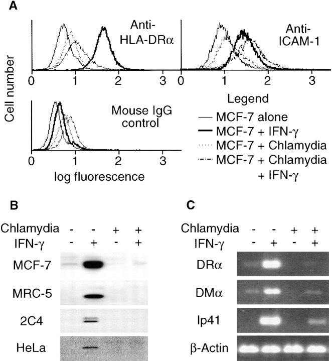
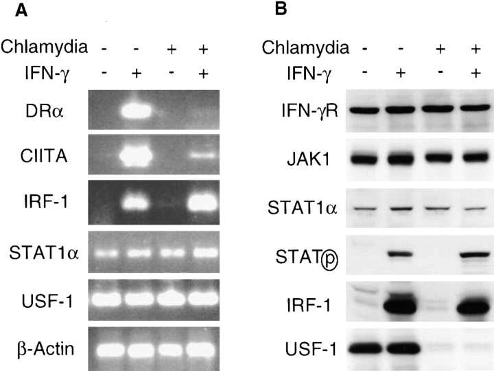
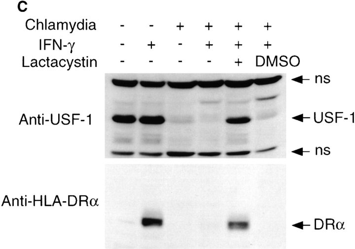
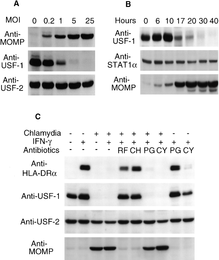
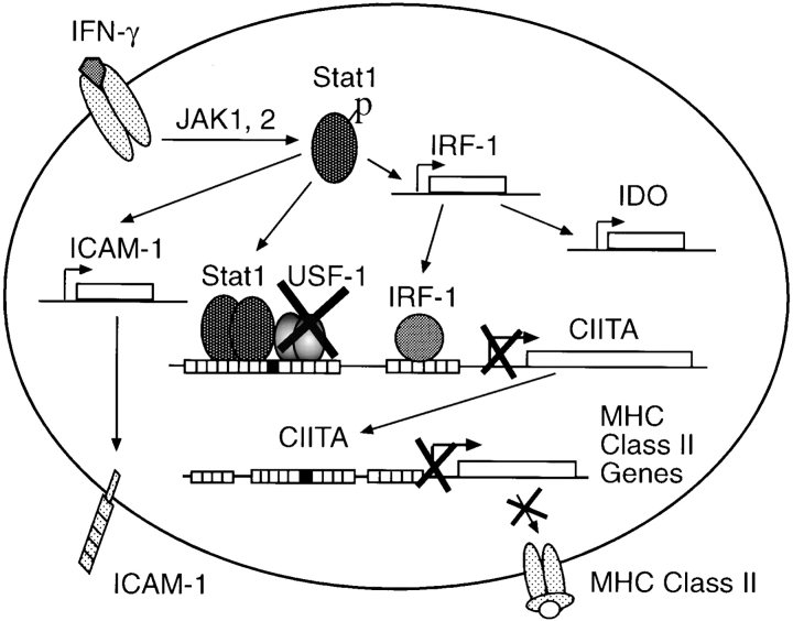
We report that chlamydiae, which are obligate intracellular bacterial pathogens, can inhibit interferon (IFN)-gamma-inducible major histocompatibility complex (MHC) class II expression. However, the IFN-gamma-induced IFN regulatory factor-1 (IRF-1) and intercellular adhesion molecule 1 (ICAM-1) expression is not affected, suggesting that chlamydia may selectively target the IFN-gamma signaling pathways required for MHC class II expression. Chlamydial inhibition of MHC class II expression is correlated with degradation of upstream stimulatory factor (USF)-1, a constitutively and ubiquitously expressed transcription factor required for IFN-gamma induction of class II transactivator (CIITA) but not of IRF-1 and ICAM-1. CIITA is an obligate mediator of IFN-gamma-inducible MHC class II expression. Thus, diminished CIITA expression as a result of USF-1 degradation may account for the suppression of the IFN-gamma-inducible MHC class II in chlamydia-infected cells. These results reveal a novel immune evasion strategy used by the intracellular bacterial pathogen chlamydia that improves our understanding of the molecular basis of pathogenesis.


Figures

Figure 1
Figure 1
Chlamydial infection selectively inhibits IFN-γ–inducible MHC class II expression. MCF-7 cells with or without chlamydial infection were stimulated with IFN-γ and collected for flow cytometry (A), Western blot (B), and RT-PCR (C) analysis. (A) Chlamydial infection prevents IFN-γ–inducible HLA-DR but not ICAM-1 surface expression. (B) Chlamydial infection suppresses the total cellular protein level of IFN-γ–inducible HLA-DRα in various human cell lines. MRC-5, 2C4, and HeLa cells were stimulated with IFN-γ at 400 U/ml. HLA-DRα was detected in a Western blot assay. (C) Chlamydial infection inhibits HLA-DRα, DMα, and Ip41 mRNA expression. Gene-specific primers were used to amplify random primer–directed cDNA templates in an RT-PCR reaction as described in Materials and Methods.
Figure 2
Figure 2
Chlamydial infection disrupts IFN-γ intracellular signaling pathways. (A) Chlamydial infection suppresses both HLA-DRα and CIITA but not IRF-1, STAT1α, or USF-1 mRNA expression in MCF-7 cells. The mRNA levels were analyzed by RT-PCR as described in Materials and Methods. (B) USF-1 protein is not detected in chlamydia-infected MCF-7 cells. The protein levels of IFN-γR, JAK-1, STAT1α, tyrosine-phosphorylated STAT1α, IRF-1, and USF-1 were detected using a Western blot assay. (C) Lactacystin prevents USF-1 degradation and preserves HLA-DRα expression in chlamydia-infected MCF-7 cells. 10 h before IFN-γ stimulation, a chlamydia-infected cell sample was treated with lactacystin (Calbiochem) at a final concentration of  75 μM or with an equivalent amount of solvent DMSO and kept in culture during IFN-γ stimulation. HLA-DRα and USF-1 were detected in a Western blot assay. ns, nonspecific binding.
Figure 2
Figure 2
Chlamydial infection disrupts IFN-γ intracellular signaling pathways. (A) Chlamydial infection suppresses both HLA-DRα and CIITA but not IRF-1, STAT1α, or USF-1 mRNA expression in MCF-7 cells. The mRNA levels were analyzed by RT-PCR as described in Materials and Methods. (B) USF-1 protein is not detected in chlamydia-infected MCF-7 cells. The protein levels of IFN-γR, JAK-1, STAT1α, tyrosine-phosphorylated STAT1α, IRF-1, and USF-1 were detected using a Western blot assay. (C) Lactacystin prevents USF-1 degradation and preserves HLA-DRα expression in chlamydia-infected MCF-7 cells. 10 h before IFN-γ stimulation, a chlamydia-infected cell sample was treated with lactacystin (Calbiochem) at a final concentration of  75 μM or with an equivalent amount of solvent DMSO and kept in culture during IFN-γ stimulation. HLA-DRα and USF-1 were detected in a Western blot assay. ns, nonspecific binding.
Figure 3
Figure 3
Chlamydial protein synthesis is required for both USF-1 degradation and inhibition of HLA-DRα expression. (A) Correlation between infection dose and USF-1 degradation. 24 h after chlamydial infection at various MOI, MCF-7 cells were analyzed for chlamydial MOMP and host USF-1 and USF-2 protein levels in a Western blot assay. As the USF-2 protein level was not altered by chlamydial infection, it served as an internal control. (B) Time course relationship between chlamydial growth and USF-1 degradation. At various time points after infection, MCF-7 cells were lysed for the detection of USF-1, STAT1α, and MOMP in a Western blot assay. (C) Inhibition of chlamydial but not host protein synthesis prevents USF-1 degradation and preserves HLA-DRα expression. Rifampin (RF; final concentration, 0.1 μg/ml), chloramphenicol (CH; 60 μg/ml), and penicillin (PG; 100 μg/ml) were added at the beginning of chlamydial infection and maintained throughout the culture. Cycloheximide (CY; 10 μg/ml) was added to the culture 10 h before IFN-γ treatment and maintained during the IFN-γ stimulation. The MCF-7 cells were analyzed for protein levels of HLA-DRα, USF-1, USF-2, and chlamydial MOMP in a Western blot assay.
Figure 4
Figure 4
Model for chlamydial inhibition of IFN-γ–inducible MHC class II expression. IFN-γ binding activates intracellular JAK/STAT pathways. USF-1 is a constitutively and ubiquitously expressed downstream transcription factor required for IFN-γ–inducible CIITA expression. Chlamydial infection degrades USF-1, which results in diminished expression of CIITA in chlamydia-infected cells. As CIITA is an obligate mediator for transcription of MHC class II genes, chlamydial degradation of USF-1 finally leads to suppression of IFN-γ–inducible MHC class II. However, because the upstream pathways of IFN-γ signaling are still intact in chlamydia-infected cells, expression of IRF-1, ICAM-1, and IDO is not affected by chlamydial infection.

Similar articles

Cited by

References

    1. Germain RN. MHC-dependent antigen processing and peptide presentation: providing ligands for T lymphocyte activation. Cell. 1994;76:287–299. - PubMed
    1. Ploegh HL. Viral strategies of immune evasion. Science. 1998;280:248–253. - PubMed
    1. Ahn K, Gruhler A, Galocha B, Jones TR, Wiertz EJ, Ploegh HL, Peterson PA, Yang Y, Fruh K. The ER-luminal domain of the HCMV glycoprotein US6 inhibits peptide translocation by TAP. Immunity. 1997;6:613–621. - PubMed
    1. Hengel H, Flohr T, Hammerling GJ, Koszinowski UH, Momburg F. Human cytomegalovirus inhibits peptide translocation into the endoplasmic reticulum for MHC class I assembly. J Gen Virol. 1996;77:2287–2296. - PubMed
    1. Lehner PJ, Karttunen JT, Wilkinson GW, Cresswell P. The human cytomegalovirus US6 glycoprotein inhibits transporter associated with antigen processing-dependent peptide translocation. Proc Natl Acad Sci USA. 1997;94:6904–6909. - PMC - PubMed

Publication types

MeSH terms