. 1999 Jun 21;189(12):1973-80.
doi: 10.1084/jem.189.12.1973.

Susceptibility of mice deficient in CD1D or TAP1 to infection with Mycobacterium tuberculosis

Affiliations

Susceptibility of mice deficient in CD1D or TAP1 to infection with Mycobacterium tuberculosis

S M Behar et al. J Exp Med. .

Abstract

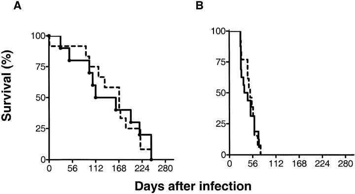
Cellular immunity against Mycobacterium tuberculosis controls infection in the majority of infected humans. Studies in mice have delineated an important role for CD4(+) T cells and cytokines including interferon gamma and tumor necrosis factor alpha in the response to infection with mycobacteria. Recently, the identification of CD8(+) CD1-restricted T cells that kill M. tuberculosis organisms via granulysin and the rapid death after infection of beta2 microglobulin deficient mice in humans has drawn attention to a critical role for CD8(+) T cells. The nature of mycobacterial-specific CD8(+) T cells has been an enigma because few have been identified in any species. Here, we delineate the contribution of class I MHC-restricted T cells in the defense against tuberculosis as transporter associated with antigen processing (TAP)1-deficient mice died rapidly, bore a greater bacterial burden, and had more severe tissue pathology than control mice. In contrast, CD1D-/- mice were not significantly different in their susceptibility to infection than control mice. This data demonstrates a critical role for TAP-dependent peptide antigen presentation and provides further evidence that class I MHC-restricted CD8(+) T cells, the major T cell subset activated by this antigen processing pathway, play an essential role in immunity to tuberculosis.


Figures

Figure 1
Figure 1
Survival of CD1D−/− mice after infection with M. tuberculosis. CD1D−/− mice on a resistant genetic background (C57BL/6) (A) or a susceptible genetic background (BALB/c) (B) were inoculated with 106 M. tuberculosis intravenously. Each group contained 12–13 mice and there were no statistically significant differences in survival of the CD1D−/− mice compared with CD1D+/+ mice using a Mann-Whitney test. A, P = 0.91; B, P = 0.95.
Figure 2
Figure 2
Peripheral blood CD8+ T cells in TAP1−/− mice and controls after infection with M. tuberculosis. 5 wk after infection, blood was obtained and stained with a CD8-specific antibody. The percentage of CD8+ cells in peripheral blood lymphocytes was determined by flow cytometry for four mice/ group. The bar represents the mean for each group. This difference was statistically significant using a Mann-Whitney test (P = 0.0022).
Figure 3
Figure 3
Survival of TAP1−/− mice infected with M. tuberculosis. TAP1−/− mice or controls were infected with 106 M. tuberculosis intravenously. The data presented are from three separate experiments and represent a total of 42 mice in each group. The survival curves were generated using the Kaplan-Meier method and the difference is statistically significant with a P < 0.0001 by the log-rank test.
Figure 4
Figure 4
Survival of TAP1−/− CD1−/− mice infected with M. tuberculosis. CD1D−/−TAP1−/− or CD1D+TAP1−/− littermate controls were infected with 106 M. tuberculosis. There were no statistically significant differences in survival (P = 0.95).
Figure 5
Figure 5
Mycobacterial burden in TAP1−/− and control mice after infection. The number of CFU recovered from the lung, spleen, and liver was determined at the time points indicated after intravenous infection with 106 M. tuberculosis. The baseline mycobacterial inoculum was determined 24 h after infection. Each data point is the mean bacterial counts from three to five mice ± SEM.
Figure 6
Figure 6
Histopathology of the lung from TAP1+/+ (A–D) and TAP1−/− (E–H) mice after infection with M. tuberculosis. Mice were killed at the indicated times after infection, tissues were harvested, and formalin-fixed, paraffin-embedded sections were stained with Fite-Faraco stain for AFB or with the hematoxylin and eosin 1 d (B and F), 3 wk (A, C, E, and G), or 7 wk (D and H) after infection. Original magnification: A and E, ×2,000; B–D and F–H, ×300.

References

    1. World Health Organization. 1998. Global Tuberculosis Programme. Tuberculosis Fact Sheet. WHO, Geneva. http://www.who.int/gtb/publications/factsheet/index.htm
    1. Russell DG, Dant J, Sturgill-Koszycki S. Mycobacterium avium- and Mycobacterium tuberculosis-containing vacuoles are dynamic, fusion-competent vesicles that are accessible to glycosphingolipids from the host cell plasmalemma. J Immunol. 1996;156:4764–4773. - PubMed
    1. Muller I, Cobbold SP, Waldmann H, Kaufmann SH. Impaired resistance to Mycobacterium tuberculosisinfection after selective in vivo depletion of L3T4+ and Lyt-2+ T cells. Infect Immun. 1987;55:2037–2041. - PMC - PubMed
    1. Orme IM. The kinetics of emergence and loss of mediator T lymphocytes acquired in response to infection with Mycobacterium tuberculosis. . J Immunol. 1987;138:293–298. - PubMed
    1. Orme IM, Collins FM. Protection against Mycobacterium tuberculosisinfection by adoptive immunotherapy. Requirement for T cell–deficient recipients. J Exp Med. 1983;158:74–83. - PMC - PubMed

Publication types

MeSH terms