

Calcium-activated potassium conductances contribute to action potential repolarization at the soma but not the dendrites of hippocampal CA1 pyramidal neurons
- PMID: 10377332
- PMCID: PMC6782335
- DOI: 10.1523/JNEUROSCI.19-13-05205.1999
Calcium-activated potassium conductances contribute to action potential repolarization at the soma but not the dendrites of hippocampal CA1 pyramidal neurons
Abstract
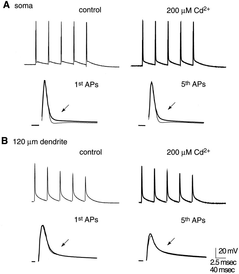
Evidence is accumulating that voltage-gated channels are distributed nonuniformly throughout neurons and that this nonuniformity underlies regional differences in excitability within the single neuron. Previous reports have shown that Ca2+, Na+, A-type K+, and hyperpolarization-activated, mixed cation conductances have varying distributions in hippocampal CA1 pyramidal neurons, with significantly different densities in the apical dendrites compared with the soma. Another important channel mediates the large-conductance Ca2+-activated K+ current (IC), which is responsible in part for repolarization of the action potential (AP) and generation of the afterhyperpolarization that follows the AP recorded at the soma. We have investigated whether this current is activated by APs retrogradely propagating in the dendrites of hippocampal pyramidal neurons using whole-cell dendritic patch-clamp recording techniques. We found no IC activation by back-propagating APs in distal dendritic recordings. Dendritic APs activated IC only in the proximal dendrites, and this activation decayed within the first 100-150 micrometer of distance from the soma. The decay of IC in the proximal dendrites occurred despite AP amplitude, plus presumably AP-induced Ca2+ influx, that was comparable with that at the soma. Thus we conclude that IC activation by action potentials is nonuniform in the hippocampal pyramidal neuron, which may represent a further example of regional differences in neuronal excitability that are determined by the nonuniform distribution of voltage-gated channels in dendrites.
Figures


Similar articles
-
Dendritic potassium channels in hippocampal pyramidal neurons.J Physiol. 2000 May 15;525 Pt 1(Pt 1):75-81. doi: 10.1111/j.1469-7793.2000.00075.x. J Physiol. 2000. PMID: 10811726 Free PMC article. Review.
-
Dendritic voltage-gated ion channels regulate the action potential firing mode of hippocampal CA1 pyramidal neurons.J Neurophysiol. 1999 Oct;82(4):1895-901. doi: 10.1152/jn.1999.82.4.1895. J Neurophysiol. 1999. PMID: 10515978
-
Voltage-gated potassium channels activated during action potentials in layer V neocortical pyramidal neurons.J Neurophysiol. 2000 Jan;83(1):70-80. doi: 10.1152/jn.2000.83.1.70. J Neurophysiol. 2000. PMID: 10634854
-
Arachidonic acid reciprocally alters the availability of transient and sustained dendritic K(+) channels in hippocampal CA1 pyramidal neurons.J Neurosci. 1999 Oct 1;19(19):8163-71. doi: 10.1523/JNEUROSCI.19-19-08163.1999. J Neurosci. 1999. PMID: 10493718 Free PMC article.
-
Control of Na+ spike backpropagation by intracellular signaling in the pyramidal neuron dendrites.Mol Neurobiol. 2000 Aug-Dec;22(1-3):129-41. doi: 10.1385/MN:22:1-3:129. Mol Neurobiol. 2000. PMID: 11414276 Review.
Cited by
-
Mice with deficient BK channel function show impaired prepulse inhibition and spatial learning, but normal working and spatial reference memory.PLoS One. 2013 Nov 26;8(11):e81270. doi: 10.1371/journal.pone.0081270. eCollection 2013. PLoS One. 2013. PMID: 24303038 Free PMC article.
-
The role of BK-type Ca2+-dependent K+ channels in spike broadening during repetitive firing in rat hippocampal pyramidal cells.J Physiol. 1999 Nov 15;521 Pt 1(Pt 1):135-46. doi: 10.1111/j.1469-7793.1999.00135.x. J Physiol. 1999. PMID: 10562340 Free PMC article.
-
Dendritic potassium channels in hippocampal pyramidal neurons.J Physiol. 2000 May 15;525 Pt 1(Pt 1):75-81. doi: 10.1111/j.1469-7793.2000.00075.x. J Physiol. 2000. PMID: 10811726 Free PMC article. Review.
-
Amplitude-dependent spike-broadening and enhanced Ca(2+) signaling in GnRH-secreting neurons.Biophys J. 2000 Sep;79(3):1310-23. doi: 10.1016/S0006-3495(00)76384-3. Biophys J. 2000. PMID: 10968994 Free PMC article.
-
Progressive dendritic HCN channelopathy during epileptogenesis in the rat pilocarpine model of epilepsy.J Neurosci. 2007 Nov 21;27(47):13012-21. doi: 10.1523/JNEUROSCI.3605-07.2007. J Neurosci. 2007. PMID: 18032674 Free PMC article.
References
-
- Callaway JC, Ross WN. Frequency-dependent propagation of sodium action potentials in dendrites of hippocampal CA1 pyramidal neurons. J Neurophysiol. 1995;74:1395–1403. - PubMed
Publication types
MeSH terms
Substances
Grants and funding
LinkOut - more resources
Full Text Sources
Other Literature Sources
Miscellaneous