. 1999 Jul 1;19(13):5332-47.
doi: 10.1523/JNEUROSCI.19-13-05332.1999.

The native rat olfactory cyclic nucleotide-gated channel is composed of three distinct subunits

Affiliations

The native rat olfactory cyclic nucleotide-gated channel is composed of three distinct subunits

W Bönigk et al. J Neurosci. .

Abstract

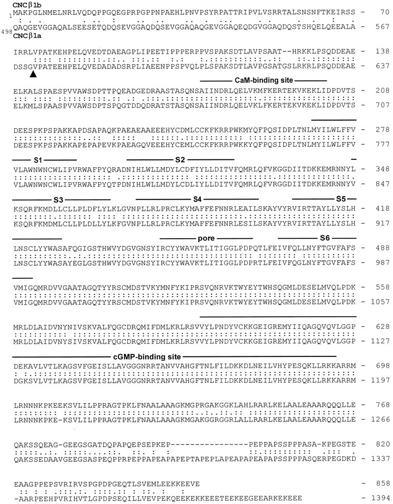
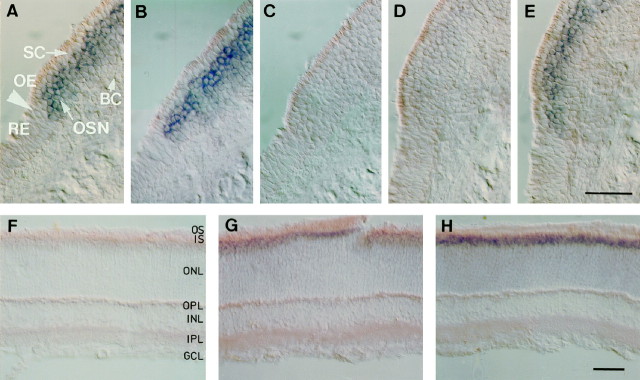
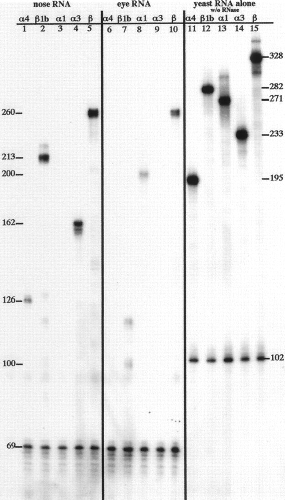
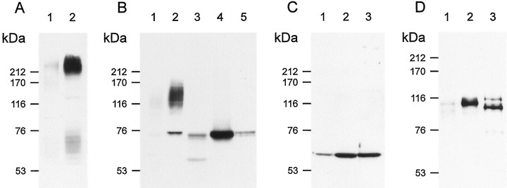
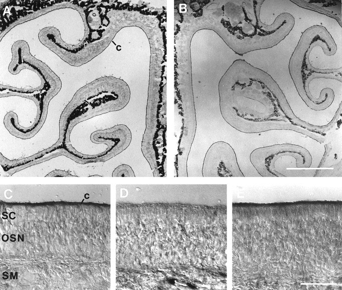
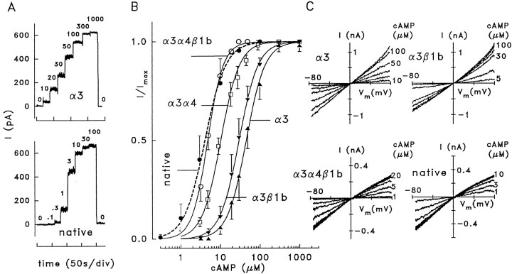
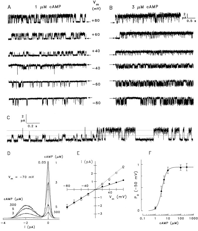
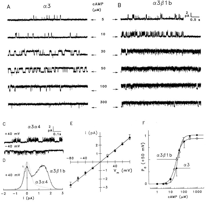
Cyclic nucleotide-gated (CNG) channels play central roles in visual and olfactory signal transduction. In the retina, rod photoreceptors express the subunits CNCalpha1 and CNCbeta1a. In cone photoreceptors, only CNCalpha2 expression has been demonstrated so far. Rat olfactory sensory neurons (OSNs) express two homologous subunits, here designated CNCalpha3 and CNCalpha4. This paper describes the characterization of CNCbeta1b, a third subunit expressed in OSNs and establishes it as a component of the native channel. CNCbeta1b is an alternate splice form of the rod photoreceptor CNCbeta1a subunit. Analysis of mRNA and protein expression together suggest co-expression of all three subunits in sensory cilia of OSNs. From single-channel analyses of native rat olfactory channels and of channels expressed heterologously from all possible combinations of the CNCalpha3, -alpha4, and -beta1b subunits, we conclude that the native CNG channel in OSNs is composed of all three subunits. Thus, CNG channels in both rod photoreceptors and olfactory sensory neurons result from coassembly of specific alpha subunits with various forms of an alternatively spliced beta subunit.


Figures

Fig. 1.
Fig. 1.
Alignment of the deduced amino acid sequence of rat olfactory CNCβ1b with bovine rod CNCβ1a. Numbersindicate positions of amino acid residues in the polypeptide. The sequence of CNCβ1a is presented starting at residue 498.Colons and periods between the two sequences indicate identical residues and conservative substitutions, respectively. Structural features similar to those of α subunits are represented by lines above the sequence.S1–S6, Membrane-spanning segments; S4, voltage sensor-like motif; P, the pore motif that lines the cavity of the channel; CaM, a nonconventional calmodulin-binding site (Weitz et al., 1998). Arrowheadindicates an exon boundary identified in human rod CNCβ1a (Ardell et al., 1996).
Fig. 2.
Fig. 2.
Cell type-specific expression of CNCα3, -α4, and -β1b mRNAs in the olfactory epithelium. In situhybridization analysis of olfactory channel subunits expressed in rat olfactory epithelium (A–E) and retina (F–H). Expression of channel mRNA was examined in 20-μm-thick sections using digoxigenin-labeled antisense RNA probes against nonconserved regions of each subunit. Visualization was achieved with an AP-conjugated anti-digoxigenin antibody. A positive signal is indicated by a purple AP reaction product. Shown are signals with probes against (A, F) the olfactory specific 5′ region of CNCβ1b, (B, G) the 3′ region in common with CNCβ1a and CNCβ1b, (C, H) CNCα1, (D) CNCα4, and (E) CNCα3. Arrows inA mark the transition zone between olfactory epithelium (OE) and respiratory epithelium (RE). Positive signals correspond to expression in olfactory sensory neurons (OSN), not supporting cells (SC) or basal cells (BC). In F–H, positive signals are restricted to the inner segment (IS) layer of photoreceptors; other layers are outer segments (OS), outer nuclear layer (ONL), outer plexiform layer (OPL), inner nuclear layer (INL), inner plexiform layer (IPL), and ganglion cell layer (GCL). Scale bars, 50 μm.
Fig. 3.
Fig. 3.
Analysis of CNC α and β mRNA expression by RNase protection. 32P-labeled antisense RNA probes were hybridized to total RNA from olfactory or eye tissue, subjected to RNase digestion, and resolved on a sequencing gel. Left panel, lanes 1–5, Protection of probes for GAPDH and CNC subunits α4, β1b, α1, α3, and β by olfactory RNA. Center panel, lanes 6–10, Protection of probes for GAPDH and CNC subunits α4, β1b, α1, α3, and β by eye RNA. Right panel, lanes 11–15, Probes for GAPDH and CNC subunits α4, β1b, α1, α3, and β. Numbers along right sideindicate length of undigested probe. Numbers alongleft side indicate length of protected products obtained with olfactory or eye RNA. See Materials and Methods for complete description of probes and protected products. Exposure shown was for 70 hr at room temperature.
Fig. 4.
Fig. 4.
Analysis by Western blot of CNCα3, -α4 and -β1b expression in sensory cilia of rat olfactory epithelium.A, Western blot of equal amounts of membrane protein prepared from either whole olfactory epithelium (OE) (lane 1) or from isolated olfactory cilia (lane 2) probed with an anti-ACIII-specific antibody produced a much stronger ∼230 kDa signal with the cilia preparations. B, Western blot probed with polyclonal anti-α3 antibody. The antibody recognized a ∼75 kDa band and a “fuzzy” smear between ∼110 and 145 kDa in the preparations from both whole OE (lane 1) and cilia (lane 2). The CNCα3 subunit expressed in HEK 293 cells displays an apparent molecular mass of ∼75 kDa (lane 5, 10 μg protein), suggesting that the 75 kDa band recognized in olfactory tissue, in fact, represents the CNCα3 subunit. Treatment of membrane proteins from whole OE (lane 3) and cilia (lane 4) with N-glycosidase F abolished the smear entirely and correspondingly increased the intensity of the 75 kDa band. C, Western blot of membranes derived from whole OE (lane 1), cilia (lane 2), and HEK 293 cells expressing CNCα4 (lane 3) probed with monoclonal antibody mAB7B11 against the CNCα4 subunit. D, Western blot as in C, probed with polyclonal antibody FP21K against CNCβ1b subunit.Lane 3 is membrane derived from HEK 293 cells expressing CNCβ1b. As seen in B with CNCα3, both the CNCα4- and -β1b antibodies produced much stronger signals with preparations of cilia membranes, indicating that the molar concentration of the respective polypeptide (relative to the total protein content) is higher in ciliary-enriched than in whole OE membranes.
Fig. 5.
Fig. 5.
Immunohistochemical staining of olfactory epithelium. A, Upper part of the nasal cavity, stained with an antibody against ACIII. Strong staining is found in the thin ciliary layer (c) covering the epithelium on the septum (right border) and turbinates. The cartilage appears dark because of an artifact of the low-power optics but is not stained. B, The anti-α3 antibody homogeneously stained the ciliary layer. C, Higher magnification of the field shown in B. D, Antibody mAB7B11 against the CNCα4 subunit. No staining above background is found in the cilia. E, Purified antiserum FPc21K against the β subunit strongly stained the cilia of OSNs. Scale bars: A, B, 1 mm; C–E, 50 μm.
Fig. 6.
Fig. 6.
Ligand sensitivity and ion selectivity of native and heterologously expressed olfactory CNG channels. A, Macroscopic current recordings from inside-out patches of HEK 293 cells transfected with CNCα3 and from a patch excised from a dendritic knob of a rat OSN containing native CNG channels. Current was recorded at +40 mV and the indicated cAMP concentrations. B, Dose–response relations for the activation of macroscopic currents by cAMP at +40 mV. Lines were constructed by fitting a Hill-type function to the normalized current (see Materials and Methods). Fitting parameters are given in Table 1. C, Macroscopic currents recorded from inside-out patches with the indicated cAMP concentrations. The main permeable ions were Na+(extracellular) and K+ (intracellular). The current shows inward rectification only with α3α4β1b and native channels, indicating that these channels conduct Na+ better than K+.
Fig. 7.
Fig. 7.
Single-channel analysis of native CNG channels from rat OSNs. A, Recording from an inside-out patch with 1 μm cAMP at the indicated membrane voltages.Arrows indicate the closed state. The channel shows a significant voltage dependence of open probability. B, The same channel as in A at 3 μm cAMP. Positive voltages favor the open state, whereas negative voltages induce continuous flickering. Single-channel recordings were obtained in symmetrical Na+ solutions, sampled at 3 kHz and filtered at 1 kHz. C, Transition of the native channel from a state of low conductance (14–16 pS) to the conductance level that is observed most of the time (35 pS); 1 μm cAMP, +60 mV. D, All-point amplitude histograms of four 40 sec recordings from a native channel at −70 mV and the indicated cAMP concentrations. Although brief, unresolved closing events result in a comparably broad current distribution in the open state (approximately −1.9 pA), the two peaks reflecting open and closed states can be clearly distinguished. The ordinate indicates the percentage of total time spent at each current level plotted. E, Voltage dependence of the single-channel current recorded in symmetrical (○, means of 5 channels), and bi-ionic conditions (●, means of 9 channels). The conductance at negative voltages (fitted to the mean values) was 27 pS in both symmetrical and bi-ionic (intracellular K+, extracellular Na+) conditions. At positive voltages the conductance was 35 pS in symmetrical and 14.5 pS in bi-ionic conditions. F, Dependence of open probability on cAMP concentration. Single-channel analysis from four patches at −50 mV yieldedK1/2 = 3.4 μm,n = 2.3.
Fig. 8.
Fig. 8.
Analysis of channels consisting of CNCα3 plus the CNCβ1b or CNCα4 subunit. A, Single-channel recording from a homomeric α3 channel at +50 mV and the indicated concentrations of cAMP. The channel shows a lower ligand sensitivity than native channels and long uninterrupted dwell periods in the open state. B, Single-channel recording from a α3β1b channel. Coexpression of the CNCβ1b subunit slightly increases ligand sensitivity and causes rapid open–closed transitions at both positive and negative membrane voltages. C, Recordings from α3α4 channels at 10 μm cAMP display extremely rapid flickering. D, All-point histograms from single-channel recordings from α3α4 channels (10 μm cAMP, +40 mV,dotted line) and α3β1b channels (30 μmcAMP, +40 mV, solid line). The skewed histogram obtained for α3α4 channels did not permit determination of single-channel current and open probability. E, Voltage dependence of single-channel current yielded an apparent conductance of 33.7 ± 2.7 pS (12 patches) for α3 (●) and 34.3 ± 1.4 pS (9 patches) for α3β1b channels (○). F, cAMP dependence of open probability measured at +50 mV. Solid lines were fitted for α3 channels (●, 4 patches) withK1/2 = 40 μm,n = 2.5, and for α3β1b channels (○, 3 patches) with K1/2 = 28 μm, n = 1.9.
Fig. 9.
Fig. 9.
Single-channel analysis of α3α4β1b channels.A, Single-channel recording from an α3α4β1b channel at +50 mV and the indicated concentrations of cAMP.B, Dependence of the open probability on cAMP concentration. Top panel, All-point amplitude histogram of 19–23 sec recordings at 1, 3, 10, and 100 μm cAMP and +50 mV. Bottom panel, Mean values obtained from four single-channel recordings were fitted with a Hill-type equation usingK1/2 = 4 μm,n = 2, and a maximal P0of 0.9. C, Voltage dependence of channel current for symmetrical Na+ solutions (21 pS inward, 27 pS outward) and for bi-ionic solutions (21 pS for Na+inward, 12 pS for K+ outward currents).D, Recording from an α3α4β1b channel at +60 mV with 3 μm cAMP, showing a 9.5 sec episode during which the channel switched from the main gating state into a substate with reduced conductance and different gating kinetics (arrows). E, Transition of an α3α4β1b channel from the main conductance state (27 pS) to a subconductance state (15 pS, top trace) and back to the main state (bottom trace); 5 μm cAMP, −50 mV; symmetrical Na+ solutions.

References

    1. Anholt RRH, Aebi U, Snyder SH. A partially purified preparation of isolated chemosensory cilia from the olfactory epithelium of the bullfrog, Rana catesbeiana. J Neurosci. 1986;6:1962–1965. - PMC - PubMed
    1. Ardell MD, Aragon I, Oliveira L, Porche GE, Burke E, Pittler SJ. The β subunit of human rod photoreceptor cGMP-gated cation channel is generated from a complex transcription unit. FEBS Lett. 1996;389:213–218. - PubMed
    1. Bakalyar HA, Reed RR. Identification of a specialized adenylyl cyclase that may mediate odorant detection. Science. 1990;250:1403–1406. - PubMed
    1. Balasubramanian S, Lynch JW, Barry PH. Calcium-dependent modulation of the agonist affinity of the mammalian olfactory cyclic nucleotide-gated channel by calmodulin and a novel endogenous factor. J Membr Biol. 1996;152:13–23. - PubMed
    1. Baumann A, Frings S, Godde M, Seifert R, Kaupp UB. Primary structure and functional expression of a Drosophila cyclic nucleotide-gated channel present in eyes and antennae. EMBO J. 1994;13:5040–5050. - PMC - PubMed

Publication types

MeSH terms

Associated data