. 1999 Jul 1;19(13):5348-59.
doi: 10.1523/JNEUROSCI.19-13-05348.1999.

The Caenorhabditis elegans unc-49 locus encodes multiple subunits of a heteromultimeric GABA receptor

Affiliations

The Caenorhabditis elegans unc-49 locus encodes multiple subunits of a heteromultimeric GABA receptor

B A Bamber et al. J Neurosci. .

Abstract

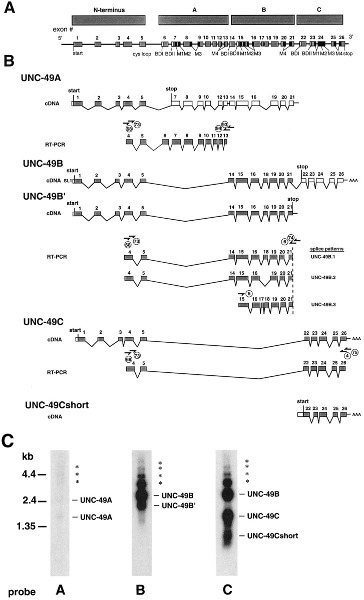
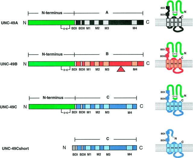
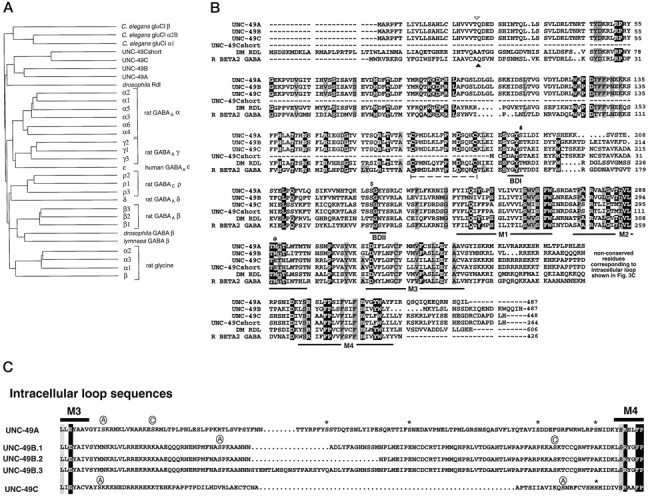
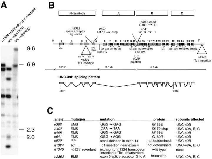
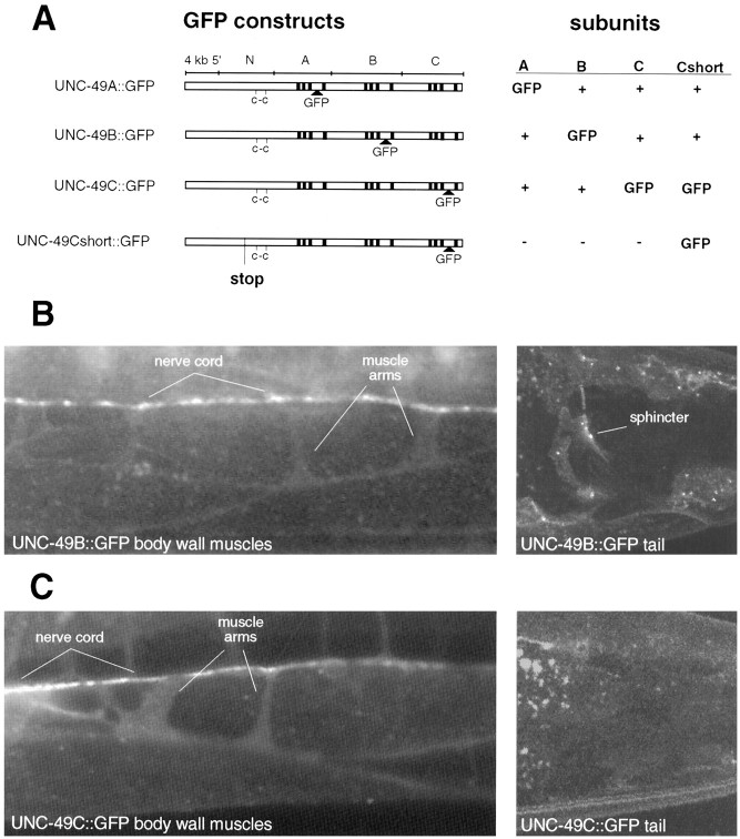
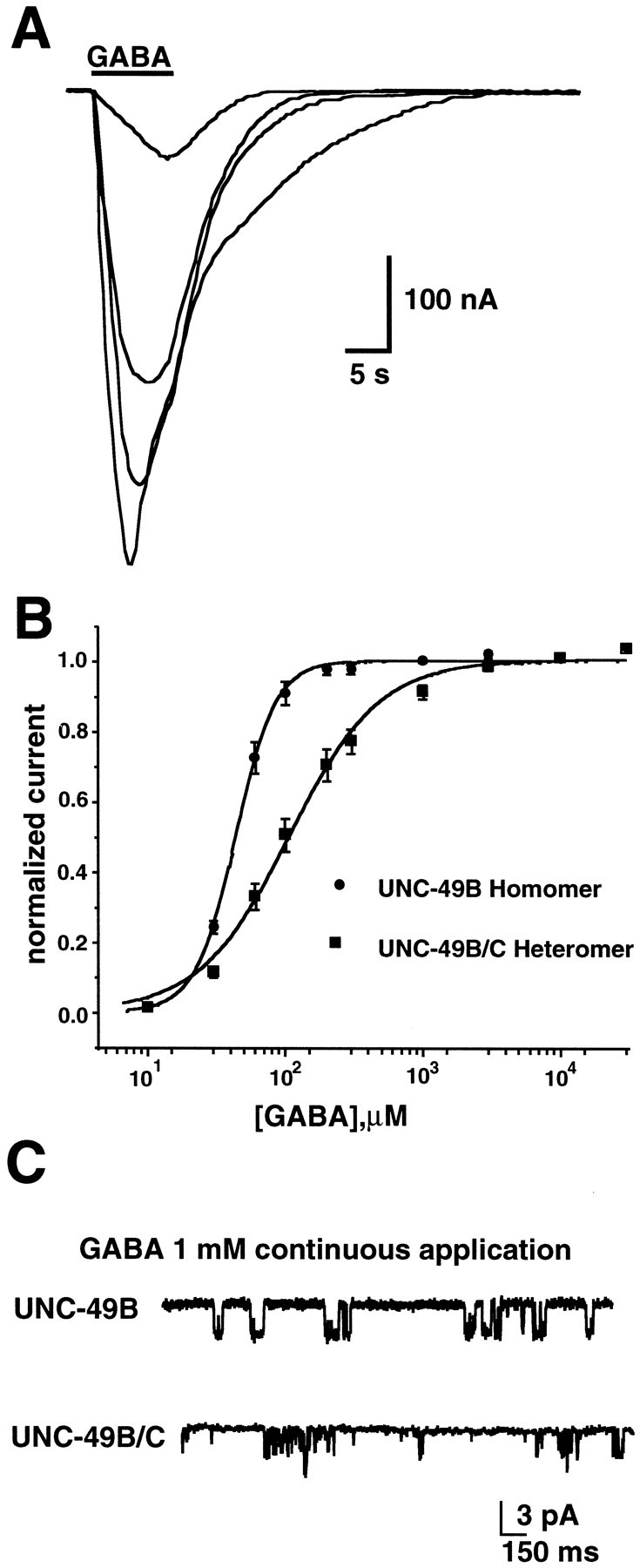
Ionotropic GABA receptors generally require the products of three subunit genes. By contrast, the GABA receptor needed for locomotion in Caenorhabditis elegans requires only the unc-49 gene. We cloned unc-49 and demonstrated that it possesses an unusual overlapping gene structure. unc-49 contains a single copy of a GABA receptor N terminus, followed by three tandem copies of a GABA receptor C terminus. Using a single promoter, unc-49 generates three distinct GABAA receptor-like subunits by splicing the N terminus to each of the three C-terminal repeats. This organization suggests that the three UNC-49 subunits (UNC-49A, UNC-49B, and UNC-49C) are coordinately rescued and therefore might coassemble to form a heteromultimeric GABA receptor. Surprisingly, only UNC-49B and UNC-49C are expressed at high levels, whereas UNC-49A expression is barely detectable. Green fluorescent protein-tagged UNC-49B and UNC-49C subunits are coexpressed in muscle cells and are colocalized to synaptic regions. UNC-49B and UNC-49C also coassemble efficiently in Xenopus oocytes and HEK-293 cells to form a heteromeric GABA receptor. Together these data argue that UNC-49B and UNC-49C coassemble at the C. elegans neuromuscular junction. Thus, C. elegans is able to encode a heteromeric GABA receptor with a single locus.


Figures

Fig. 1.
Fig. 1.
unc-49 produces three distinct GABA receptor subunits. A, Structure of theunc-49 locus showing the positions of conserved GABA receptor structural motifs. Domain structure of the locus is indicated by bars at the top (see Results).B, unc-49 mRNA structure. Transcripts were isolated both from cDNA libraries and from RT-PCR experiments. Multiple UNC-49A cDNA clones were identified with different splicing patterns, all resulting in premature stops. One representative example is shown here. Properly spliced RNAs were identified by RT-PCR; theshort arrows and circled numbersrepresent PCR primers. Two superimposed primers (for example, 68 and 73) represent a set of nested PCR primers. Theshaded boxes represent coding exons, and the open boxes represent untranslated regions. The SL1 splice leader was found at the 5′ ends of the mRNA species where indicated.C, Northern analysis of poly(A+) RNA. The probes, indicated below each lane, correspond to the C-terminal repeats. Labels to the right of each lane indicate the probable identity of each band. In the UNC-49C lane the UNC-49B mRNA is visible because it contains the UNC-49C open reading frame in its 3′ UTR. Asterisks indicate higher molecular weight bands that may correspond to partially splicedunc-49 pre-mRNA. All lanes were exposed for the same length of time.
Fig. 2.
Fig. 2.
Structural overlap among unc-49subunits. UNC-49A, UNC-49B, and UNC-49C are identical over the N-terminal 40% of their length, but they contain different putative GABA-binding domains and transmembrane domains. The left panel shows an alignment of each subunit mRNA (thebar at the top indicates the origin of exons encoding each portion). The triangle indicates the position of the alternative splice site in UNC-49B. Note that the UNC49Cshort subunit is identical to the unique C-terminal portion of UNC-49C but that it lacks the entire N terminus common to the other subunits; in its place are four unique N-terminal amino acids (gray box). The right paneldepicts the predicted unc-49 subunit proteins.
Fig. 3.
Fig. 3.
GABA receptor family. A, Dendrogram of GABA receptor subunits. The three unc-49 subunits do not correspond to any of the vertebrate classes of GABAAreceptor subunits. Alignments were performed with the Pileup program in the Genetics Computer Group analysis package. B, Sequence alignment of unc-49 subunits. Residues inblack boxes are conserved in all members of a set of seven representative non-C. elegans GABA receptor subunits, and residues in gray boxes are conserved in six of seven members of this set (see Materials and Methods). The rat β2 GABAA and Drosophila rdl receptor subunits are included for comparison. The dashed lineindicates the disulfide-bonded loop motif (CX13C) conserved in all ligand-gated ion channel subunits. The barslabeled BDI and BDII indicate putative GABA-binding domains, and the bars labeledM1–M4 indicate membrane-spanning domains. Residues in BDI and BDII, which are functionally important in the ρ and β GABA receptor subunits but are divergent in the C. eleganssubunits, are denoted by # and $, respectively. The unusual glutamic acid residue in UNC-49C M2 is denoted by @. Arrowheadsindicate predicted sites of signal peptide cleavage for UNC-49B and UNC-49C and the rat β2 subunit. Residues are numbered from the predicted start of translation, except for the rat β2 subunit, which is numbered from the predicted signal peptide cleavage site according to convention. UNC-49B is numbered according to the UNC-49B.1 sequence.C, Residues comprising the M3–M4 intracellular loops of the unc-49-encoded subunits. Sequences of the three UNC-49B isoforms are shown also. Intracellular loop sequences have not been aligned. The symbols above each intracellular loop indicate potential regulatory phosphorylation sites (Aindicates a PKA site, C indicates a PKC site, and anasterisk indicates a CKII site).
Fig. 4.
Fig. 4.
All unc-49 mutations affect UNC-49B. A, Southern blot ofEcoRV-digested genomic DNA probed with T21C12 insert DNA. The numbers at the right indicate the positions of DNA size standards. B, Positions of mutations in the unc-49 alleles are shown.e382, e468, e641,and e929 affect only UNC-49B, wherease407, n1324, and n2392affect UNC-49A, UNC-49B, and UNC-49C. The bars at thetop represent unc-49 domains.C, Summary of unc-49 mutations.
Fig. 5.
Fig. 5.
UNC-49B and UNC-49C are coexpressed and colocalized. A, Structure of UNC-49:: GFP transgenes. The left panel shows the site at which GFP was inserted, in frame, into the unc-49 rescuing fragment. Vertical bars represent transmembrane domains. The right panel shows the subunits that are produced by the transgene. GFP indicates subunits tagged with GFP; + indicates wild-type subunits; − indicates inactivated subunits. B, Fluorescence micrographs of UNC-49B:: GFP transgenic worms. Left panel, Bright GFP fluorescence is visible in a punctate pattern along the nerve cord, where neuromuscular junctions are located. Fainter GFP fluorescence is also visible outlining the muscle cell bodies (lens-shaped bodies beneath the nerve cord) and muscle arms (narrow processes extending from the muscle cell bodies to the nerve cord).Right panel, Tail region of an UNC-49B:: GFP worm showing bright fluorescence in the sphincter muscle. C, Fluorescence micrographs of UNC-49C:: GFP transgenic worms. The pattern of fluorescence is similar to that observed in the UNC-49B:: GFP transgenic animals in the body wall muscles and nerve cord (left panel). However, no fluorescence is visible in the sphincter muscle (right panel).
Fig. 6.
Fig. 6.
UNC-49B and UNC-49C coassemble in heterologous cells. A, Response of a representative UNC-49B.1-injected oocyte to 10 sec pulses of GABA at 10, 30, 60, and 100 μm. B, GABA dose–response curves obtained from Xenopus oocytes injected with UNC-49B (circles) or UNC-49B plus UNC-49C (squares). Error bars represent SEM. C, Single-channel recordings from HEK-293 cells expressing UNC-49B alone (top trace) or UNC-49B plus UNC-49C (bottom trace).

Similar articles

Cited by

References

    1. Alfonso A, Grundahl K, McManus JR, Asbury JM, Rand JB. Alternative splicing leads to two cholinergic proteins in Caenorhabditis elegans. J Mol Biol. 1994;241:627–630. - PubMed
    1. Amin J, Weiss DS. Homomeric ρ1 GABA channels: activation properties and domains. Receptors Channels. 1994;2:227–236. - PubMed
    1. Amin J, Weiss DS. GABAA receptor needs two homologous domains of the beta-subunit for activation by GABA but not by pentobarbital. Nature. 1993;366:565–569. - PubMed
    1. Andres AJ, Thummel CS. Methods for quantitative analysis of transcription in larvae and prepupae. Methods Cell Biol. 1994;44:565–573. - PubMed
    1. Ausubel FM, Brent R, Kingston RE, Moore DD, Seidman JG, Smith JA, Struhl K. Current protocols in molecular biology. Wiley; New York: 1995.

Publication types

MeSH terms

LinkOut - more resources