

Brain mechanisms of propofol-induced loss of consciousness in humans: a positron emission tomographic study
- PMID: 10377359
- PMCID: PMC6782309
- DOI: 10.1523/JNEUROSCI.19-13-05506.1999
Brain mechanisms of propofol-induced loss of consciousness in humans: a positron emission tomographic study
Abstract
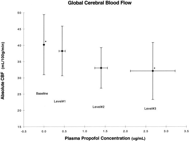
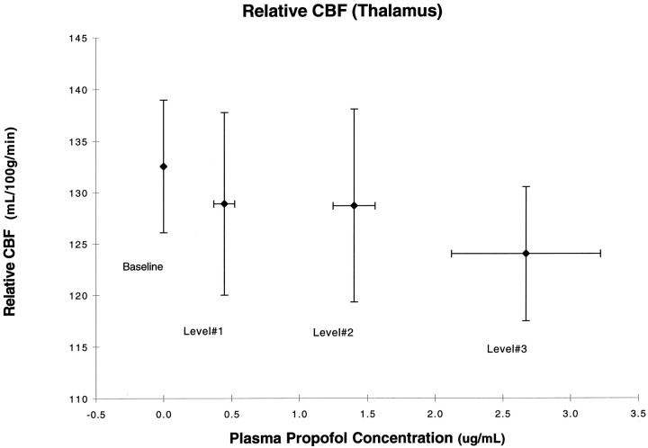
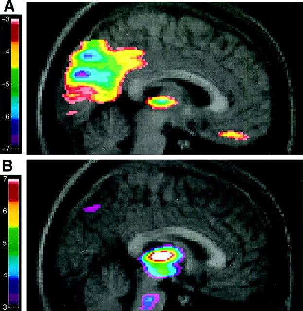
In the present study, we used positron emission tomography to investigate changes in regional cerebral blood flow (rCBF) during a general anesthetic infusion set to produce a gradual transition from the awake state to unconsciousness. Five right-handed human volunteers participated in the study. They were given propofol with a computer-controlled infusion pump to achieve three stable levels of plasma concentrations corresponding to mild sedation, deep sedation, and unconsciousness, the latter defined as unresponsiveness to verbal commands. During awake baseline and each of the three levels of sedation, two scans were acquired after injection of an H215O bolus. Global as well as regional CBF were determined and correlated with propofol concentrations. In addition, blood flow changes in the thalamus were correlated with those of the entire scanned volume to determine areas of coordinated changes. In addition to a generalized decrease in global CBF, large regional decreases in CBF occurred bilaterally in the medial thalamus, the cuneus and precuneus, and the posterior cingulate, orbitofrontal, and right angular gyri. Furthermore, a significant covariation between the thalamic and midbrain blood flow changes was observed, suggesting a close functional relationship between the two structures. We suggest that, at the concentrations attained, propofol preferentially decreases rCBF in brain regions previously implicated in the regulation of arousal, performance of associative functions, and autonomic control. Our data support the hypothesis that anesthetics induce behavioral changes via a preferential, concentration-dependent effect on specific neuronal networks rather than through a nonspecific, generalized effect on the brain.
Figures
References
-
- Alkire MT, Haier RJ, Barker SJ, Shah NK, Wu JC, Kao J. Cerebral metabolism during propofol anesthesia in humans studied with positron emission tomography. Anesthesiology. 1995;82:393–403. - PubMed
-
- Angel A. Central neuronal pathways and the process of anaesthesia. Br J Anaesth. 1993;71:148–163. - PubMed
-
- Casati A, Fanelli G, Casaletti E, Colnaghi E, Cedrati V, Torri G. Clinical assessment of target-controlled infusion of propofol during monitored anesthesia care. Can J Anaesth. 1999;46:235–239. - PubMed
-
- Cavazzuti M, Porro CA, Barbieri A, Galetti A. Brain and spinal cord metabolic activity during propofol anaesthesia. Br J Anaesth. 1991;66:490–495. - PubMed
Publication types
MeSH terms
Substances
LinkOut - more resources
Full Text Sources
Other Literature Sources