. 1999 Jun 22;96(13):7214-9.
doi: 10.1073/pnas.96.13.7214.

NFAT5, a constitutively nuclear NFAT protein that does not cooperate with Fos and Jun

Affiliations

NFAT5, a constitutively nuclear NFAT protein that does not cooperate with Fos and Jun

C Lopez-Rodríguez et al. Proc Natl Acad Sci U S A. .

Abstract

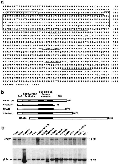
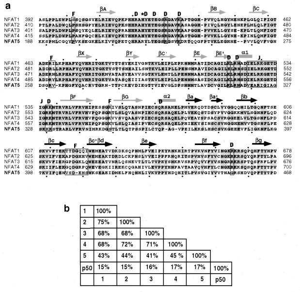
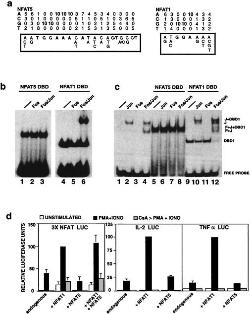
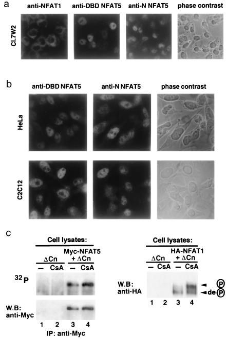
NFAT transcription factors are related to NF-kappaB/Rel proteins and form cooperative complexes with Fos and Jun on DNA. We have identified an NFAT-related protein, NFAT5, which differs from the conventional NFAT proteins NFAT1-4 in its structure, DNA binding, and regulation. NFAT5 contains a NFAT-like Rel homology domain, conserves the DNA contact residues of NFAT1-4, and binds DNA sequences similar to those found in the regulatory regions of well-characterized NFAT-dependent genes. However, it lacks the majority of Fos/Jun contact residues and does not bind cooperatively with Fos and Jun to DNA. Unlike NFAT1-4, whose nuclear import is tightly regulated by calcineurin-mediated dephosphorylation, NFAT5 is a constitutively nuclear phosphoprotein regardless of calcineurin activation. These features suggest that unlike the conventional NFAT proteins, NFAT1-4, which activate gene transcription by integrating inputs from calcium/calcineurin and protein kinase C/mitogen-activated protein kinase signaling pathways, NFAT5 participates in as-yet-unidentified signaling pathways in diverse immune and nonimmune cells.


Figures

Figure 1
Figure 1
Sequence and expression pattern of NFAT5. (a) Predicted amino acid sequence of human NFAT5. The Rel homology region and a bipartite nuclear localization sequence are indicated by large and small boxes, respectively. Two polyglutamine stretches and a putative protein interaction motif (41) in the C-terminal region are singly and doubly underlined. (b) Schematic comparison of the five NFAT family members. Selected splice variants of NFAT1–4 are shown. The DNA-binding domain (DBD) conserved in NFAT1–5 and the regulatory domain (REG) represented in NFAT1–4 are indicated. TAD, transactivation domain. (c) Widespread expression of NFAT5 mRNA. The single NFAT5 transcript (≈13 kb) is expressed at highest levels in skeletal muscle, brain, heart, and peripheral blood leukocytes (PBL), and at moderate to detectable levels in other tissues. The blots were rehybridized with a human β-actin cDNA probe as a control for RNA loading.
Figure 2
Figure 2
Comparison of the DNA-binding domains of NFAT1–5. (a) Alignment of the Rel homology regions of human NFAT1–5. Residues that are identical in at least three of the five proteins are shown in gray. The arrows and bars above the sequence indicate the secondary structure elements deduced from the crystal structure of the NFAT1:Fos:Jun:DNA complex (25). Light and dark arrows are used for the N- and C-terminal portions of the Rel homology domain. The residues implicated in DNA (D), Fos (F), and Jun (J) contacts are boxed. The asterisk indicates alteration of the invariant His residue in the DNA-recognition loops of NFAT1–4 to Arg in NFAT5. (b) Pairwise sequence comparison of the Rel homology regions of p50 and NFAT1–5. Numbers indicate the percentage of identical residues.
Figure 3
Figure 3
DNA binding, Fos-Jun interaction, and transactivation by NFAT5. (a) Identification of the optimal binding site for NFAT5. The boxed sequences summarize the results of aligning 45 NFAT5-selected and 21 NFAT1-selected PCR clones, obtained from four independent selections. At each position, the probability of occurrence of each of the four nucleotides is normalized to an overall value of 10, with the numbers rounded off to the nearest integer. Bases showing a greater than 60% probability of occurrence are indicated in bold. (b) NFAT5 does not bind cooperatively with Fos and Jun to the murine IL-2 promoter ARRE-2 element. DBD, DNA-binding domain. (c) NFAT5 does not bind cooperatively with Fos and Jun to the ARRE-2 element modified to contain a strong consensus AP-1 site. (d) NFAT5 lacks the ability to activate luciferase (LUC) expression driven by three copies of the ARRE-2 site, the human IL-2 promoter, or the human tumor necrosis factor α (TNF-α) promoter. PMA, phorbol 12-myristate 13-acetate; IONO, ionomycin.
Figure 4
Figure 4
Nuclear localization and phosphorylation status of NFAT5. (a) Nuclear localization of endogenous NFAT5 in a resting murine T cell clone. The first three panels show immunocytochemical staining of Cl.7W2 murine T cells with anti-NFAT1, or antisera against the N-terminal region (anti-N) or DNA-binding domain (anti-DBD) of NFAT5. (Right) Phase-contrast photomicrograph of the field stained with anti-N of NFAT5. (b) Nuclear localization of endogenous NFAT5 in resting HeLa (fibroblast, Upper) and C2C12 (myoblast, Lower) cells. (Left and Middle) Antisera against the NFAT5 DNA-binding domain (anti-DBD) or N-terminal region (anti-N) of NFAT5. (Right) Phase-contrast photomicrograph of the same field stained with anti-N. (c) Analysis of NFAT5 phosphorylation. (Upper) 293 cells expressing ΔCn or Myc-NFAT5-GFP and ΔCn were metabolically labeled with 32P-orthophosphate in the absence or presence of 1 μM CsA. NFAT5 was immunoprecipitated (IP) with anti-Myc and detected by autoradiography (32P) and Western blotting (W.B.). (Lower) In a parallel experiment, activity of ΔCn and inhibition by CsA were monitored by expressing hemagglutinin (HA)-tagged NFAT1 in 293 cells and evaluating its phosphorylation status in SDS-lysates by Western blotting with anti-HA. Arrows indicate the dephosphorylated and phosphorylated forms of NFAT1.

References

    1. McCaffrey P G, Luo C, Kerppola T K, Jain J, Badalian T M, Ho A M, Burgeon E, Lane W S, Lambert J N, Curran T, et al. Science. 1993;262:750–754. - PubMed
    1. Northrop J P, Ho S N, Chen L, Thomas D J, Timmerman L A, Nolan G P, Admon A, Crabtree G R. Nature (London) 1994;369:497–502. - PubMed
    1. Park J, Takeuchi A, Sharma S. J Biol Chem. 1996;271:20914–20921. - PubMed
    1. Chuvpilo C, Zimmer M, Kerstan A, Glockner J, Avots A, Escher C, Fischer C, Jankevikcs E, Berberich-Siebelt F, Schmitt E, Serfling E. Immunity. 1999;10:261–269. - PubMed
    1. Hoey T, Sun Y L, Williamson K, Xu X. Immunity. 1995;2:461–472. - PubMed

Publication types

Associated data