. 1999 Jun 22;96(13):7220-5.
doi: 10.1073/pnas.96.13.7220.

Identification of human prostaglandin E synthase: a microsomal, glutathione-dependent, inducible enzyme, constituting a potential novel drug target

Affiliations

Identification of human prostaglandin E synthase: a microsomal, glutathione-dependent, inducible enzyme, constituting a potential novel drug target

P J Jakobsson et al. Proc Natl Acad Sci U S A. .

Abstract

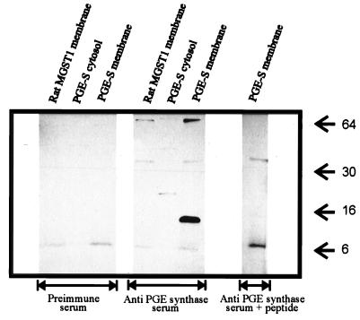
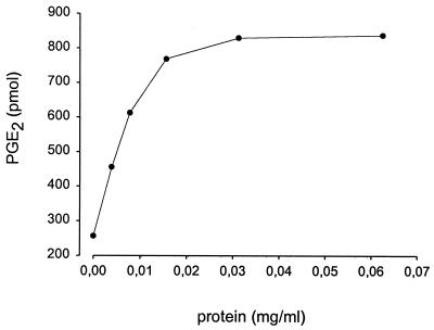
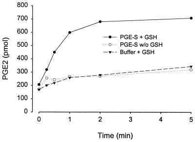
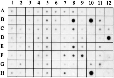
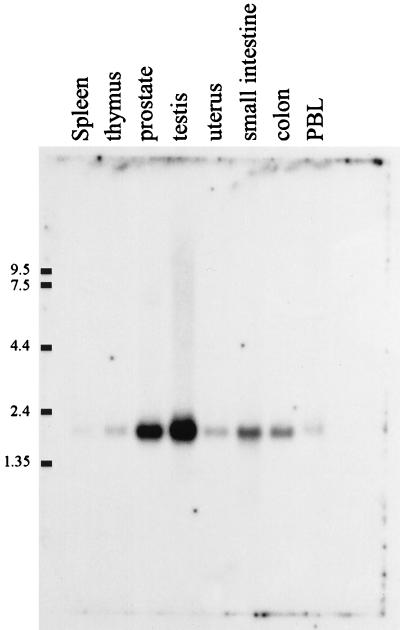
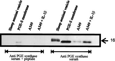
Human prostaglandin (PG) E synthase (EC 5.3.99.3) is a member of a recently recognized protein superfamily consisting of membrane associated proteins involved in eicosanoid and glutathione metabolism (the MAPEG family). Previous designations of the protein are PIG12 and MGST1-L1. PGE synthase was expressed in Escherichia coli, and both cytosolic and membrane fractions were prepared. Western blot analysis specifically detected a 15- to 16-kDa protein in the membrane fraction. Both fractions were incubated with prostaglandin H2 in the presence or absence of reduced glutathione. The membrane but not the cytosolic fraction was found to possess high glutathione-dependent PGE synthase activity (0.25 micromol/min/mg). The human tissue distribution was analyzed by Northern blot analysis. High expression of PGE synthase mRNA was detected in A549 and HeLa cancer cell lines. Intermediate level of expression was demonstrated in placenta, prostate, testis, mammary gland, and bladder whereas low mRNA expression was observed in several other tissues. A549 cells have been used as a model system to study cyclooxygenase-2 induction by IL-1beta. If A549 cells were grown in the presence of IL-1beta, a significant induction of the PGE synthase was observed by Western blot analysis. Also, Western blot analysis specifically detected a 16-kDa protein in sheep seminal vesicles. In summary, we have identified a human membrane bound PGE synthase. The enzyme activity is glutathione-dependent, and the protein expression is induced by the proinflammatory cytokine IL-1beta. PGE synthase is a potential novel target for drug development.


Figures

Figure 1
Figure 1
Predicted amino acid sequence of PGE synthase. This ORF was used for subsequent protein expression in bacteria. Underlined letters represent amino acids aligning together with MGST1 after running gap within the wisconsin package 9.0 (Genetics Computer Group, Madison, WI).
Figure 2
Figure 2
Western blot analysis of PGE synthase expression. Both the membrane and cytosolic fractions from bacteria expressing PGE synthase were analyzed by SDS/PAGE and Western blotting. As control, the membrane fraction from bacteria expressing ratMGST1 was included. In all lanes, 5 μg of total protein were analyzed. The middle blot demonstrates the results obtained by using antipeptide antiserum against PGE synthase whereas the left blot shows the results using the corresponding preimmune serum. For the right blot, the antipeptide antiserum was diluted in the presence of 10−6 M peptide antigen. Arrows denote the migration of protein size markers. The exposure time was 2 min.
Figure 3
Figure 3
RP-HPLC chromatogram of the products formed after incubations with PGH2. (A) PGE synthase membrane fraction premixed with stop solution before addition of 3H-PGH2 (10 μM). (B) Buffer control. (C) PGE synthase membrane fraction. B and C were incubated with 3H-PGH2 (10 μM) for 2 min. Products were detected by using radioactivity detection. The first 20 min represents isocratic elution using water, acetonitrile, and trifluoroacetic acid (70:30:0.007, by vol) as mobile phase with a flow rate of 1 ml/min. Then, a linear gradient was applied from 100% mobile phase to 100% methanol over a 10-min period, which was sustained for the rest of the run. CPM, counts per minute.
Figure 4
Figure 4
Dependency of PGE2 formation on membrane protein concentration. Various dilutions of the membrane fraction isolated from bacteria expressing PGE synthase were incubated with PGH2 (10 μM) in the presence of glutathione (2.5 mM) for 2 min. Product formation was analyzed by RP-HPLC, and PGE2 was detected and quantified by using UV absorbance at 195 nm.
Figure 5
Figure 5
Time course of PGE2 formation. The membrane fraction obtained from bacteria expressing PGE synthase (0.02 mg/ml) was incubated with PGH2 (10 μM) for the indicated times in the presence (filled circles) or absence (open circles) of glutathione (2.5 mM). Filled triangles represent nonenzymatic (buffer only) PGE2 formation after incubation with PGH2. The product formation was analyzed by RP-HPLC, and PGE2 was detected and quantified by using UV absorbance at 195 nm.
Figure 6
Figure 6
Northern dot blot analysis of PGE synthase in human tissues. A human multiple tissue expression array was hybridized with a radiolabeled cDNA probe specific for PGE synthase. Because the amount of mRNA on each dot has been adjusted to produce normalized signals for various housekeeping genes, the array allows for comparisons of gene expression. The exposure time was 66 h. The various dots represent the following: A1, whole brain; B1, cerebral cortex; C1, frontal lobe; D1, parietal lobe; E1, occipital lobe; F1, temporal lobe; G1, paracentral gyrus of cerebral cortex; H1, pons; A2, cerebellum, left; B2, cerebellum, right; C2, corpus callosum; D2, amygdala; E2, caudate nucleus; F2, hippocampus; G2, medulla oblongata; H2, putamen; A3, substantia nigra; B3, accumbens nucleus; C3, thalamus; D3, pituitary gland; E3, spinal cord; F-H3, empty; A4, heart; B4, aorta; C4, atrium, left; D4, atrium, right; E4, ventricle, left; F4, ventricle, right; G4, interventricular septum; H5, apex of the heart; A5, esophagus; B5, stomach; C5, duodenum; D5, jejunum; E5, ileum; F5, ilocecum; G5, appendix; H5, colon, ascending; A6, colon, transverse; B6, colon, desending; C6, rectum; D-H6, empty; A7, kidney; B7, skeletal muscle; C7, spleen; D7, thymus; E7, peripheral blood leukocytes; F7, lymph node; G7, bone marrow; H7, trachea; A8, lung; B8, placenta; C8, bladder; D8, uterus; E8, prostate; F8, testis; G8, ovary; H8, empty; A9, liver; B9, pancreas; C9, adrenal gland; D9, thyroid gland; E9, salivary gland; F9, mammary gland; G-H9, empty; A10, HL-60; B10, HeLa S3; C10, K-562; D10, Molt-4; E10, Burkitt’s lymphoma, Raji; F10, Burkitt’s lymphoma, Daudi; G10, colorectal adenocarcinoma, SW480; H10, lung carcinoma, A549; A11, fetal brain; B11, fetal heart; C11, fetal kidney; D11, fetal liver; E11, fetal spleen; F11, fetal thymus; G11, fetal lung; H11, empty; A12, yeast, total RNA; B12, yeast tRNA; C12, E. coli rRNA; D12, E. coli DNA; E12, Poly r(A); F12, human CoT-1 DNA; G12, human DNA 100 ng; H12, human DNA 500 ng.
Figure 7
Figure 7
Northern blot analysis of PGE synthase in human tissues. This human multiple tissue mRNA blot was hybridized with a radiolabeled cDNA probe specific for PGE synthase. The migration of various RNA size markers is indicated. Each lane represent 2 μg of mRNA from the indicated tissues (no normalization for different amounts of housekeeping genes). The exposure time was 32 h.
Figure 8
Figure 8
Western blot analysis of PGE synthase in A549 cells and sheep seminal vesicles. The microsomal fractions (10 μg) from cells grown for 24 h in the presence (1 ng/ml) or absence of IL-1β were fractionated by SDS/PAGE and were transferred to poly(vinylidene difluoride) membrane. The membranes were incubated by using either PGE synthase antiserum or PGE synthase antiserum containing the antigenic peptide (10−6 M). Also analyzed was the commercially available, partly purified PGE synthase from sheep seminal vesicles (6 μg) as well as the membrane fraction from bacteria expressing human PGE synthase (1 μg).

References

    1. Smith W. Adv Exp Med Biol. 1997;400B:989–1011. - PubMed
    1. Herschman H R. Biochim Biophys Acta. 1996;1299:125–140. - PubMed
    1. Dubois R, Abramson S, Crofford L, Gupta R, Simon L, Van de Putte L, Lipsky P. FASEB J. 1998;12:1063–1073. - PubMed
    1. Smith W L. Am J Physiol. 1992;263:F181–F191. - PubMed
    1. Hamberg M, Samuelsson B. J Biol Chem. 1967;242:5336–5343. - PubMed

Publication types