. 1999 Jun 22;96(13):7250-5.
doi: 10.1073/pnas.96.13.7250.

The active form of the steroidogenic acute regulatory protein, StAR, appears to be a molten globule

Affiliations

The active form of the steroidogenic acute regulatory protein, StAR, appears to be a molten globule

H S Bose et al. Proc Natl Acad Sci U S A. .

Abstract

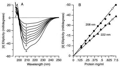
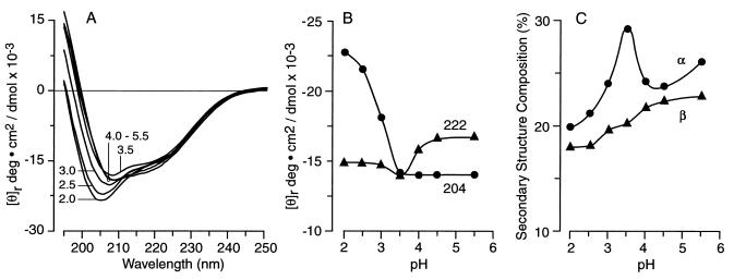
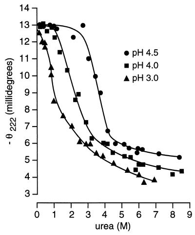
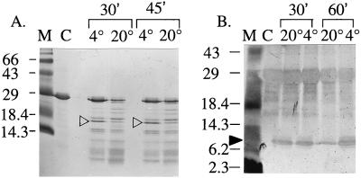
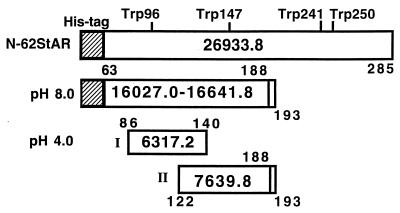
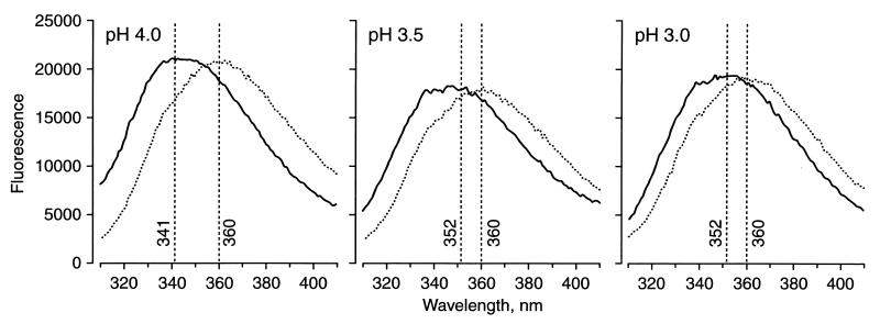
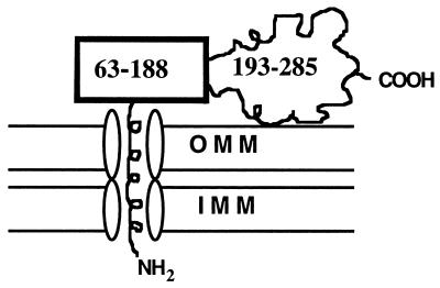
The steroidogenic acute regulatory protein (StAR) increases the movement of cholesterol from the outer to the inner membrane of adrenal and gonadal mitochondria, thus providing the substrate for steroid hormone biosynthesis. Deletion of 62 amino-terminal aa produces a cytoplasmic form of StAR (N-62 StAR) that lacks the mitochondrial leader sequence but retains full activity and appears to act at the outer mitochondrial membrane. At neutral pH the native state of bacterially expressed N-62 StAR protein displays cooperative unfolding under the influence of urea with DeltaGH2O = -4.1 kcal/mol, and it remains correctly folded down to pH 4. Limited proteolysis at different pHs shows that the biologically essential C-terminal region is accessible to solvent, and that the N-terminal domain is compact at pH 8 and partially unfolds below pH 4. Secondary structural analysis of CD curves suggests that the unfolding may coincide with an increase in alpha-helical character at pH 3.5. Fluorescence spectroscopy at pH 3-8 and at 0-6 M urea is consistent with two distinct domains, a compact N-terminal domain containing tryptophans 96 and 147 and a more solvent-accessible C-terminal domain containing tryptophans 241 and 250. These observations suggest that StAR forms a molten globule structure at pH 3.5-4.0. As the mitochondrial proton pump results in an electrochemical gradient, and as StAR must unfold during mitochondrial entry, StAR probably undergoes a similar conformational shift to an extended structure while interacting with the mitochondrial outer membrane, allowing this apparent molten globule form to act as an on/off switch for cholesterol entry into the mitochondria.


Figures

Figure 1
Figure 1
Effect of protein concentration on conformation. (A) Unsmoothed far-UV CD spectra of wild-type N-62 StAR recorded in a 1-mm path-length cuvette between 195 nm and 250 nm for protein concentrations of 0.125 mg/ml (wave 1), 0.187 mg/ml (wave 2), 0.250 mg/ml (wave 3), 0.312 mg/ml (wave 4), 0.387 mg/ml (wave 5), 0.449 mg/ml (wave 6), 0.512 mg/ml (wave 7), and 0.574 mg/ml (wave 8), and in a 0.1-mm path-length cuvette for a protein concentration of 7.5 mg/ml (wave 9), all at pH 4.5. (B) Correlation between protein concentration and ellipticity (Θ) at 208 and 222 nm, indicating that the protein conformation is independent of concentration.
Figure 2
Figure 2
Effect of pH on protein conformation. (A) Unsmoothed far-UV CD spectrum of N-62 StAR protein, recorded between 190 nm and 250 nm and equilibrated at the pH values shown. (B) Correlation between [θ]204 and [θ]222 and pH, indicating the increase in random coil below pH 4 and the retention of secondary structure, even down to pH 2.0. (C) Correlation between secondary structure as represented by the percentage α-helix and β-sheet compositions and pH, suggesting an increase in helicity at pH 3.5.
Figure 3
Figure 3
Urea denaturation. Equivalent amounts of wild-type N-62 StAR were equilibrated overnight with different concentrations of urea buffered with 10 mM Tris or 20 mM Na2HPO4 at pH values ranging from 2.0 to 8.3. The ellipticity at 222 nm was recorded in a 1-mm path-length cuvette at 20°C and plotted against the urea concentration, as determined in a refractometer. For clarity only data obtained at pH 4.5 (●), 4.0 (■), and 3.0 (▴) are presented.
Figure 4
Figure 4
Proteolytic digestion of N-62 StAR. (A) Trypsin digestion of 5 μg samples of N-62 StAR for 30 or 45 min at 4°C or 20°C as indicated, displayed on 20% acrylamide and stained with Coomassie blue. The ≈16-kDa band (open arrowheads) was excised for mass spectrometric analysis. This photograph understates the differences in staining intensity in the original gels; the indicated bands were decidedly more prominent than any others. (B) Pepsin digestion of 1 μg samples of N-62 StAR for 30 or 60 min at 20°C or 4°C, as indicated, displayed on 20% acrylamide and stained with silver nitrate. The ≈8.5-kDa band (solid arrowhead) appeared as a doublet and hence was excised as two separate bands for mass spectrometric analysis. Peptides smaller than 10 kDa do not stain well.
Figure 5
Figure 5
Interpretation of the data in Table 1 showing regions of N-62 StAR that are protected from proteolysis at pH 4.0 and pH 8.0. (Top) The N-62 StAR construct has four Trp residues in the indicated positions and retains the 6 His-tag and 11 other amino-terminal aa (cross-hatched area); its total molecular mass is 26,933.8. (Middle) The truncated 16-kDa species resulting from limited trypsin digestion at pH 8.0 is shown with two possible carboxyl termini because resides 189–193 are not amenable to mass spectrometric analysis. Thus masses of either 16,027.0 or 16,641.8 are possible. (Bottom) The ≈6.5-kDa band isolated from pepsin digestion at pH 4 contains two partially overlapping peptides of 6,317.2 and 7,639.8, assuming the 189–193 peptide is retained.
Figure 6
Figure 6
Fluorescence spectroscopy. Samples of N-62 StAR at pH 8.3, 4.0, 3.5, and 3.0 were excited at 295 nm in the absence or presence of 2.0, 4.0, and 6.0 M urea. Only the data without urea and with 6.0 M urea at pH 4.0, 3.5, and 3.0 are shown. The pH 8.3 and 4.0 data in the absence of urea were equivalent. The effects of 2.0 and 4.0 M urea were intermediate between those of 0.0 and 6.0 M urea. The wavelengths of the emission maxima in each condition are given in Table 2.
Figure 7
Figure 7
Model for StAR’s entry into the mitochondria. The amphipathic helical mitochondrial leader peptide transverses the outer mitochondrial membrane (OMM) and inner mitochondrial membrane (IMM) by using standard mitochondrial protein-import machinery. The protease-resistant domain comprising residues 63–188 slows protein unfolding and mitochondrial entry, permitting the biologically active protease-accessible carboxyl-terminal domain (residues 189–285) to have more opportunity to interact with the OMM.

References

    1. Anfinsen C B. Biochem J. 1972;128:737–749. - PMC - PubMed
    1. Anfinsen C B. Science. 1973;181:222–225. - PubMed
    1. Brooks C L I, Greubele M, Onuchic J N, Wolynes P G. Proc Natl Acad Sci USA. 1998;95:11037–11038. - PMC - PubMed
    1. Privalov P L. J Mol Biol. 1996;258:707–725. - PubMed
    1. Kuwajima K. Proteins Struct Funct Genet. 1989;6:87–103. - PubMed

Publication types