. 1999 Jun 22;96(13):7300-5.
doi: 10.1073/pnas.96.13.7300.

Ezrin, a plasma membrane-microfilament linker, signals cell survival through the phosphatidylinositol 3-kinase/Akt pathway

Affiliations

Ezrin, a plasma membrane-microfilament linker, signals cell survival through the phosphatidylinositol 3-kinase/Akt pathway

A Gautreau et al. Proc Natl Acad Sci U S A. .

Abstract

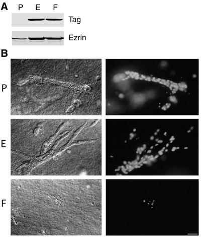
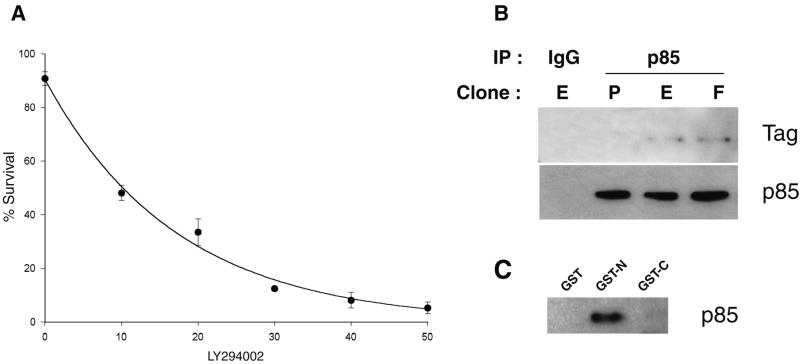
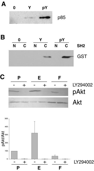
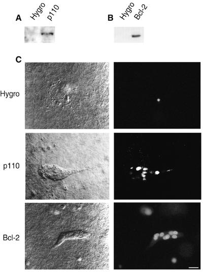
ERM (Ezrin-Radixin-Moesin) proteins function as plasma membrane-actin cytoskeleton linkers and participate in the formation of specialized domains of the plasma membrane. We have investigated ezrin function in tubulogenesis of a kidney-derived epithelial cell line, LLC-PK1. Here we show that cells overproducing a mutant form of ezrin in which Tyr-353 was changed to a phenylalanine (Y353F) undergo apoptosis when assayed for tubulogenesis. While investigating the mechanism responsible for this apoptosis, we found that ezrin interacts with p85, the regulatory subunit of phosphatidylinositol 3-kinase (PI 3-kinase). Two distinct sites of ezrin are involved in this interaction, the amino-terminal domain containing the first 309 aa and the phosphorylated Tyr-353 residue, which binds to the carboxyl-terminal SH2 domain of p85. Cells producing Y353F ezrin are defective in activation of the protein kinase Akt, a downstream target of PI 3-kinase that protects cells against apoptosis. Furthermore, the apoptotic phenotype of these cells is rescued by production of a constitutively activated form of PI 3-kinase. Taken together, these results establish a novel function for ezrin in determining survival of epithelial cells by activating the PI 3-kinase/Akt pathway.


Figures

Figure 1
Figure 1
Apoptosis of LLC-PK1 cells producing Y353F ezrin in a tubulogenesis assay. (A) Immunodetection of endogenous and overproduced tagged ezrin in stable LLC-PK1 clones. P, E, and F cells were transfected, respectively, with the empty vector, the same vector encoding VSV-G tagged wild-type ezrin, or VSV-G tagged Y353F ezrin. Total cellular lysates were analyzed with either P5D4 mAb (Tag) or an anti-ezrin serum. (B) Cells were cultured for 7 days in presence of HGF in a collagen type I gel. Cultures were examined with Nomarski optics (Left) and for Hoechst 33258 staining (Right). In a collagen type I gel, only apoptotic bodies, as highlighted by pyknotic nuclei, were observed in F cell cultures, whereas P and E cells were able to form tubules in presence of HGF. (Bar = 20 μm.)
Figure 2
Figure 2
PI 3-kinase, which is required for survival, interacts with ezrin. (A) LY294002 induces apoptosis of LLC-PK1 cells in the tubulogenesis assay. Mean ± SD of an experiment performed in triplicate is plotted. LY294002 concentration is given in μM. (B) Coimmunoprecipitation of either wild-type or Y353F ezrin with the p85 subunit of PI 3-kinase. Lysates from P, E, or F clones were immunoprecipitated with p85 affinity-purified antibodies or rabbit IgG. The immunoprecipitates were analyzed with P5D4 mAb (Tag) or polyclonal anti p85 antibodies. (C) p85 binding to the amino-terminal domain of ezrin. A lysate from placenta was incubated with immobilized GST, GST–ezrin1–309 (GST-N), or GST–ezrin310–585 (GST-C). After washing, bound material was analyzed with polyclonal anti-p85 antibodies.
Figure 3
Figure 3
Activation of the PI 3-kinase/Akt pathway requires ezrin phosphorylation at residue Tyr-353. (A) Binding of p85 to a phosphorylated peptide corresponding to amino acids 348–358 of ezrin. A lysate from LLC-PK1 cells was incubated with either the beads alone (0), the immobilized peptide with the nonphosphorylated residue Tyr-353 (Y), or phosphorylated residue Tyr-353 (pY). Bound material was analyzed by Western blotting with polyclonal anti-p85 antibodies. (B) Binding of the carboxyl-terminal SH2 domain of p85 to the peptide containing the phosphorylated Tyr-353. The p85 amino-terminal SH2 (N) and the carboxyl-terminal SH2 (C) domains fused to GST were incubated with either the beads alone (O), the peptide with the nonphosphorylated residue Tyr-353 (Y), or the peptide with the phosphorylated residue Tyr-353 (pY). Bound material was analyzed by Western blotting with an anti-GST serum. (C) Analysis of Akt activity in three-dimensional cultures. Serum-starved LLC-PK1 clones were embedded in collagen gels. Lysates from P, E, and F cells treated with either LY294002 (+) or DMSO (−) were analyzed with either anti-phosphospecific Akt (Ser-473) (pAkt) or anti-Akt (Akt) polyclonal antibodies. Signals were analyzed by densitometry. The pAkt/Akt ratio was calculated and set to 100% for untreated P cells. Mean ± SEM of three independent experiments are presented in the histogram.
Figure 4
Figure 4
Rescue of the F cell survival defect by constitutively active PI 3-kinase and Bcl-2. One F clone was stably transfected with either the control hygromycin-resistance plasmid (Hygro), a cDNA encoding a CD2-p110 chimera (p110), or a cDNA encoding human Bcl-2. (A) Immunodetection of the chimeric CD2-p110 protein with anti-CD2 mAb Ox34. (B) Immunodetection of exogenous Bcl-2 with a human-specific anti-Bcl-2 mAb. (C) Survival and tubulogenesis were assessed as described in Fig. 1. (Bar = 15 μm.)

References

    1. Bretscher A, Reczek D, Berryman M. J Cell Sci. 1997;110:3011–3018. - PubMed
    1. Takeuchi K, Sato N, Kasahara H, Funayama N, Nagafuchi A, Yonemura S, Tsukita S. J Cell Biol. 1994;125:1371–1384. - PMC - PubMed
    1. Crepaldi T, Gautreau A, Comoglio P M, Louvard D, Arpin M. J Cell Biol. 1997;138:423–434. - PMC - PubMed
    1. Chen J, Cohn J A, Mandel L J. Proc Natl Acad Sci USA. 1995;92:7495–7499. - PMC - PubMed
    1. Kondo T, Takeuchi K, Doi Y, Yonemura S, Nagata S, Tsukita S, Tsukita S. J Cell Biol. 1997;139:749–758. - PMC - PubMed

Publication types