

Rapid p53 sequence analysis in primary lung cancer using an oligonucleotide probe array
- PMID: 10377423
- PMCID: PMC22094
- DOI: 10.1073/pnas.96.13.7382
Rapid p53 sequence analysis in primary lung cancer using an oligonucleotide probe array
Abstract
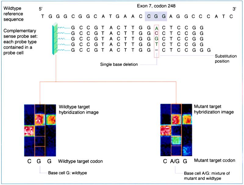
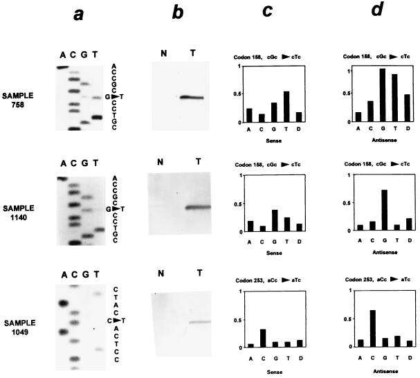
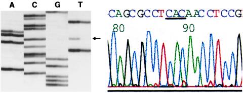
The p53 gene was sequenced in 100 primary human lung cancers by using direct dideoxynucleotide cycle sequencing and compared with sequence analysis by using the p53 GeneChip assay. Differences in sequence analysis between the two techniques were further evaluated to determine the accuracy and limitations of each method. p53 mutations were either detected by using both techniques or, if only detected by one technique, were confirmed by using mutation-specific oligonucleotide hybridization. Dideoxynucleotide sequencing of the conserved regions of the p53 gene (exons 5-9) detected 76% of the mutations within this region of the gene. The GeneChip p53 assay detected 81% of all (exons 2-11) mutations, including 80% of the mutations within the conserved regions of the gene. The GeneChip assay detected 46 of 52 missense mutations (88%), but 0 of 5 frameshift mutations. The specificity of direct sequencing and of the p53 GeneChip assay at detecting p53 mutations were 100% and 98%, respectively. The GeneChip p53 assay is a rapid and reasonably accurate approach for detecting p53 mutations; however, neither direct sequencing nor the p53 GeneChip are infallible at p53 mutation detection.
Figures
References
-
- Greenblatt M S, Bennett W P, Hollstein M, Harris C C. Cancer Res. 1994;54:4855–4878. - PubMed
-
- Sidransky D, Hollstein M. Annu Rev Med. 1996;47:285–301. - PubMed
-
- Harris C C, Hollstein M. N Engl J Med. 1993;329:1318–1327. - PubMed
-
- Rusch V, Klimstra D, Venkatraman E, Oliver J, Martini N, Gralla R, Kris M, Dmitrovsky E. Cancer Res. 1995;55:5038–5042. - PubMed
-
- Lowe S W, Bodis S, McClatchey A, Remington L, Ruley H E, Fisher D E, Housman D E, Jacks T. Science. 1994;266:807–810. - PubMed
Publication types
MeSH terms
Substances
Grants and funding
LinkOut - more resources
Full Text Sources
Other Literature Sources
Medical
Research Materials
Miscellaneous
