

The natriuretic peptide clearance receptor locally modulates the physiological effects of the natriuretic peptide system
- PMID: 10377427
- PMCID: PMC22098
- DOI: 10.1073/pnas.96.13.7403
The natriuretic peptide clearance receptor locally modulates the physiological effects of the natriuretic peptide system
Abstract
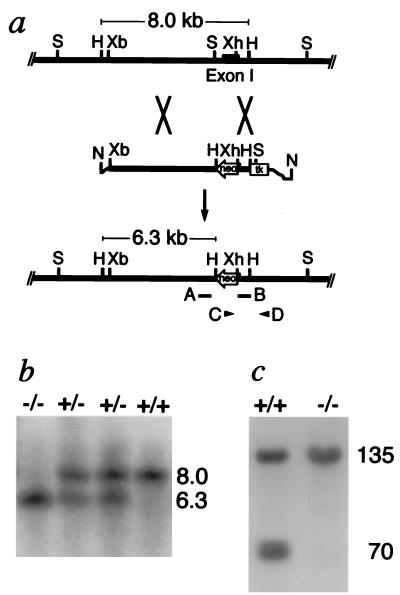
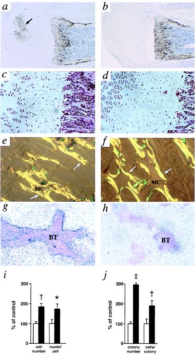
Natriuretic peptides (NPs), mainly produced in heart [atrial (ANP) and B-type (BNP)], brain (CNP), and kidney (urodilatin), decrease blood pressure and increase salt excretion. These functions are mediated by natriuretic peptide receptors A and B (NPRA and NPRB) having cytoplasmic guanylyl cyclase domains that are stimulated when the receptors bind ligand. A more abundantly expressed receptor (NPRC or C-type) has a short cytoplasmic domain without guanylyl cyclase activity. NPRC is thought to act as a clearance receptor, although it may have additional functions. To test how NPRC affects the cardiovascular and renal systems, we inactivated its gene (Npr3) in mice by homologous recombination. The half life of [125I]ANP in the circulation of homozygotes lacking NPRC is two-thirds longer than in the wild type, although plasma levels of ANP and BNP in heterozygotes and homozygotes are close to the wild type. Heterozygotes and homozygotes have a progressively reduced ability to concentrate urine, exhibit mild diuresis, and tend to be blood volume depleted. Blood pressure in the homozygotes is 8 mmHg (1 mmHg = 133 Pa) below normal. These results are consistent with the sole cardiovascular/renal function of NPRC being to clear natriuretic peptides, thereby modulating local effects of the natriuretic peptide system. Unexpectedly, Npr3 -/- homozygotes have skeletal deformities associated with a considerable increase in bone turnover. The phenotype is consistent with the bone function of NPRC being to clear locally synthesized CNP and modulate its effects. We conclude that NPRC modulates the availability of the natriuretic peptides at their target organs, thereby allowing the activity of the natriuretic peptide system to be tailored to specific local needs.
Figures


Similar articles
-
Exercise performance is not improved in mice with skeletal muscle deletion of natriuretic peptide clearance receptor.PLoS One. 2023 Nov 2;18(11):e0293636. doi: 10.1371/journal.pone.0293636. eCollection 2023. PLoS One. 2023. PMID: 37917630 Free PMC article.
-
Discovery of another mechanism for the inhibition of particulate guanylyl cyclases by the natriuretic peptide clearance receptor.Proc Natl Acad Sci U S A. 2023 Jul 11;120(28):e2307882120. doi: 10.1073/pnas.2307882120. Epub 2023 Jul 3. Proc Natl Acad Sci U S A. 2023. PMID: 37399424 Free PMC article.
-
The genetic contribution of the natriuretic peptide system to cardiovascular diseases.Endocr J. 2005 Feb;52(1):11-21. doi: 10.1507/endocrj.52.11. Endocr J. 2005. PMID: 15758553 Review.
-
Guanylyl cyclase/natriuretic peptide receptor-A signaling antagonizes phosphoinositide hydrolysis, Ca(2+) release, and activation of protein kinase C.Front Mol Neurosci. 2014 Aug 22;7:75. doi: 10.3389/fnmol.2014.00075. eCollection 2014. Front Mol Neurosci. 2014. PMID: 25202235 Free PMC article. Review.
-
Cardiac and intestinal natriuretic peptides: insights from genetically modified mice.Peptides. 2005 Jun;26(6):1078-85. doi: 10.1016/j.peptides.2004.08.031. Epub 2005 Apr 14. Peptides. 2005. PMID: 15911075 Review.
Cited by
-
The receptor guanylyl cyclase Npr2 is essential for sensory axon bifurcation within the spinal cord.J Cell Biol. 2007 Oct 22;179(2):331-40. doi: 10.1083/jcb.200707176. J Cell Biol. 2007. PMID: 17954614 Free PMC article.
-
Defining the molecular character of the developing and adult kidney podocyte.PLoS One. 2011;6(9):e24640. doi: 10.1371/journal.pone.0024640. Epub 2011 Sep 8. PLoS One. 2011. PMID: 21931791 Free PMC article.
-
Npr3 regulates neural crest and cranial placode progenitors formation through its dual function as clearance and signaling receptor.Elife. 2023 May 10;12:e84036. doi: 10.7554/eLife.84036. Elife. 2023. PMID: 37162198 Free PMC article.
-
Opposite effects of ANP receptors in attenuation of LPS-induced endothelial permeability and lung injury.Microvasc Res. 2012 Mar;83(2):194-9. doi: 10.1016/j.mvr.2011.09.012. Epub 2011 Oct 6. Microvasc Res. 2012. PMID: 22001395 Free PMC article.
-
The biological impact of blood pressure-associated genetic variants in the natriuretic peptide receptor C gene on human vascular smooth muscle.Hum Mol Genet. 2018 Jan 1;27(1):199-210. doi: 10.1093/hmg/ddx375. Hum Mol Genet. 2018. PMID: 29040610 Free PMC article.
References
-
- Flynn T G. In: Contemporary Endocrinology: Natriuretic Peptides in Health and Disease. Samson W K, Levin E R, editors. Totowa, NJ: Humana; 1997. pp. 1–19.
-
- Maack T. Kidney Int. 1996;49:1732–1737. - PubMed
-
- Chinkers M, Garbers D L, Chang M S, Lowe D G, Chin H M, Goeddel D V, Schulz S. Nature (London) 1989;338:78–83. - PubMed
-
- Schulz S, Singh S, Bellet R A, Singh G, Tubb D J, Chin H, Garbers D L. Cell. 1989;58:1155–1162. - PubMed
-
- Maack T, Suzuki M, Almeida F A, Nussenzveig D, Scarborough R M, McEnroe G A, Lewicki J A. Science. 1987;238:675–678. - PubMed
Publication types
MeSH terms
Substances
Grants and funding
LinkOut - more resources
Full Text Sources
Other Literature Sources
Molecular Biology Databases
Miscellaneous
