

doi: 10.1073/pnas.96.13.7415.
Efficient export of the glucose transporter Hxt1p from the endoplasmic reticulum requires Gsf2p
Affiliations
- PMID: 10377429
- PMCID: PMC22100
- DOI: 10.1073/pnas.96.13.7415
Item in Clipboard
Efficient export of the glucose transporter Hxt1p from the endoplasmic reticulum requires Gsf2p
Proc Natl Acad Sci U S A.
.
Abstract
Mutations in the GSF2 gene cause glucose starvation phenotypes in Saccharomyces cerevisiae. We have isolated the HXT1 gene, which encodes a low-affinity, high-capacity glucose transporter, as a multicopy suppressor of a gsf2 mutation. We show that gsf2 mutants accumulate Hxt1p in the endoplasmic reticulum (ER) and that Gsf2p is a 46-kDa integral membrane protein localized to the ER. gsf2 mutants also display a galactose growth defect and abnormal localization of the galactose transporter Gal2p but are not defective in function or localization of the high-affinity glucose transporter Hxt2p. These findings suggest that Gsf2p functions in the ER to promote the secretion of certain hexose transporters.
Figures
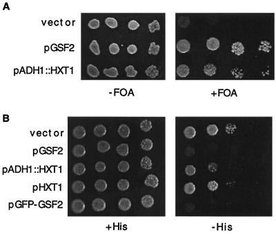
Hxt1p glucose transporter overexpression suppresses gsf2. (A) A gsf2Δ snf1Δ strain (PS4342) containing pSNF1–URA3 was transformed with the vector pSK134, pGSF2, or pADH1∷HXT1. Transformants were grown on medium containing uracil to allow loss of pSNF1–URA3, and 10-fold serial dilutions were spotted onto SC-Leu medium containing 5-fluoroorotic acid (5-FOA) (+FOA) or control medium lacking 5-FOA (−FOA). (B) A gsf2−1 strain (PS4343–11B) containing pSNF1–URA3 and pSUC2∷HIS3 was transformed with the vector pRS315 or the indicated expression plasmids, and 10-fold serial dilutions were spotted onto SD + 5% glucose medium containing histidine (+His) or 1 mM 3-aminotriazole (−His).
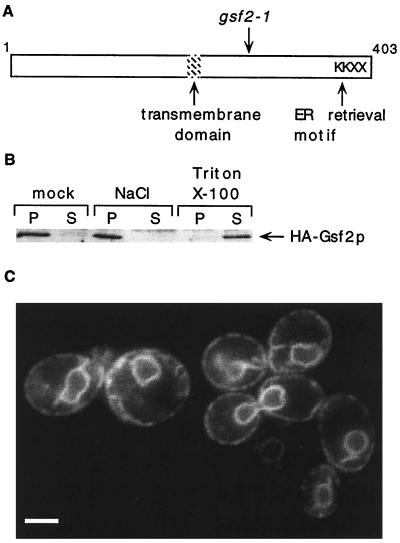
Localization of Gsf2p to the ER. (A) Schematic diagram of Gsf2p indicating the position of a putative transmembrane domain (residues 177–198, LVAQWLFFVMHIFKVGIITLFL), the gsf2-1 mutation (Q260 CAG codon → TAG stop codon), and a C-terminal dilysine (KKXX) ER-retrieval motif (10, 11). (B) Immunoblot analysis of HA–Gsf2p. A spheroplast lysate was prepared from a gsf2Δ strain (PS352) carrying pHA–GSF2 and was centrifuged at 13,000 × g. The P13 fraction was resuspended in buffer. Aliquots were washed with buffer (mock) or with buffer adjusted to 1 M NaCl or 1% Triton X-100 and centrifuged at 13,000 × g to generate pellet (P) and supernatant (S) fractions. Proteins were resolved by SDS/10% PAGE, and HA–Gsf2p was detected by immunoblotting with anti-HA antibody. (C) Fluorescence microscopy of GFP–Gsf2p. The gsf2Δ strain PS352 was transformed with pGFP–GSF2, and cultures were grown to mid-logarithmic phase in SC-Leu + 4% glucose. A 0.5-ml aliquot was centrifuged briefly at low speed, and 1 μl of a concentrated cell suspension was visualized by using fluorescence microscopy. Exposure time was 2 sec. (Bar = 2.5 μm.)
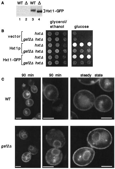
Function and localization of Hxt1p. (A) Isogenic wild-type (WT, PS350) and gsf2Δ (Δ, PS352) strains containing p(U)HXT1 (lanes 1 and 2) or p(U)HXT1–GFP (lanes 3 and 4) were grown in SC − Ura + 5%glycerol/2%ethanol, glucose was added to 4% to induce HXT1–GFP expression, and aliquots were harvested 90 min after glucose addition. Whole-cell extracts were prepared by glass bead lysis of cells resuspended in Laemmli sample buffer plus protease inhibitors, proteins were resolved by SDS/10% PAGE, and Hxt1–GFP was detected by using immunoblot analysis with anti-GFP antibody. No prominent proteolytic fragments were detected. (B) Isogenic hxtΔ and gsf2Δ hxtΔ strains lacking all major hexose transporter genes (HY133 and PS1332) were transformed with the vector pRS316, p(U)HXT1, or p(U)HXT1–GFP. Transformants were grown in SC − Ura + 5%glycerol/2%ethanol, and 10-fold serial dilutions were spotted onto SC − Ura + 5%glycerol/2%ethanol and SC − Ura + 5%glucose media. The glucose plate was incubated anaerobically to inhibit respiration. Pregrowth of transformants in glucose for several generations did not improve the growth of gsf2Δ mutants relative to wild-type on glucose (data not shown). (C) Wild-type (PS350) and gsf2Δ (PS352) strains carrying pHXT1–GFP were grown in SC-Leu + 5%glycerol/2%ethanol, glucose was added to 4%, and aliquots were harvested 90 min after glucose addition (Left and Center), or cells were grown for many generations in SC-Leu + 4%glucose (Right). Cells were prepared for fluorescence microscopy as described in Fig. 2. Exposure times for all images were 2 sec. (Bar = 2.5 μm.)
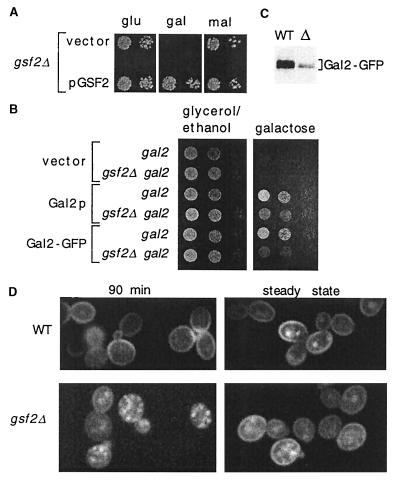
Function and localization of Gal2p. (A) The gsf2Δ strain PS352 was transformed with pMAL63-YCp50 (14) to provide Mal activator function and pGSF2 or the vector pRS315. Serial dilutions (10-fold) were spotted onto SC-Leu-Ura medium containing 1 μg/ml antimycin A to inhibit respiration and 2% glucose, 2% galactose, or 2% maltose as carbon source. Plates were incubated at 24°. (B) Isogenic gal2 hxtΔ and gsf2Δ gal2 hxtΔ strains (HY133 and PS1332) were transformed with the vector pRS316 or plasmids expressing Gal2p (pAK104) or Gal2–GFP (pAK166). Transformants were grown in SC − Ura + 5%glycerol/2%ethanol, and 10-fold serial dilutions were spotted onto SC − Ura + 5%glycerol/2%ethanol and SC − Ura + 2%galactose media. The galactose plate was incubated anaerobically. Pregrowth of transformants in galactose for several generations did not improve the growth of gsf2Δ mutants relative to wild-type on galactose (data not shown). (C) Wild-type and gsf2Δ strains (PS350, PS352) containing pAK166 were grown in SC − Ura + 5%glycerol/2%ethanol, galactose was added to 4% to induce Gal2–GFP expression, and aliquots were harvested 90 min later. Immunoblot analysis was carried out as described in Fig. 3. No prominent proteolytic fragments were detected. (D) Cultures were prepared as in C (Left) or were grown for many generations in SC − Ura + 4%galactose (Right), and cells were prepared for fluorescence microscopy as described in Fig. 2. Exposure times were 0.6 sec.
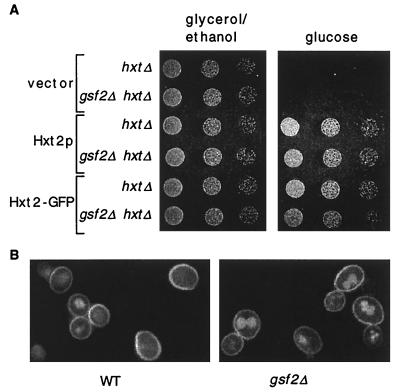
Function and localization of Hxt2p. (A) Strains HY133 (hxtΔ) and PS1332 (gsf2Δ hxtΔ) containing the vector pRS316 or plasmids expressing Hxt2p (pAK145) or Hxt2–GFP (pAK146) were grown in SC − Ura + 5%glycerol/2%ethanol, and 10-fold serial dilutions were spotted onto SC − Ura + 5%glycerol/2%ethanol and SC − Ura + 2%glucose media. The glucose plate was incubated anaerobically. (B) Wild-type (PS350) and gsf2Δ (PS352) strains containing pAK146 were grown in SC − Ura + 4%glucose and shifted to SC − Ura + 0.1%glucose to induce Hxt2–GFP expression. Cells were harvested 90 min after the shift to low glucose and were prepared for fluorescence microscopy as described in Fig. 2. Exposure times were 4 sec.
References
-
- Bisson L F, Coons D M, Kruckeberg A L, Lewis D A. Crit Rev Biochem Mol Biol. 1993;28:259–308. - PubMed
-
- Kruckeberg A L. Arch Microbiol. 1996;166:283–292. - PubMed
-
- Boles E, Hollenberg C P. FEMS Microbiol Rev. 1997;21:85–111. - PubMed
-
- Nelissen B, De Wachter R, Goffeau A. FEMS Microbiol Rev. 1997;21:113–134. - PubMed
Publication types
MeSH terms
Substances
Grants and funding
LinkOut - more resources
Full Text Sources
Molecular Biology Databases
