. 1999 Jun 22;96(13):7421-6.
doi: 10.1073/pnas.96.13.7421.

Protein kinase B/Akt-mediated phosphorylation promotes nuclear exclusion of the winged helix transcription factor FKHR1

Affiliations

Protein kinase B/Akt-mediated phosphorylation promotes nuclear exclusion of the winged helix transcription factor FKHR1

W H Biggs 3rd et al. Proc Natl Acad Sci U S A. .

Abstract

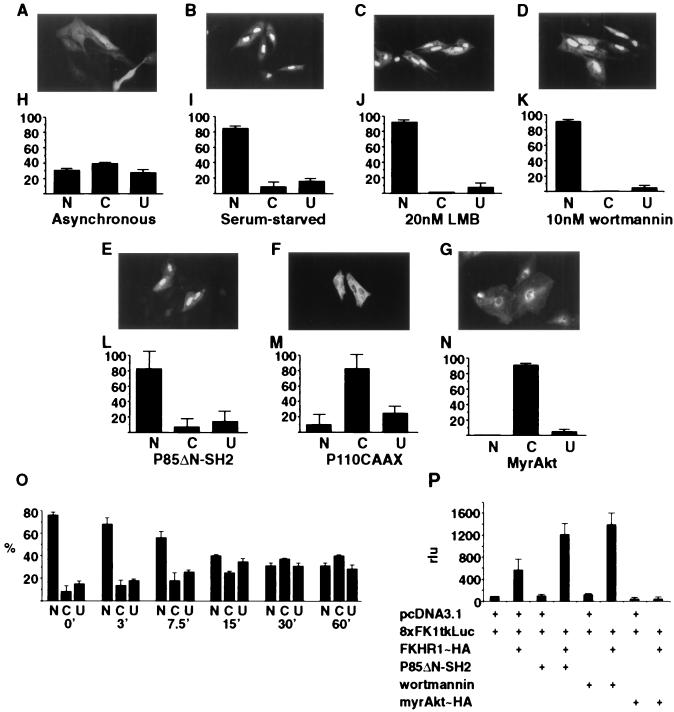
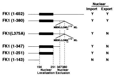
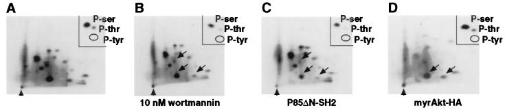
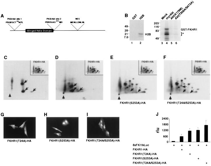
Although genetic analysis has demonstrated that members of the winged helix, or forkhead, family of transcription factors play pivotal roles in the regulation of cellular differentiation and proliferation, both during development and in the adult, little is known of the mechanisms underlying their regulation. Here we show that the activation of phosphatidylinositol 3 (PI3) kinase by extracellular growth factors induces phosphorylation, nuclear export, and transcriptional inactivation of FKHR1, a member of the FKHR subclass of the forkhead family of transcription factors. Protein kinase B (PKB)/Akt, a key mediator of PI3 kinase signal transduction, phosphorylated recombinant FKHR1 in vitro at threonine-24 and serine-253. Mutants FKHR1(T24A), FKHR1(S253A), and FKHR1(T24A/S253A) were resistant to both PKB/Akt-mediated phosphorylation and PI3 kinase-stimulated nuclear export. These results indicate that phosphorylation by PKB/Akt negatively regulates FKHR1 by promoting export from the nucleus.


Figures

Figure 1
Figure 1
Nuclear/cytoplasmic shuttling and transcriptional activity of FKHR1-HA in transiently transfected cells. (AG) Detection of FKHR1-HA by anti-HA immunofluorescent or epifluorescent staining. (HN) Quantification of the nuclear (N), cytoplasmic (C), and uniform (U) localization of FKHR1-HA or FKHR1-GFP. Immunolocalization of FKHR1-HA in CV1 cells. (A and H) Asynchronous culture. (B and I) Culture in the absence of serum for 24 hr. (C and J) Treatment with 20 nM leptomycin B (LMB) for 6 hr before fixation and staining. (D and K) Treatment with 10 nM wortmannin for 3 hr before fixation. (E and L) Cotransfection of FKHR1-HA with pSG5-P85ΔN-SH2. (F and M) Cotransfection of FKHR1-HA with pcDNA3.1-P110CAAX. (G and N) Cotransfection of FKHR1-GFP with pCMV6-myrAkt-HA. (O) Kinetic analysis of FKHR1-HA relocalization induced by 50 nM insulin-like growth factor I. (P) Localization of FKHR1-HA directly affects the expression of a FKHR1-responsive reporter. CV1 cells transfected with the FKHR1-responsive reporter 8XFK1tkLuc and cytomegalovirus (CMV)-β-gal, together with FKHR1-HA, P85ΔN-SH2, and myrAkt-HA as indicated. Cells transfected with 8XFK1tkLuc, CMV-β-gal, and FKHR1-HA also were treated with 10 nM wortmannin. Luciferase acivities of the individual samples were normalized to the activity of the 8XFK1tkLuc reporter in the absence of any other construct.
Figure 2
Figure 2
Immunolocalization of FKHR1-GFP fusion proteins. CV1 cells were transiently transfected with a series of FKHR1-GFP fusion proteins with carboxyl-terminal deletions of FKHR1 and examined by fluorescence microscopy. The regions identified as being required for either nuclear localization (amino acids 147–251) or nuclear exclusion (amino acids 347–380) are indicated. The sequence of the putative leucine-rich NES (M368ENLLNLNL377) is indicated.
Figure 3
Figure 3
Phosphorylation of FKHR1-HA in asynchronously growing CV1 cells. (AD) Two-dimensional tryptic phosphopeptide maps of 32P-labeled wild-type FKHR1-HA immunoprecipitated from metabolically labeled CV1 cells either treated or cotransfected as indicated. Thin layer cellulose plates were run as indicated (horizontal: electrophoresis anode to left; vertical: chromatography thin layer analysis). The position of the origin is indicated (arrowhead). (Insets) Phosphoamino acid content of each of the corresponding 32P-labeled FKHR1-HA immunoprecipitates. The locations of 32P-labeled phospho-serine (P-ser) and phospho-threonine (P-thr) present on FKHR1-HA are indicated. No phospho-tyrosine (P-tyr) was detected. (A) FKHR1-HA. (B) FKHR1-HA immunoprecipitated from metabolically labeled CV1 cells treated with 10 nM wortmannin. Arrows indicate those phosphopeptides that were reduced. (C) FKHR1-HA immunoprecipitated from metabolically labeled CV1 cells cotransfected with FKHR1-HA and P85ΔN-SH2. Arrows indicate those phosphopeptides that were reduced. (D) FKHR1-HA immunoprecipitated from metabolically labeled CV1 cells cotransfected with FKHR1-HA and myrAkt-HA. Arrows indicate those phosphopeptides that were increased.
Figure 4
Figure 4
Regulation of FKHR1-HA localization by PKB/AKT-mediated phosphorylation. (A) Relative location of the two consensus AKT phosphorylation sites (threonine-24 and serine-253), as well as the putative leucine-rich NES, within FKHR1. The winged helix DNA binding domain also is indicated. (B) PKB/AKT phosphorylates GST-FKHR1 in vitro. Lane 1, GST incubated with myrAkt-HA; lane 2, H2B incubated with myrAkt-HA; lane 3, GST-FKHR1 incubated with immunoprecipitate from mock-transfected cells; lane 4, GST-FKHR1 incubated with myrAkt-HA; lane 5, GST-FKHR1 incubated with Akt(K67A)-HA; lane 6, GST-FKHR1 incubated with Akt(T308A/S473A)-HA. Degradation products of GST-FKHR1 that copurified with the full fusion protein are indicated (∗). These fragments also were phosphorylated by myrAkt-HA. Molecular mass (kDa) markers (×1,000) are indicated. (C) Two-dimensional tryptic phosphopeptide map of GST-FKHR1 phosphorylated in vitro by myrAkt-HA. (Inset) Two-dimensional tryptic phosphopeptide map of FKHR1-HA immunoprecipitated from 32P-labeled CV1 cells. Phosphopeptides from in vitro and in vivo maps, which comigrate, are indicated by arrows. Position of the origin is indicated (arrowhead). (D) Two-dimensional tryptic phosphopeptide map of a mixture of FKHR1 immunoprecipitated from asynchronous CV1 cells metabolically labeled with 32P-inorganic phosphate and FKHR1-HA phosphorylated in vitro by myrAkt-HA. (Inset) Two-dimensional tryptic phosphopeptide map of FKHR1-HA immunoprecipitated from 32P-labeled CV1 cells. Phosphopeptides shared between the in vivo and in vitro maps are indicated by arrows. Position of the origin is indicated (arrowhead). (E) Two-dimensional tryptic phosphopeptide map of FKHR1(S253)-HA immunoprecipitated from asynchronous CV1 cells metabolically labeled with 32P-inorganic phosphate. (Inset) Two-dimensional tryptic phosphopeptide map of FKHR1-HA immunoprecipitated from 32P-labeled CV1 cells. Phosphopeptides absent in the FKHR1(S253A)-HA map are indicated by arrows. Position of the origin is indicated (arrowhead). (F) Two-dimensional tryptic phosphopeptide map of FKHR1(T24A/S253)-HA immunoprecipitated from asynchronous CV1 cells metabolically labeled with 32P-inorganic phosphate. (Inset) Two-dimensional tryptic phosphopeptide map of FKHR1-HA immunoprecipitated from 32P-labeled CV1 cells. Phosphopeptides absent in the FKHR1(T24A/S253A)-HA map are indicated by arrows. Position of the origin is indicated (arrowhead). (GI) Mutation of PKB/AKT phosphorylation sites alters the localization of FKHR1-HA. (G) Nuclear localization of FKHR1(T24A)-HA in CV1 cells. (H) Nuclear localization of FKHR1(S253A)-HA in CV1 cells. (I) Nuclear localization of FKHR1(T24A/S253A)-HA in CV1 cells. (J) Mutation of the PKB/Akt phosphorylation sites, threonine-24, and/or serine-253 increased the transcriptional activity of FKHR1-HA. Luciferase activities of the individual samples were normalized to the activity of the 8XFK1tkLuc reporter in the absence of any other construct.

Similar articles

Cited by

References

    1. Kaufmann E, Knöchel W. Mech Dev. 1996;57:3–20. - PubMed
    1. Hromas R, Costa R. Crit Rev Oncol Hematol. 1995;20:129–140. - PubMed
    1. Vogt P K, Li J, Freyaldenhoven B S. Virology. 1997;238:1–7. - PubMed
    1. Barr F G. Curr Top Microbiol Immunol. 1997;220:113–129. - PubMed
    1. Hillion J, Le Coniat M, Jonveaux P, Berger R, Bernard O A. Blood. 1997;90:3714–3719. - PubMed

Publication types

MeSH terms