. 1999 Jun 22;96(13):7445-50.
doi: 10.1073/pnas.96.13.7445.

Conformational variants of class II MHC/peptide complexes induced by N- and C-terminal extensions of minimal peptide epitopes

Affiliations

Conformational variants of class II MHC/peptide complexes induced by N- and C-terminal extensions of minimal peptide epitopes

O Rötzschke et al. Proc Natl Acad Sci U S A. .

Abstract

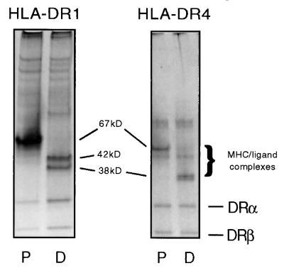
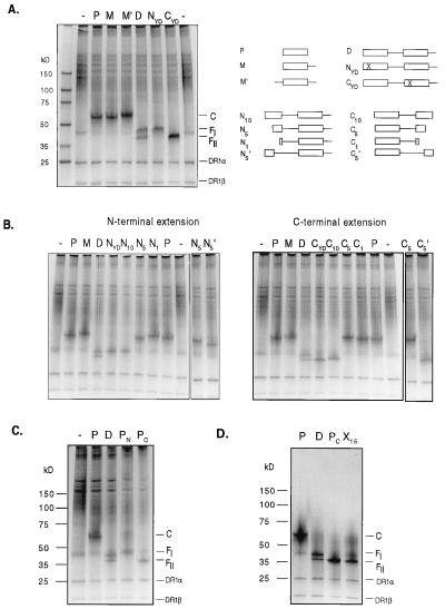
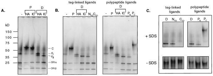
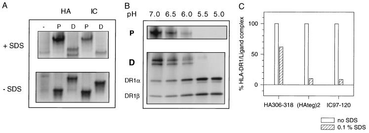
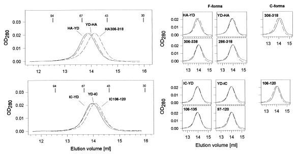
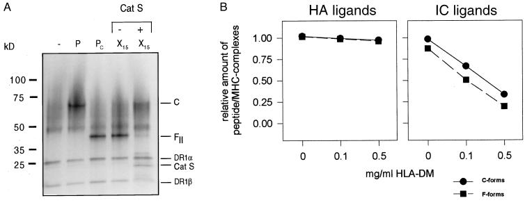
Class II MHC molecules are known to exist in conformational variants. "Floppy" and "compact" forms of murine MHC molecules, for example, are discriminated by their migration behavior on SDS/PAGE and represent empty and ligand-loaded forms. Here we show that formation of distinctly faster-migrating ligand complexes (F-forms) rather than the normal compact (C-) forms of HLA-DR1 or -DR4 results from extensions of minimal peptide epitopes (such as HA306-318 or IC106-120) by approximately 10 amino acids at either the N or the C terminus. Two similar but distinct F-forms (FI and FII) were detected, depending on the site of the extension. Both F-forms were characterized by increased surface hydrophobicity and reduced SDS-stability. Native gel separations and size exclusion chromatography indicated that the F-forms had increased hydrodynamic radii compared with the C-form and an apparent size similar to that of empty MHC molecules. The regions on the ligand overhangs responsible for the effect began at a distance of approximately 5 amino acids on either side of the epitopes, comprised 4-8 amino acids (i.e., a total overhang of 9-14), and did not have a particular sequence preference. The possible functional significance of these forms is discussed.


Figures

Figure 1
Figure 1
Apparent molecular weight (MWapp) of class II MHC/ligand complexes loaded with monomeric or dimeric ligand constructs. (A) Soluble HLA-DR1 or -DR4 molecules loaded with HA306-318 peptide (P lanes) or dimeric ligand constructs consisting of two HA306-318 epitope units linked by a tetraethylene glycol spacer (D lanes) were separated by SDS/PAGE. The MWapp indicated in the figure refers to bands representing distinct MHC/ligand complexes. Gels were visualized by silver staining.
Figure 2
Figure 2
Induction of FI- and FII-complexes by amino acid overhangs of the ligand. Soluble HLA-DR1 molecules were loaded with ligand constructs containing the epitope HA306-318. The structure of the ligands is depicted schematically on the upper right panel. The box represents the HA306-318 binding epitope, and the line represents a tetraethylene glycol unit used to connect the epitopes. The cross in the NYD- and the CYD-ligand marks a substitution of the main anchor residue Y308 with D in one of the two HA306-318 epitopes, which abrogates binding to the HLA-DR1 molecule. N10-N1 and C10-C1 represent dimeric ligand constructs with truncations in the N- or C-terminal epitope. (A) Formation of FI- and FII-ligand complexes with teg-linked peptide constructs with defined N- or C-terminal polypeptide extensions. HLA-DR1/ligand complexes, loaded with monomeric (lanes P, M, and M′) and dimeric HA306-318 ligand constructs (lanes D, NYD, and CYD), were separated by SDS/PAGE. The position of bands representing the compact C-form (associated with the monomeric ligands) as well as of the two distinct FI- and FII-forms (associated with the dimeric ligands) are indicated. (B) To estimate distance and size of the polypeptide region responsible for the formation of the FI- and FII-complexes, HLA-DR1 molecules were loaded with truncated forms of the dimeric HA306-318 construct. The series of N-terminal truncations (Left) consists of HA306-318 peptides linked by a teg spacer with the extension VKQNTLKLAT (lane N10), LKLAT (lane N5), and T (lane N1). In addition, a ligand also was used in which the LKLAT overhang was connected by two instead of one teg-units (lane N5′, shown on the right side of the gel). For the series of C-terminal truncations (Right), overhangs were used with the sequence GPKYVKQNTL (lane C10), GPKYV (lanes C5 and C5′), and G (lane C1). On both gels, empty HLA-DR1 molecules (−) and ligand complexes preformed with the HA306-318 peptide (P lanes), the monomer (M lanes), and the dimer (D lanes) are shown as a reference. (C) Formation of F-complexes with naturally extended polypeptide ligands. HLA-DR1 molecules were loaded with peptides extended by 20 amino acids of the natural sequence of the hemagglutinin protein on the N-terminal side (HA286-318; lane PN) and on the C-terminal side (HA306-338; lane PC) of the HA binding epitope. SDS/PAGE separation is shown in comparison to the HA306-318-loaded complex (lane P) and the complex loaded with the dimer (lane D). (D) Formation of an FII-complex by a ligand with randomized overhang. HLA-DR1 molecules were loaded with an HA306-318 epitope, which (through a teg-spacer on the C-terminal side) was connected to a 15-aa-long peptide with random sequence (HA-X15). The overhang was produced by a sequential synthesis using an amino acid pool (G, A, P, V, I, L, M, Y, F, S, T, N, Q, D, E, K, R, H). The HLA-DR1/HA-X15 ligand complex (lane X15) is shown in comparison to the complex loaded with the HA306-318-peptide (lane P), the dimer (lane D), and the HA306-338 peptide (lane PC).
Figure 3
Figure 3
Formation of HLA-DR1/ligand complexes with ligand constructs containing the IC106-120 epitope. (A) Comparison of HLA-DR1/ligand complexes formed with the HA306-318 and IC106-120 ligand constructs. The MHC molecules were loaded with the peptides HA306-318 and IC106-120 (P lanes) or with dimeric teg-linked tandem constructs HA-HA or IC-IC (D lanes). The position of bands representing the C-form or the FI- or FII-form is indicated. (B) SDS/PAGE separation of ligand complexes loaded with IC106-120 binding epitopes with defined N- and C-terminal overhangs. For the left gel, IC ligands were used, which, through the teg-spacer, were extended by the nonbinding Y308D-substituted HA306-318 epitope [N-terminal, YD-IC (lane NYD); C-terminal, IC-YD (lane CYD)]. The right gel shows complexes formed with natural polypeptide ligands extended by the amino acid sequence of the invariant chain protein [N-terminal, IC97-120 (lane PN); C-terminal, IC106-135 (lane PC)]. (C) Comparison of the migration of extended IC106-120 ligands in the absence of SDS. Gel separations are shown that were carried out in the presence (Upper) or absence (Lower) of SDS.
Figure 4
Figure 4
Characterization of the F- and C-forms of HLA-DR1/ligand complexes. (A) Surface hydrophobicity. The migration behavior of HLA-DR1/ligand complexes was compared under native conditions and in the presence of SDS. Soluble HLA-DR1 molecules were loaded with the HA306-318 or the IC106-120 peptide (P lanes) or with dimeric constructs HA-HA or IC-IC (D lanes). Gels of the MHC/ligand complexes are shown that were separated in the presence (Upper) or in the absence (Lower) of SDS. (B) pH stability. HLA-DR1/ligand complexes were loaded with HA306-318 peptide (P lanes) or the dimer (D lanes) at a pH ranging from 5.0–7.0 (indicated above the lanes) and were analyzed by SDS/PAGE. (C) SDS stability. HLA-DR1 molecules, preloaded with the biotinylated ligands HA306-318, HA-HA, or IC97-120, were incubated for 3 h at 37°C in the absence (open bars) or presence (hatched bars) of 0.1% SDS. The amount of stable MHC/ligand complex was determined after the incubation by ELISA.
Figure 5
Figure 5
Determination of the apparent size of F- and C-forms of HLA-DR1/ligand complexes by size exclusion chromatography. The elution profiles of HLA-DR1/ligand complexes formed with ligands containing the HA306-318 (Upper) and IC106-120 epitope (Lower) are shown. In the left panels, the elution of peptide-loaded C-forms is compared with the elution of the F-forms generated with the teg-linked YD hybrid dimers (indicated in the figure). As a reference, elution volumes of marker proteins and their molecular weights are indicated. The right panels show a comparison of the elution of the ligand-loaded forms (solid lines) to the elution of empty HLA-DR1 preparations (dashed lines).
Figure 6
Figure 6
Effect of cathepsin S and HLA-DM on C- and on F-forms of HLA-DR1/ligand complexes. HLA-DR1/ligand complexes were incubated with cathepsin S or soluble HLA-DM. (A) Conversion of an F-complex into the C-form by cathepsin S digest. FII-complexes, preformed by loading the HA-X15 ligand onto the HLA-DR1 molecules, were subjected to cathepsin S digest to remove the random peptide overhang. The gel shows the SDS/PAGE analysis of HLA-DR1/HA-X15 ligand complexes (lanes X15) before (−) and after (+) the treatment with cathepsin S. Also shown are the complexes formed with the HA306-318 peptide (lane P) and the HA306-338 peptide (PC). (B) HLA-DM mediated release of ligands from C- or F-ligand complexes. F- (closed square) or C-HLA-DR1/ligand complexes (closed circle) were formed with biotinylated ligands containing the HA306-318 (Left) or the IC106-120 binding epitope (Right). HA306-318 and IC106-120 were used for the formation of C-complexes, and the F complexes were generated with the HA teg-dimer or the IC97-120 peptide. The ligand complexes were incubated with the indicated amounts of soluble HLA-DM, and the release of ligands was measured by ELISA by determining the amount of stable MHC ligand complexes left after the incubation.

Similar articles

Cited by

References

    1. Trowbridge I S. Curr Opin Cell Biol. 1991;3:634–641. - PubMed
    1. Unwin N. J Struct Biol. 1998;121:181–190. - PubMed
    1. Bluestone J A, Jameson S, Miller S, Dick R D. J Exp Med. 1992;176:1757–1761. - PMC - PubMed
    1. Chervonsky A V, Medzhitov R M, Denzin L K, Barlow A K, Rudensky A Y, Janeway C A., Jr Proc Natl Acad Sci USA. 1998;95:10094–10099. - PMC - PubMed
    1. Reich Z, Altman J D, Boniface J J, Lyons D S, Kozono H, Ogg G, Morgan C, Davis M M. Proc Natl Acad Sci USA. 1997;94:2495–2500. - PMC - PubMed

Publication types