. 1999 Jun 22;96(13):7456-61.
doi: 10.1073/pnas.96.13.7456.

Cancer-specific chromosome alterations in the constitutive fragile region FRA3B

Affiliations

Cancer-specific chromosome alterations in the constitutive fragile region FRA3B

K Mimori et al. Proc Natl Acad Sci U S A. .

Erratum in

  • Proc Natl Acad Sci U S A 1999 Sep 14;96(19):10944

Abstract

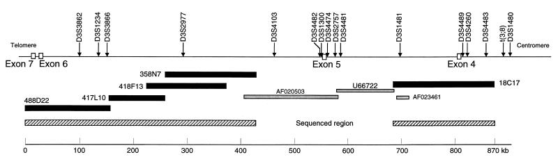
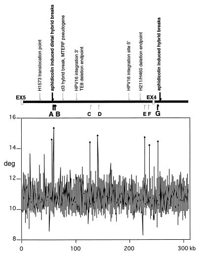
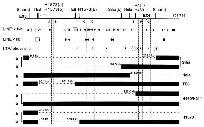
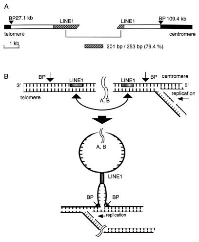
We have sequenced 870 kilobases of the FHIT/FRA3B locus, from FHIT intron 3 to intron 7. The locus is AT rich (61.5%) and Alu poor (6. 2%), and it apparently does not harbor other genes. In a detailed analysis of the 308-kilobase region between FHIT exon 5 and the telomeric end of intron 3, a region known to encompass a human papillomavirus-16 integration site and two clusters of aphidicolin-induced chromosome 3p14.2 breakpoints, we have precisely mapped 10 deletion and translocation endpoints in cancer-derived cell lines relative to positions of specific repetitive elements, regions of high genome flexibility and aphidicolin-induced breakpoints. Conclusions are (i) that aphidicolin-induced breakpoint clusters fall close to high-flexibility sequences, suggesting that these sequences contribute directly to aphidicolin-induced fragility; (ii) that 9 of the 10 FHIT allelic deletions in cancer cell lines resulted in loss of exons, with 7 deletion endpoints near long interspersed nuclear elements or long terminal repeat elements; and (iii) that cancer-specific deletions encompass multiple high-flexibility genomic regions, suggesting that fragile breaks may occur at these regions, whereas repair of the breaks involves homologous pairing of flanking sequences with concomitant deletion of the damaged fragile sequence.


Figures

Figure 1
Figure 1
The FHIT locus: intron 3 through intron 7. The hatched bar on the right represents the 180-kb sequenced region that covers 120 kb of intron 4 and 60 kb of intron 3 but not the t(3;8) chromosome translocation break. The hatched bar on the left shows the 400-kb sequence that covers 360.9 kb of intron 5, 2.6 kb of intron 6, and 36.5 kb of intron 7. The closed bars represent the five BACs that were sequenced to assemble the complete sequence. AF020503, U66722, and AF023461 are GenBank accession numbers for the previously sequenced portions of FRA3B. The loci shown across the top were placed on the map by sequence homology.
Figure 2
Figure 2
Relationship between fragile-region landmarks and flexible regions. (Upper) Previously identified landmarks are indicated above the line representing the FHIT intron 4 locus. Small arrows indicate nucleotide positions of landmarks in intron 4 and intron 3. They designate sequences of a lung cancer cell line translocation point (H1573) at 26.7 kb; a spontaneous hybrid break and the MTERF pseudogene at 79 kb; HPV16 integration site endpoints at 101 and 198 kb; and a deletion endpoint of lung carcinoma cell line (H460/H211) at 222 kb. Bold arrows indicate the distal and proximal aphidicolin-induced hybrid breaks at positions 55 and 257 kb. Helix flexibility was evaluated by using a previously developed computer program (flexstab). This program measures the flexibility parameter, which is expressed as fluctuation in the twist angle. Regions of helix flexibility may be fragile points within the FRA3B, which includes introns 4 and 5. (Lower) The vertical axis shows degrees of inclination in the twist angle, and the horizontal axis indicates nucleotide position of each 100-bp window. The peak values relative to the average were considered as potential flexible regions and drawn as spikes. Positions of the seven spikes, A to G, are shown by bold and small arrows below the locus. The thick arrows representing spikes A, B, and G emphasize the observation that the aphidicolin-induced distal and proximal hybrid breaks are located close to flexibility spikes (0.35 kb from spike A and 7.1–7.6 kb from spike G, respectively).
Figure 3
Figure 3
Fragile region topography near cancer cell breakpoints. Depiction of 10 deletion endpoints relative to L1 elements (L1 sequences of >1 kb, bold arrows; L1 sequences of <1 kb, small arrowheads) and LTR/retroviral elements. The four squares enclosing L1 elements emphasize that the TE8 breakpoints, the telomeric Siha breakpoint, the translocation point, and the telomeric breakpoint of H1573 are located within or near L1 elements. The two circles enclosing LTR arrowheads stress that the H460/H211 breakpoints and the centromeric breakpoint of Siha allele a are near LTR/retroviral elements. The dashed vertical lines represent the flexibility spikes A–G (shown in Fig. 2) and show the position of flexibility/fragility spikes relative to cancer cell deletions. For the HeLa and TE8 cell lines, the exact structure of the b alleles in this region is not yet known.
Figure 4
Figure 4
Model for the role of homologous sequence elements in repair of double-strand breaks within the fragile region. Carcinogen-induced damage in the fragile region may cause double- or single-strand breaks at flexibility peaks, necessitating repair during replication. (A) The H1573 allele b is shown with black bars representing sequences retained in the cell, BP representing positions of deletion endpoints, and hatched regions representing the 79.4% homologous LINE1 elements near the breakpoints. (B) This simple model suggests how the homologous LINE1 elements might participate in a repair mechanism that would allow replication of the deleted H1573b allele.
Figure 5
Figure 5
Absence of Fhit protein in H1573 lung cancer cells. The H1573 lung cancer cell line allele a has a translocation between chromosome 3 and 4 (11) at 26.7 kb centromeric to exon 5 so that exons 5–9 are present (with a homozygous deletion within intron 5, ref. 11). Allele b, as shown in Fig. 3, has sustained a deletion within intron 4 (and another in intron 5, ref. 11). Thus, none of the protein-coding exons have been lost through deletion; however, the H1573 cells make very little Fhit protein. (A) Immunostaining with Fhit-specific antiserum of a section of a nude mouse H1573 tumor. (B) Immunostaining (the brown chromagen) of a rare human lung cancer that is positive for Fhit expression.

Similar articles

Cited by

References

    1. Sinden R R. Am J Hum Genet. 1999;64:346–353. - PMC - PubMed
    1. Sutherland G R, Richards R I. Am J Hum Genet. 1999;64:354–359. - PMC - PubMed
    1. Selig S, Okumura K, Ward D C, Cedar H. EMBO J. 1992;11:1217–1225. - PMC - PubMed
    1. Le Beau M M, Rassool F V, Neilly M E, Espinosa R, III, Glover T W, Smith D I, McKeithan T W. Hum Mol Genet. 1998;7:755–761. - PubMed
    1. Wang L, Darling J, Zhang J-S, Huang H, Liu W, Smith D I. Hum Mol Genet. 1999;8:431–437. - PubMed

Publication types

Associated data

LinkOut - more resources