

Mice lacking complex gangliosides develop Wallerian degeneration and myelination defects
- PMID: 10377449
- PMCID: PMC22120
- DOI: 10.1073/pnas.96.13.7532
Mice lacking complex gangliosides develop Wallerian degeneration and myelination defects
Abstract
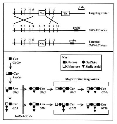
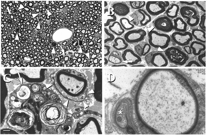
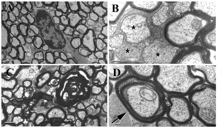
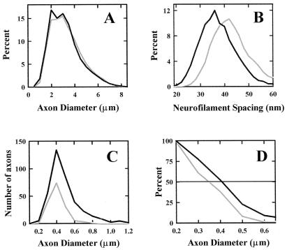
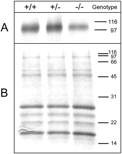
Gangliosides are a family of sialic acid-containing glycosphingolipids highly enriched in the mammalian nervous system. Although they are the major sialoglycoconjugates in the brain, their neurobiological functions remain poorly defined. By disrupting the gene for a key enzyme in complex ganglioside biosynthesis (GM2/GD2 synthase; EC 2.4.1.92) we generated mice that express only simple gangliosides (GM3/GD3) and examined their central and peripheral nervous systems. The complex ganglioside knockout mice display decreased central myelination, axonal degeneration in both the central and peripheral nervous systems, and demyelination in peripheral nerves. The pathological features of their nervous system closely resemble those reported in mice with a disrupted gene for myelin-associated glycoprotein (MAG), a myelin receptor that binds to complex brain gangliosides in vitro. Furthermore, GM2/GD2 synthase knockout mice have reduced MAG expression in the central nervous system. These results indicate that complex gangliosides function in central myelination and maintaining the integrity of axons and myelin. They also support the theory that complex gangliosides are endogenous ligands for MAG. The data extend and clarify prior observations on a similar mouse model, which reported only subtle conduction defects in their nervous system [Takamiya, K., Yamamoto, A., Furukawa, K., Yamashiro, S., Shin, M., Okada, M., Fukumoto, S., Haraguchi, M., Takeda, N., Fujimura, K., et al. (1996) Proc. Natl. Acad. Sci. USA 93, 10662-10667].
Figures
Similar articles
-
A functional role for complex gangliosides: motor deficits in GM2/GD2 synthase knockout mice.Exp Neurol. 2000 Dec;166(2):227-34. doi: 10.1006/exnr.2000.7504. Exp Neurol. 2000. PMID: 11085888
-
Mice with disrupted GM2/GD2 synthase gene lack complex gangliosides but exhibit only subtle defects in their nervous system.Proc Natl Acad Sci U S A. 1996 Oct 1;93(20):10662-7. doi: 10.1073/pnas.93.20.10662. Proc Natl Acad Sci U S A. 1996. PMID: 8855236 Free PMC article.
-
Myelin-associated glycoprotein and complementary axonal ligands, gangliosides, mediate axon stability in the CNS and PNS: neuropathology and behavioral deficits in single- and double-null mice.Exp Neurol. 2005 Sep;195(1):208-17. doi: 10.1016/j.expneurol.2005.04.017. Exp Neurol. 2005. PMID: 15953602 Free PMC article.
-
Beta1,4-N-acetylgalactosaminyltransferase--GM2/GD2 synthase: a key enzyme to control the synthesis of brain-enriched complex gangliosides.Biochim Biophys Acta. 2002 Dec 19;1573(3):356-62. doi: 10.1016/s0304-4165(02)00403-8. Biochim Biophys Acta. 2002. PMID: 12417418 Review.
-
Brain gangliosides: functional ligands for myelin stability and the control of nerve regeneration.Biochimie. 2001 Jul;83(7):677-82. doi: 10.1016/s0300-9084(01)01308-6. Biochimie. 2001. PMID: 11522397 Review.
Cited by
-
Ganglioside biochemistry.ISRN Biochem. 2012 Dec 19;2012:506160. doi: 10.5402/2012/506160. eCollection 2012. ISRN Biochem. 2012. PMID: 25969757 Free PMC article. Review.
-
Functional evaluation of novel variants of B4GALNT1 in a patient with hereditary spastic paraplegia and the general population.Front Neurosci. 2024 Jul 31;18:1437668. doi: 10.3389/fnins.2024.1437668. eCollection 2024. Front Neurosci. 2024. PMID: 39145292 Free PMC article.
-
Cerebellar neurons lacking complex gangliosides degenerate in the presence of depolarizing levels of potassium.Proc Natl Acad Sci U S A. 2001 Jan 2;98(1):307-12. doi: 10.1073/pnas.98.1.307. Proc Natl Acad Sci U S A. 2001. PMID: 11134519 Free PMC article.
-
Impaired ganglioside metabolism in Huntington's disease and neuroprotective role of GM1.J Neurosci. 2010 Mar 17;30(11):4072-80. doi: 10.1523/JNEUROSCI.6348-09.2010. J Neurosci. 2010. PMID: 20237277 Free PMC article.
-
Exposure to 3'Sialyllactose-Poor Milk during Lactation Impairs Cognitive Capabilities in Adulthood.Nutrients. 2021 Nov 23;13(12):4191. doi: 10.3390/nu13124191. Nutrients. 2021. PMID: 34959743 Free PMC article.
References
-
- Svennerholm L. Prog Brain Res. 1994;101:xi–xiv. - PubMed
-
- Yu R K, Saito M. In: Neurobiology of Glycoconjugates. Margolis R U, Margolis R K, editors. New York: Plenum; 1989. pp. 1–42.
-
- Tettamanti G, Bonali F, Marchesini S, Zambotti V. Biochim Biophys Acta. 1973;296:160–170. - PubMed
-
- van Echten G, Sandhoff K. J Biol Chem. 1993;268:5341–5344. - PubMed
Publication types
MeSH terms
Substances
Grants and funding
LinkOut - more resources
Full Text Sources
Other Literature Sources
Molecular Biology Databases
Research Materials
