. 1999 Jun 22;96(13):7558-62.
doi: 10.1073/pnas.96.13.7558.

Somatotopy of the lateral line projection in larval zebrafish

Affiliations

Somatotopy of the lateral line projection in larval zebrafish

D Alexandre et al. Proc Natl Acad Sci U S A. .

Abstract

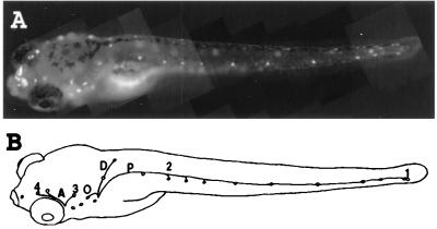
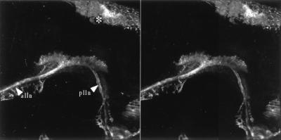
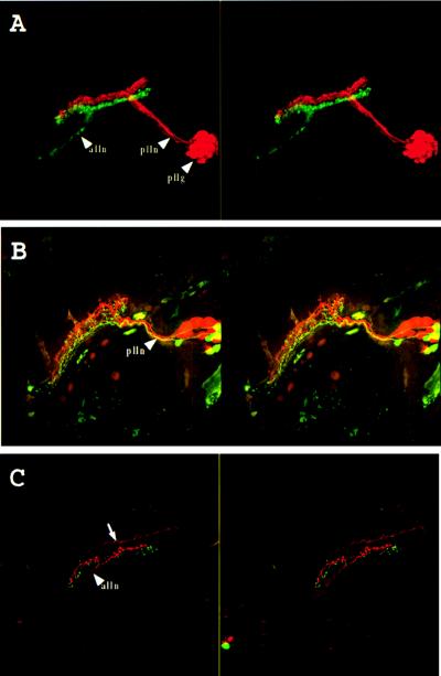
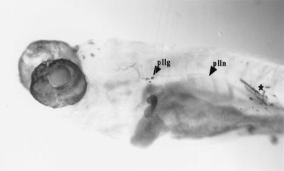
We examined the topography of the lateral line primary projection in zebrafish larvae by double labeling. The projections of two identified neuromasts of the posterior lateral line are seen as two separate sets of fibers that show reproducible spatial relationships: the projection of the anterior neuromast is always ventrolateral to that of a more posteriorly located neuromast. The same rule applies to the projection of anterior lateral line neuromasts. The position of the neuromasts along the antero posterior axis of the fish therefore is represented in the central projection of the sensory neurons. This somatotopy is similar to, and may be at the origin of, the tonotopic projection of the cochlear hair cells in mammals.


Figures

Figure 1
Figure 1
(A) Vitally labeled neuromast hair cells in a 6-day-old zebrafish larvae. The dye also is taken up by the nasal epithelium. (B) Neuromasts and lateral line nerve branches whose projection was examined in the present work. Neuromasts on one side of the fish are drawn as small circles. The nerve branches that were not back-filled in this work are not shown. A, anterior lines; P, midbody posterior line; D, dorsal line; O, occipital line; 1, last two neuromasts of the posterior midbody line; 2, second neuromast of the posterior midbody line; 3, neuromast located dorso anterior to the ear vesicle; 4, last neuromast of the supra orbital line. Neuromasts 1 and 2 are innervated by the posterior lateral line ganglion, and neuromasts 3 and 4 are innervated by the anterior lateral line ganglion. Magnification: ×20.
Figure 2
Figure 2
Vitally labeled lateral line primary projection. Stereo pair of a dorso lateral view seen from the outside of the animal. Anterior is to the left and dorsal to the top. alln, anterior lateral line nerve; plln, posterior lateral line nerve. ∗ indicates dorsal head skin. Magnification: ×160.
Figure 3
Figure 3
Double labeling of the lateral line system. (A) Labeling of the midbody posterior lateral line nerve (plln) with rhodamine-dextran (red) and of the anterior lateral line nerve (alln) with fluoresceine-dextran (green). Stereo pair of a side view seen from the outside of the animal. Anterior is to the left and dorsal to the top. pllg, posterior lateral line ganglion. The anterior lateral line ganglion could not be imaged because of the limited working distance of the lens. (B) Labeling of the afferents of the second neuromast of the midbody posterior lateral line (2 in Fig. 1) with fluoresceine-dextran and the last two neuromasts of the same line (1 in Fig. 1) with rhodamine-dextran. Stereo pair of a dorsolateral view seen from the outside of the animal. Anterior is to the left and dorsal to the top. plln, posterior lateral line nerve. (C) Labeling of the afferents of a neuromast located dorso anteriorly to the ear (3 in Fig. 1) with rhodamine-dextran and the last neuromast of the supra orbital line (4 in Fig. 1) with fluoresceine-dextran. Stereo pair of a dorsolateral view seen from the outside of the animal. Anterior is to the left and dorsal to the top. alln, anterior lateral line nerve. The arrow indicates trigeminal descending fibers, which often were labeled in these preparations. Magnification: ×160.
Figure 4
Figure 4
Labeling of the afferents of the second neuromast of the midbody posterior lateral line (numbered 2 in Fig. 1 and Table 1). ∗ indicates site of dye application. Magnification: ×60.
Figure 5
Figure 5
(A) Schematic cross-section of the lateral line somatotopy in the hindbrain. The dotted line represents the midline. 1, 2, 3, 4, and D refer to the neuromasts labeled on Fig. 1B. The upper ellipse (1 and 2) represents the cross-section of the ribbon-like projection of the posterior lateral line; the lower ellipse (3 and 4) that of the anterior lateral line, D stands for the projection of the dorsal neuromast. Arrows: D, dorsal; V, ventral; M, medial; L: lateral. (B) Somatotopic projection of the spiral ganglion neurons in the cochlear complex of the cat. Side view, anterior to the left, dorsal to the top. The projection of hair cells located at the apex (A) of the Corti organ is ventrolateral to that of cells located more basalward (B). After ref. . Magnification: ×0.5.

References

    1. Rose J. In: Neural Mechanisms of the Auditory and Vestibular Systems. Rasmussen G, Windle W, editors. Springfield, IL: Thomas; 1960. pp. 116–136.
    1. Hofer B. Ber Kgl Bayer Biol Versuchsstation München. 1908;1:115–164.
    1. Schulze F E. Arch Mikrosk Anat. 1870;6:62–68.
    1. Dijkgraaf S. In: The Mechanosensory Lateral Line: Neurobiology and Evolution. Coombs S, Görner P, Münz H, editors. New York: Springer; 1989. pp. 7–14.
    1. Will U. In: The Mechanosensory Lateral Line: Neurobiology and Evolution. Coombs S, Görner P, Münz H, editors. New York: Springer; 1989. pp. 365–386.

Publication types

Substances

LinkOut - more resources