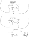
Photoinduced covalent binding of frusemide and frusemide glucuronide to human serum albumin
- PMID: 10383564
- PMCID: PMC2014883
- DOI: 10.1046/j.1365-2125.1999.00970.x
Photoinduced covalent binding of frusemide and frusemide glucuronide to human serum albumin
Abstract
Aims: To study reaction of photoactivated frusemide (F) and F glucuronide (Fgnd metabolite) with human serum albumin in order to find a clue to clarify a mechanism of phototoxic blisters from high frusemide dosage.
Methods: F was exposed to light in the presence of human serum albumin (HSA). HSA treated with this method (TR-HSA) was characterized by fluorescence spectroscopic experiment, alkali treatment and reversible binding experiment.
Results: Less 4-hydroxyl-N-furfuryl-5-sulphamoylanthranilic acid (4HFSA, a photodegradation product of F) was formed in the presence of HSA than in the absence of HSA. A new fluorescence spectrum excited at 320 nm was observed for TR-HSA. Alkali treatment of TR-HSA released 4HFSA. Quenching of the fluorescence due to the lone tryptophan near the warfarin-binding site of HSA was observed in TR-HSA. The reversible binding of F or naproxen to the warfarin-binding site of TR-HSA was less than to that of native HSA. These results indicate the photoactivated F was covalently bound to the warfarin-binding site of HSA. The covalent binding of Fgnd, which is also reversibly bound to the warfarin-binding site of HSA, was also induced by exposure to sunlight. Fgnd was more photoactive than F, indicating that F could be activated by glucuronidation to become a more photoactive compound.
Conclusions: The reactivity of photoactivated F and Fgnd to HSA and/or to other endogenous compounds may cause the phototoxic blisters that result at high F dosage.
Figures


References
-
- Weiner IM, Mudge GH. Diuretics and other agents employed in the mobilization of edema fluid. In: Gilman AG, Goodman LS, Rall TW, Murad F, editors. The Pharmacological Basis of Therapeutics. Seventh. New York: Macmillan Publishing Company; 1985. pp. 887–907.
-
- Burry JN, Lawrence JR. Phototoxic blisters from high frusemide dosage. Br J Dermatol. 1976;94:495–499. - PubMed
-
- Heydenreich G, Pindborg T, Schmidt H. Bullous dermatosis among patients with chronic renal failure on high dose frusemide. Acta Med Scand. 1977;202:61–64. - PubMed
-
- Moore DE, Sithipitaks VJ. Photolytic degradation of frusemide. J Pharm Pharmacol. 1983;35:489–493. - PubMed
-
- Bundgaard H, Norgaard T, Nielsen NM. Photodegradation and hydrolysis of furosemide and furosemide esters in aqueous solutions. Int J Pharm. 1988;42:217–224.
MeSH terms
Substances
LinkOut - more resources
Full Text Sources
Other Literature Sources

