. 1999 Jun;112(6):835-42.
doi: 10.1046/j.1523-1747.1999.00609.x.

The Nf1 tumor suppressor regulates mouse skin wound healing, fibroblast proliferation, and collagen deposited by fibroblasts

Affiliations

The Nf1 tumor suppressor regulates mouse skin wound healing, fibroblast proliferation, and collagen deposited by fibroblasts

R P Atit et al. J Invest Dermatol. 1999 Jun.

Abstract

Neurofibromatosis type 1 patients develop peripheral nerve tumors (neurofibromas) composed mainly of Schwann cells and fibroblasts, in an abundant collagen matrix produced by fibroblasts. Trauma has been proposed to trigger neurofibroma formation. To test if loss of the neurofibromatosis type 1 gene (Nf1) compromises fibroblast function in vivo following trauma, skin wounding was performed in Nf1 knockout mice. The pattern and amount of collagen-rich granulation bed tissue, manufactured by fibroblasts, was grossly abnormal in 60% of Nf1+/- wounds. Nf1 mutant fibroblasts showed cell autonomous abnormalities in collagen deposition in vitro that were not mimicked by Ras activation in fibroblasts, even though some Nf1 effects are mediated through Ras. Nf1+/- skin wound fibroblasts also proliferated past the normal wound maturation phase; this in vivo effect was potentiated by muscle injury. In vitro, Nf1+/- fibroblasts showed higher proliferation in 10% serum than Nf1+/+ fibroblasts. Macrophage-conditioned media or epidermal growth factor potentiated Nf1+/- fibroblast proliferation in vitro, demonstrating abnormal response of mutant fibroblasts to wound cytokines. Thus Nf1 is a key regulator of fibroblast responses to injury, and Nf1 mutation in mouse fibroblasts causes abnormalities characteristic of human neurofibromas.


Figures
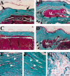
Figure 1
Figure 1. Abnormal wound repair in Nf1+/− mouse excisional skin wounds
Gomori's trichrome stained paraffin sections of Nf1+/+ (A, C) and Nf1+/− (B, D) skin wounds were analyzed at 4 wk postwounding. Collagen appears blue–green. In (C) and (D) skin was wounded, and muscle injured. Black arrows indicate the interface between normal skin containing hair follicles and glands and the regenerated dermal scar (A, B). In Nf1+/− mice, the fibrotic tissue of the granulation bed (series of black arrowheads in B, D) penetrates the underlying muscle fascicles (white letter M). Higher magnification of the dermal scar from asterisked regions in (A, B) are shown in (E, F), respectively. White arrowheads (E, F) point to elongated nuclei within the scar, suggesting that these are fibroblast nuclei. (G) Higher magnification of the double asterisked region in (B) shows the less compact fibrotic tissue penetrating muscle. Scale bar: (AD) 50 μm; (EG) 10 μm.
Figure 2
Figure 2. Rare phenotypes in the Nf1+/− excisional skin wounds
(AC) Gomori's trichrome stained sections of 4 wk old excisional Nf1+/− skin wound sections were analyzed. (A) Collagen matrix and the wound bed erupted above the skin layer that failed to re-epithelialize. In (B, C), there was a delay in normal wound healing. A thick and highly cellular granulation bed with iNflammatory cells (visible only at higher magnification), prolonged angiogenesis (C, black arrow points to blood vessels filled with red blood cells), and lack of wound closure (A, C region between black arrowheads) are seen. Keratinocyte hyperplasia (B, C, white arrowheads) and regeneration of the panniculus carnosus muscle layer (B, C, white arrows) are clearly evident in (B, C). Scale bar: 100 μm.
Figure 3
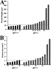
Figure 3. Increase in depth and area of some Nf1+/− mouse skin scars
Wound depth (A) and area (B) from Nf1+/+ and Nf1+/− of 4 wk old wounds. The depth and area of the Nf1+/− skin wound group were significantly higher than the Nf1+/+ group, p < 0.0001 and p < 0.05, t = −2.4, respectively, as assessed by the Student's t-test. Each bar represents the average of at least four measurements from random sections spanning an individual scar's length. “N” indicates wounds with muscle injury. Depth and area measurements were performed as noted in the text. Dotted line shows maximum height (A) and area (B) determined in +/+ wounds.
Figure 4
Figure 4. Fibroblasts proliferate past the normal wound maturation phase in Nf1+/− wounds
Anti-PCNA immunostaining of 4 wk old Nf1+/+ (A) and Nf1+/− (B) wounds with muscle injury. PCNA-positive (brown color) basal layer keratinocytes in the epidermis (black arrows) and fibroblasts (white arrows) are present in the granulation bed of the regenerated dermal scar. Hematoxylin counterstaining shows unlabeled nuclei in blue. Scale bar:10 μm. (C) Quantitation of anti-PCNA-labeled nuclei in wounds with and without muscle injury. “N” indicates wounds with muscle injury.
Figure 5
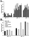
Figure 5. Increased proliferation of Nf1+/− fibroblasts to serum factors, macrophage-conditioned media, or EGF
(A) Fibroblast proliferation was analyzed in media with 10% FBS (solid bars) or media with 10% FBS + 50% macrophage-conditioned media (MCM; hatched bars). Each set of bars represents data from fibroblasts from an individual embryo. (B) Fibroblast proliferation stimulated by EGF, but not MCM, is inhibited by the EGF receptor antagonist AG1478. Data show results from a single +/+ and +/− embryo and are representative of three individual experiments using cells from different embryos. Data are presented as the mean fold increase in cell number after 8 d in duplicate cultures. Error bars show SEM.
Figure 6
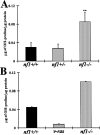
Figure 6. Ras-independent increases in collagen deposition by Nf1−/− embryonic fibroblasts
(A) Levels of hydroxyproline in fibroblast cultures of various genotypes. Collagen deposition was inferred by measurement of hydroxyproline in hydrolysates of 14 d fibroblast cultures. Data are presented as micrograms of hydroxyproline per microgram of detergent-soluble protein. Each bar represents the mean of triplicate cultures in five to seven separate experiments with fibroblasts using six to 13 individual embryos. Error bars show SD and **p < 0.0001 (Student's t-test) between Nf1+/+ and Nf1−/− fibroblast values. (B) Levels of hydroxyproline in cultures of Nf1+/+ fibroblasts as compared with Nf1+/+ fibroblasts infected with oncogenic v-H-ras (v-ras). Collagen deposition by cells from a single Nf1−/− embryo is shown for comparison. The data were coNfirmed in three independent experiments. Each bar represents the average of duplicate measurements; error bars show SEM.

References

    1. Basu TN, Gutmann DH, Fletcher JA, Glover TW, Collins FS, Downward J. Aberrant regulation of ras proteins in malignant tumour cells from type 1 neurofibromatosis patients. Nature. 1992;356:713–715. - PubMed
    1. Berg RA. Determination of 3- and 4-hydroxyproline. Methods Enzymol. 1982;82:372–398. - PubMed
    1. Brannan CI, Perkins AS, Vogel KS, et al. Targeted disruption of the neurofibromatosis type-1 gene leads to developmental abnormalities in heart and various neural crest-derived tissues. Genes Dev. 1994;8:1019–1029. - PubMed
    1. Brown RL, Breeden MP, Greenhalgh DG. PDW and TGFalpha act synergistically to improve wound healing in genetically diabetic mouse. J Surg Res. 1994;56:562–570. - PubMed
    1. Clark RA. Mechanisms of cutaneous wound repair. In: Fitzpatrick TB, Eisen AZ, Wolff K, Freedberg IM, Austen KF, editors. Dermatology in General Medicine. McGraw-Hill; New York: 1993. pp. 473–486.

Publication types