

Review
doi: 10.1128/JB.181.13.3869-3879.1999.
How photosynthetic bacteria harvest solar energy
Affiliations
- PMID: 10383951
- PMCID: PMC93873
- DOI: 10.1128/JB.181.13.3869-3879.1999
Item in Clipboard
Review
How photosynthetic bacteria harvest solar energy
J Bacteriol.
1999 Jul.
Free PMC article
No abstract available
Figures
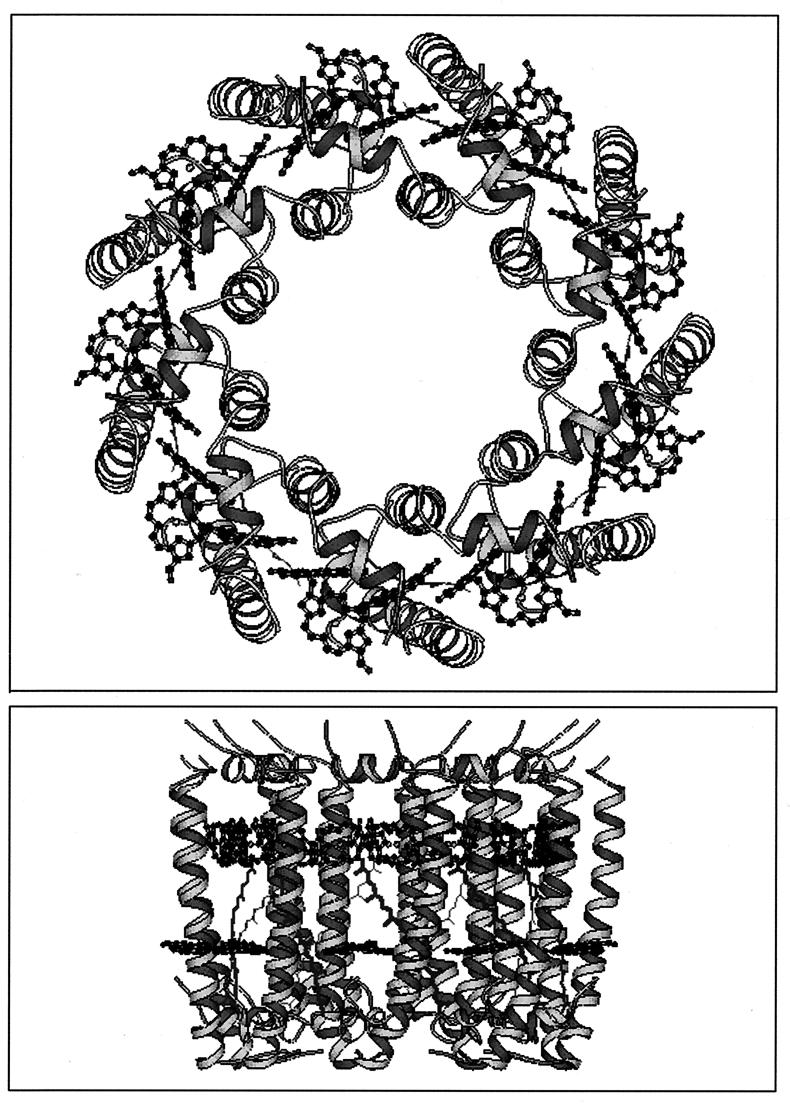
Schematic representation of the LH2 holocomplex
from Rhodopseudomonas acidophila. (Top) View from the
cytoplasmic side of the membrane, looking down the central ninefold
axis of symmetry. (Bottom) View from within the membrane. The
organization of the two rings of Bchla molecules, arranged
between the transmembrane α-helices, are shown; the 9 B800
Bchla molecules parallel to the plane of the membrane and
the second ring of 18 B850 Bchla molecules, with their
bacteriochlorin rings perpendicular to the plane of the membrane. The
bottom view also shows the carotenoid (rhodopin-glucoside) which spans
the membrane and comes into van der Waal’s contact with both groups of
Bchla. Only the chromophoric portions of the pigments are
shown. This and most of the other figures in this minireview were
produced with the Molscript program (38).
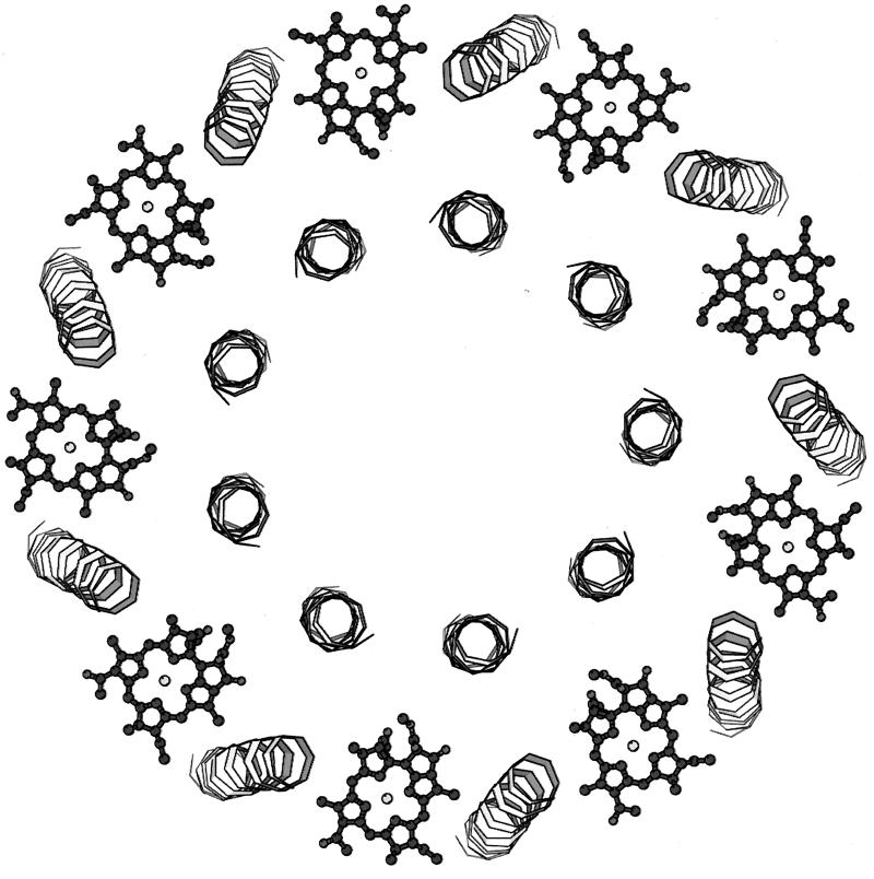
Location and organization of the B800 Bchlas
in the LH2 from Rhodopseudomonas acidophila. The B800
Bchlas (nine Bchla molecules) can be seen
arranged peripherally between the β-apoprotein α-helices.
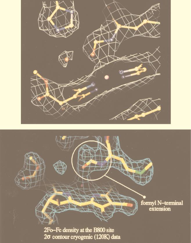
Comparison of the electron density in the region of the
B800 Bchla binding pocket of LH2 from Rhodopseudomonas
acidophila at a resolution of 2.5 Å (top) and 2.0 Å (bottom).
(Top) With a resolution of 2.5 Å, the extension of the N-terminal
methionine residue of the α-apoprotein is clearly seen in the
electron density map, together with the “modelled” formyl group.
(Bottom) At this improved resolution of 2.0 Å, the N-terminal
extension is also clearly seen. Now, however, it can be seen to
bifurcate. The modelled formyl group no longer gives a satisfactory fit
to this higher-resolution data. The electron density is shown as the
white or blue cage.
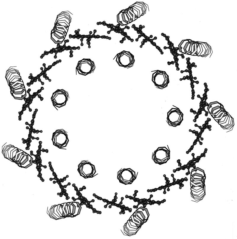
Organization of B850 Bchlas in the LH2 from
Rhodopseudomonas acidophila. The 18 Bchla
molecules, which from the B850 ring can be seen, edge on, are arranged
between the transmembrane α-helices of the α-apoprotein (inner) and
β-apoprotein (outer).
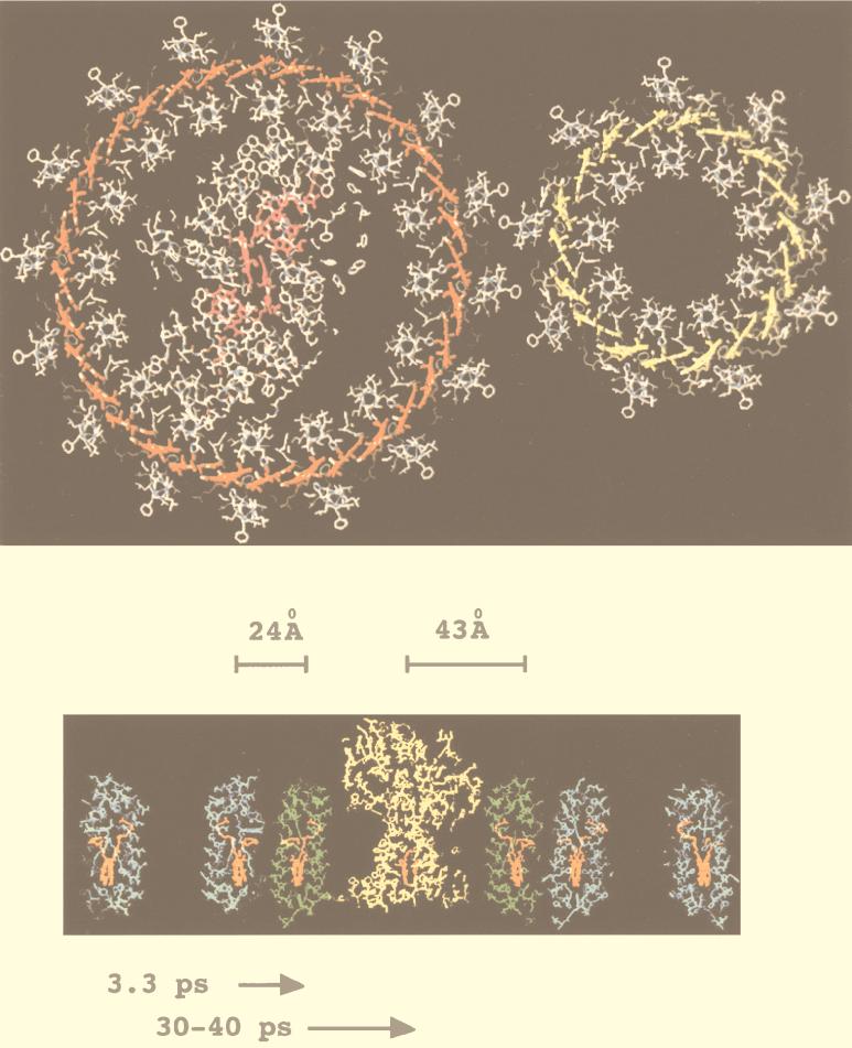
Two views of a model of the purple bacterial PSU. (Top)
A top view, looking perpendicular to the assumed plane of the membrane.
This section is taken at a point in the LH complexes where the tightly
coupled rings of Bchlas are located. This figure was adapted
from reference with permission from Elsevier
Science. The reaction center is located in the center of the LH1
complex. The smaller LH2 sits outside the large LH1 complex. (Bottom) A
side view, looking from within the assumed plane of the membrane (blue,
LH2; green, LH1; yellow, RC). This section is taken exactly
perpendicular to the view shown in the top panel. The distances shown
between the different pigment groups (shown in orange) are calculated
assuming a space-filling model and the closest possible organization.
The times shown are energy transfer times as measured in intact
membranes (31, 61, 69, 72).
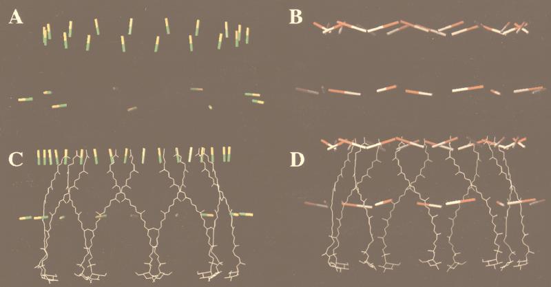
Relative orientations of the Qx and
Qy transition dipoles in the B800 and B850
Bchlas and the carotenoid in the LH2 complex from
Rhodopseudomonas acidophila. (A) Alignment of the
Qx dipoles (yellow-green). (B) Alignment of the
Qy dipoles (red-white). (C) Alignment of the carotenoid to
the Qx dipoles (the transition dipole movement of the
S2 state of the carotenoid runs up and down the long axis
of the conjugated double bands). (D) Alignment of the carotenoid with
respect to the Qy dipoles. The figure was adapted from
reference with permission from Elsevier Science
and produced with O (32). The distances between the center
of a B800 Bchla and the α-bound and β-bound B850 Bchlas
in the same αβ-apoprotein pair are 17.4 and 18.2 Å, respectively.
Qx and Qy are labels for the two
Bchla absorption bands at ∼590 nm and 800 or 850 nm,
respectively. The transition dipoles, which correspond to these
absorption bands, lie within the plane of the bacteriochlorin rings, at
right angles to each other, diagonally between the nitrogen atoms,
which coordinate the central Mg2+. One of the factors which
controls the rate of excitation energy transfer between two molecules
is the angle between the transition dipoles involved. When the dipoles
are parallel, energy transfer is favorable; when they are orthoganal,
energy transfer is much less favorable.
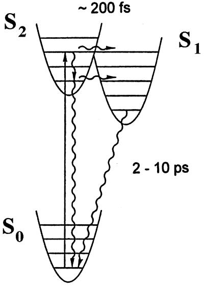
Schematic representation of the two low-lying excited
singlet states of S1 and S2 carotenoids. The
approximate positions of the S1 and S2 excited
singlet states are shown. The S0→S2
represents the optically allowed (one-photon) transition that gives
rise to the carotenoid’s well-known, strong absorption spectrum.
The approximate times for the S2→S1 and
S1→S0 transitions are also shown.
References
-
- Aagaard J, Sistrom W R. Control of the synthesis of reaction centre bacteriochlorophylls in photosynthetic bacteria. Photochem Photobiol. 1972;15:209–225. - PubMed
-
- Barz W P, Francia F, Venturoli G, Melandri B A, Verméglio A, Oesterhelt D. Role of PufX protein in photosynthetic growth of Rhodobacter sphaeroides. 1. PufX is required for efficient light-driven electron transfer and photophosphorylation under anaerobic conditions. Biochemistry. 1995;34:15235–15247. - PubMed
-
- Barz W P, Verméglio A, Francia F, Verturoli G, Melandri B A, Oesterhelt D. Role of PufX protein in photosynthetic growth of Rhodobacter sphaeroides. 2. PufX is required of efficient ubiquinone/ubiquinol exchange between the reaction centre Q(B) site and the cytochrome b/c1complex. Biochemistry. 1995;34:15248–15258. - PubMed
-
- Bauer C E, Bird T H. Regulatory circuits controlling photosynthesis gene expression. Cell. 1996;85:5–8. - PubMed
-
- Blankenship R E, Madigan M T, Bauer C E. Anoxygenic photosynthetic bacteria. Dordrecht, The Netherlands: Kluwer Academic Publishers; 1995.
Publication types
MeSH terms
Substances
LinkOut - more resources
Full Text Sources
