

doi: 10.1128/JB.181.13.4137-4141.1999.
Molecular cloning and characterization of a locus responsible for O acetylation of the O polysaccharide of Legionella pneumophila serogroup 1 lipopolysaccharide
Affiliations
- PMID: 10383989
- PMCID: PMC93911
- DOI: 10.1128/JB.181.13.4137-4141.1999
Item in Clipboard
Molecular cloning and characterization of a locus responsible for O acetylation of the O polysaccharide of Legionella pneumophila serogroup 1 lipopolysaccharide
J Bacteriol.
1999 Jul.
Abstract
Complementation experiments, Tn5 mutagenesis, and DNA sequencing were used to identify a locus (lag-1) that participates in acetylation of Legionella pneumophila serogroup 1 lipopolysaccharide. Nuclear magnetic resonance analyses of lipopolysaccharides from mutant and complemented strains suggest that lag-1 is responsible for O acetylation of serogroup 1 O polysaccharide.
Figures
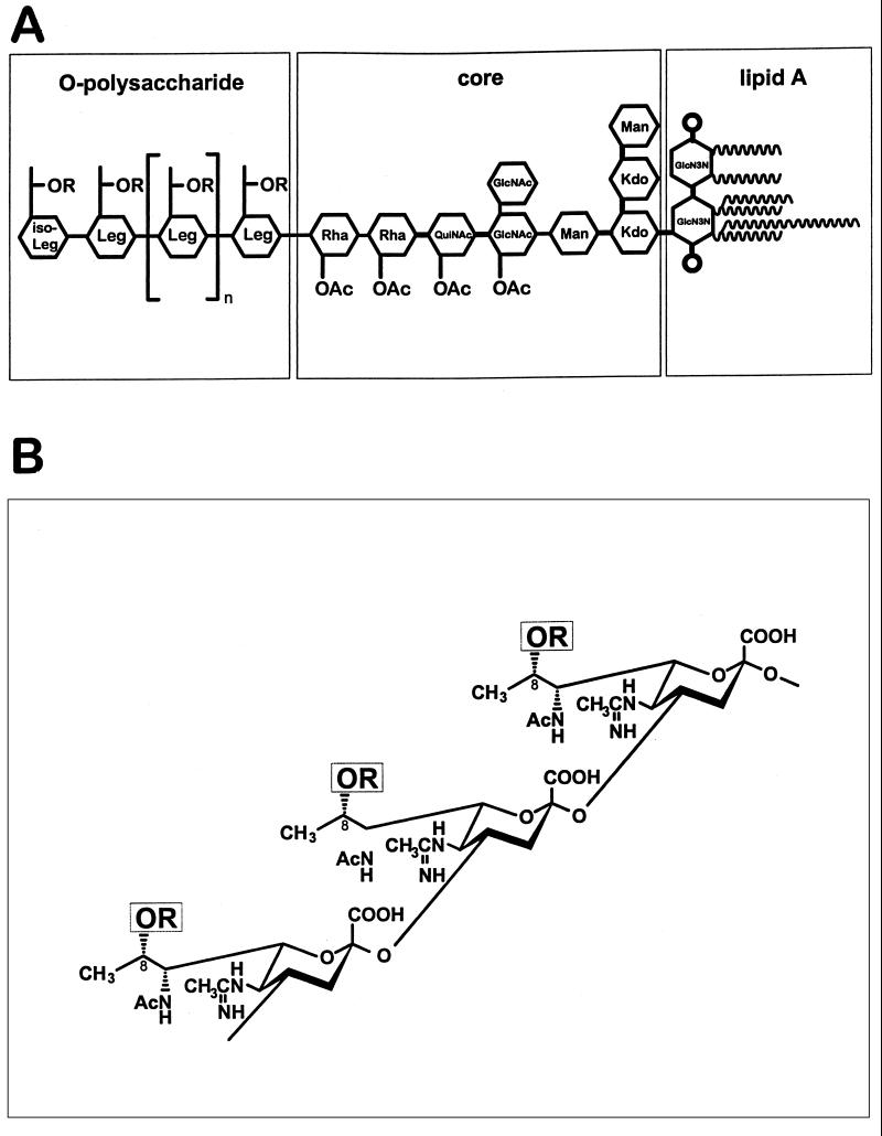
Structure of L. pneumophila serogroup 1 LPS adopted from Knirel et al. (–11), Zähringer et al. (24), and Moll et al. (18). (A) Schematic representation of the whole LPS structure. (B) Structure of OPS. Sugar abbreviations: GlcN3N, 2,3-diamino-2,3-dideoxy-d -glucose; Kdo, 3-deoxy-d -manno-octulosonic acid; Leg and iso-Leg, derivatives of legionaminic acid and its C4 epimer, respectively; Rha, l -rhamnose; QuiNAc, 2-acetamido-2,6-dideoxy-d -glucose (N-acetylquinovosamine); R = Ac in strains Philadelphia 1 and CS338 or R = H in strain CS332.

Sodium dodecyl sulfate-polyacrylamide gel electrophoresis (A) and Western blot (B) analyses of LPSs from Philadelphia 1 (wild-type) and lag-1 mutant strains. (A) LPS was isolated as described previously (19), resolved on a sodium dodecyl sulfate–14% polyacrylamide gel, and visualized by silver staining. Equal amounts of LPS (∼1.0 μg) were added to each lane. Lanes: 1, S. enterica serovar Typhimurium; 2, Philadelphia 1; 3, CS332 (lag-1 negative); 4, CS334 (CS332/pLPS16 [lag-1 positive]). (B) Equal amounts of LPS from strains Philadelphia 1, CS332, and CS338 (CS332/pLPS17 [lag-1 positive]) were resolved on sodium dodecyl sulfate–14% polyacrylamide gels, transferred to nitrocellulose paper, and probed with MAB2 as described by Mintz and Zou (15). Lanes: 1, Philadelphia 1; 2, CS332; 3, CS338.
Localization of lag-1 by Tn5 mutagenesis. Tn5 insertions were introduced into pLPS16.1 and mapped according to the methods of de Bruijn and Lupinski (2). Each triangle represents the position of a Tn5 insertion in pLPS16.1. Plasmids containing each of the Tn5 insertions were electroporated into strain CS332, and transformants were tested for the ability to bind MAB2, 33G2, and 144C2. (−), insertions eliminating binding of MAB2 and 33G2; (+), insertions having no effect on the binding of MAB2 and 33G2. Wild-type LPS has the following MAb binding pattern: MAB2 (+), 33G2 (+), 144C2 (−). Restriction sites: C, ClaI; E, EcoRI; H, HindIII; S, SphI; X, XhoI. The black box defines the physical location of the lag-1 locus as determined by DNA sequencing experiments.
13C-NMR spectra and corresponding structures of OPSs from LPSs produced by wild-type, mutant, and complemented strains. (A) CS338 (CS332/pLPS17). (B) CS339 (CS332/pLPS20). (C) CS333 (AM511/pLAW300). Signals for the 8-O-acetyl group (Me at δ 22.0 and CO at δ 174.4) are marked by stars. Numbers refer to carbons C1 to C9 of legionaminic acid.
Similar articles
-
Distribution of lag-1 Alleles, ORF7, and ORF8 Genes of Lipopolysaccharide and Sequence-Based Types Among Legionella pneumophila Serogroup 1 Isolates in Japan and China.Front Cell Infect Microbiol. 2019 Aug 5;9:274. doi: 10.3389/fcimb.2019.00274. eCollection 2019. Front Cell Infect Microbiol. 2019. PMID: 31448241 Free PMC article.
-
Complex O-acetylation in Legionella pneumophila serogroup 1 lipopolysaccharide. Evidence for two genes involved in 8-O-acetylation of legionaminic acid.Biochemistry. 2001 Jun 26;40(25):7630-40. doi: 10.1021/bi002946r. Biochemistry. 2001. PMID: 11412117
-
A point mutation in the active site of Legionella pneumophila O-acetyltransferase results in modified lipopolysaccharide but does not influence virulence.Int J Med Microbiol. 2001 Nov;291(5):345-52. doi: 10.1078/1438-4221-00140. Int J Med Microbiol. 2001. PMID: 11727818
-
The lipopolysaccharide of Legionella pneumophila serogroup 1 (strain Philadelphia 1): chemical structure and biological significance.Prog Clin Biol Res. 1995;392:113-39. Prog Clin Biol Res. 1995. PMID: 8524918 Review.
-
Genetics of Legionella pneumophila virulence.Annu Rev Genet. 1992;26:51-69. doi: 10.1146/annurev.ge.26.120192.000411. Annu Rev Genet. 1992. PMID: 1482124 Review. No abstract available.
Cited by
-
A hospital-associated outbreak of Legionnaires' disease caused by Legionella pneumophila serogroup 1 is characterized by stable genetic fingerprinting but variable monoclonal antibody patterns.J Clin Microbiol. 2003 Jun;41(6):2503-8. doi: 10.1128/JCM.41.6.2503-2508.2003. J Clin Microbiol. 2003. PMID: 12791873 Free PMC article.
-
Genomic heterogeneity differentiates clinical and environmental subgroups of Legionella pneumophila sequence type 1.PLoS One. 2018 Oct 18;13(10):e0206110. doi: 10.1371/journal.pone.0206110. eCollection 2018. PLoS One. 2018. PMID: 30335848 Free PMC article.
-
Distribution of lag-1 Alleles, ORF7, and ORF8 Genes of Lipopolysaccharide and Sequence-Based Types Among Legionella pneumophila Serogroup 1 Isolates in Japan and China.Front Cell Infect Microbiol. 2019 Aug 5;9:274. doi: 10.3389/fcimb.2019.00274. eCollection 2019. Front Cell Infect Microbiol. 2019. PMID: 31448241 Free PMC article.
-
A structural comparison of lipopolysaccharide biosynthesis loci of Legionella pneumophila serogroup 1 strains.BMC Microbiol. 2013 Sep 4;13:198. doi: 10.1186/1471-2180-13-198. BMC Microbiol. 2013. PMID: 24069939 Free PMC article.
-
Current and emerging Legionella diagnostics for laboratory and outbreak investigations.Clin Microbiol Rev. 2015 Jan;28(1):95-133. doi: 10.1128/CMR.00029-14. Clin Microbiol Rev. 2015. PMID: 25567224 Free PMC article. Review.
References
-
- de Bruijn F J, Lupinski J R. The use of transposon Tn5 mutagenesis in the rapid generation of correlated physical and genetic maps of DNA segments cloned into multicopy plasmids. Gene. 1984;27:131–149. - PubMed
-
- Dournon E, Bibb W F, Rjagopalan P, Desplaces N, McKinney R M. Monoclonal antibody reactivity as a virulence marker for Legionella pneumophila serogroup 1 strains. J Infect Dis. 1988;157:496–501. - PubMed
MeSH terms
Substances
Associated data
- Actions
LinkOut - more resources
Full Text Sources
Other Literature Sources
Molecular Biology Databases
