. 1999 May;127(2):343-8.
doi: 10.1038/sj.bjp.0702555.

Parallel modulation of receptor for activated C kinase 1 and protein kinase C-alpha and beta isoforms in brains of morphine-treated rats

Affiliations

Parallel modulation of receptor for activated C kinase 1 and protein kinase C-alpha and beta isoforms in brains of morphine-treated rats

P V Escribá et al. Br J Pharmacol. 1999 May.

Abstract

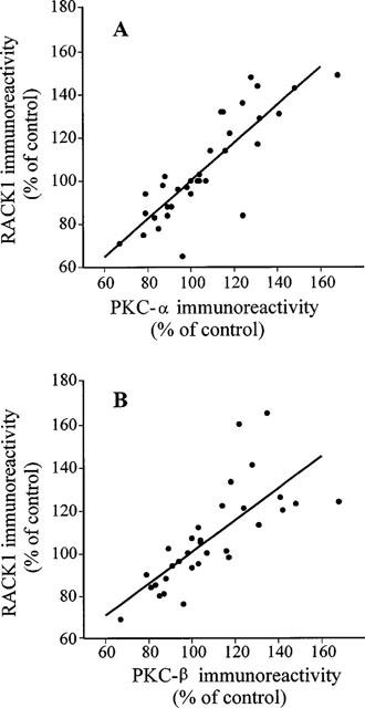
1. Receptor for activated C kinase 1 (RACK1) is an intracellular receptor for protein kinase C (PKC) that regulates the cellular enzyme localization. Because opiate drugs modulate the levels of brain PKC (Ventayol et al., 1997), the aim of this study was to assess in parallel the effects of morphine on RACK1 and PKC-alpha and beta isozymes densities in rat brain frontal cortex by immunoblot assays. 2. Acute morphine (30 mg kg(-1), i.p., 2 h) induced significant increases in the densities of RACK1 (33%), PKC-alpha (35%) and PKC-beta (23%). In contrast, chronic morphine (10-100 mg kg(-1), i.p., 5 days) induced a decrease in RACK1 levels (22%), paralleled by decreases in the levels of PKC-alpha (16%) and PKC-beta (16%). 3. Spontaneous (48 h) and naloxone (2 mg kg(-1), i.p., 2 h)-precipitated morphine withdrawal after chronic morphine induced marked up-regulations in the levels of RACK1 (38-41%), PKC-alpha (51-52%) and PKC-beta (48-62%). 4. In the same brains and for all combined treatments, there were significant positive correlations between the density of RACK1 and those of PKC-alpha (r=0.85, n = 35) and PKC-beta (r=0.75, n=32). 5. These data indicate that RACK1 is involved in the short- and long-term effects of morphine and in opiate withdrawal, and that RACK1 modulation by morphine or its withdrawal is parallel to those of PKC-alpha and beta isozymes. Since RACK1 facilitates the PKC substrate accessibility, driving its cellular localization, the coordinate regulation of the PKC/RACK system by morphine could be a relevant molecular mechanism in opiate addiction.


Figures

Figure 1
Figure 1
RACK1 immunoreactivity. Representative immunoblotting assay of RACK1 in the rat brain (cerebral cortex), using a specific monoclonal anti-RACK1 antibody. Under the reported conditions, a unique peptide of ∼36 kDa was detected. The electrophoretical mobility of this protein was identical to that of a control provided by the antibody manufacturer (data not shown). This figure also shows the concentration-dependence of the signals obtained by immunoblotting, corresponding to a standard curve. Total protein loaded on the gel and IOD values for these bands were: 12 μg and 7.32 IOD units (lane 1); 24 μg and 17.80 IOD units (lane 2); 30 μg and 27.37 IOD units (lane 3); 36 μg and 36.08 IOD units (lane 4) and 48 μg and 53.59 IOD units (lane 5), respectively. The amount of total protein loaded for problem samples was around 30 μg.
Figure 2
Figure 2
Effects of acute and chronic morphine treatment and naloxone-precipitated withdrawal on RACK1 immunoreactivity in the rat cerebral cortex. Rats were treated i.p. with saline vehicle (S, n=10), acute morphine (A, n=8; 30 mg kg−1, 2 h), chronic morphine (C, n=8; 10–100 mg kg−1, 5 days) or chronic morphine plus naloxone (N, n=6; 2 mg kg−1, 2 h). Upper panel: data are mean±s.e.mean (bars) percentages of immunoreactivities in naive rats (untreated group). One-way ANOVA [F(3,28)=37.15, P<0.0001] followed by a multiple comparison test detected significant increases in RACK1 immunoreactivity after acute morphine and naloxone-precipitated withdrawal in morphine-dependent rats and a significant decrease after chronic morphine treatment: *P<0.05; **P<0.01; ***P<0.001, when compared with saline (S). Lower panel: representative immunoblot for the effects of acute morphine (A), chronic morphine (C) and chronic morphine plus naloxone (N) on RACK1 levels. Total protein loaded on the gel and IOD values were: 32.6 μg and 32.97 IOD units (S), 33.1 μg and 48.2 IOD units (A), 27.4 μg and 9.86 IOD units (C) and 30.3 μg and 37.1 IOD units (N).
Figure 2
Figure 2
Effects of acute and chronic morphine treatment and naloxone-precipitated withdrawal on RACK1 immunoreactivity in the rat cerebral cortex. Rats were treated i.p. with saline vehicle (S, n=10), acute morphine (A, n=8; 30 mg kg−1, 2 h), chronic morphine (C, n=8; 10–100 mg kg−1, 5 days) or chronic morphine plus naloxone (N, n=6; 2 mg kg−1, 2 h). Upper panel: data are mean±s.e.mean (bars) percentages of immunoreactivities in naive rats (untreated group). One-way ANOVA [F(3,28)=37.15, P<0.0001] followed by a multiple comparison test detected significant increases in RACK1 immunoreactivity after acute morphine and naloxone-precipitated withdrawal in morphine-dependent rats and a significant decrease after chronic morphine treatment: *P<0.05; **P<0.01; ***P<0.001, when compared with saline (S). Lower panel: representative immunoblot for the effects of acute morphine (A), chronic morphine (C) and chronic morphine plus naloxone (N) on RACK1 levels. Total protein loaded on the gel and IOD values were: 32.6 μg and 32.97 IOD units (S), 33.1 μg and 48.2 IOD units (A), 27.4 μg and 9.86 IOD units (C) and 30.3 μg and 37.1 IOD units (N).
Figure 3
Figure 3
Effects of chronic morphine treatment and spontaneous withdrawal on RACK1, PKC-α and PKC-β immunoreactivities in the rat cerebral cortex. Rats were treated i.p. with morphine (10–100 mg kg−1, 5 days) and killed 2 h (n=3), 12 h (n=3) or 48 h (n=3) after the last morphine injection. Data are mean±s.e.mean (bars) percentages of immunoreactivity of saline-treated rats (n=3). One-way ANOVA [F(3, 8)=26.3–29.3, P<0.001] followed by a multiple comparison test detected significant changes in RACK1 and PCK isozymes immunoreactivities during withdrawal: *P<0.05, when compared with saline.
Figure 4
Figure 4
Scatterlots and linear regression lines showing the relation between RACK1 and PKC-α (A) or PKC-β (B) in cerebral cortices of the same rats after various treatments (saline vehicle, acute and chronic morphine, chronic morphine plus naloxone and spontaneous morphine withdrawal). Values are expressed as percentage immunoreactivity in naive rats. See Figures 2 and 3 for further details. Data were best described by the expressions: PKC-α, y=12+0.88x (r=0.85, n=35, P<0.001) and PKC-β, y=26+0.77x (r=0.75, n=32, P<0.001).

Similar articles

Cited by

References

    1. AMMER H., SCHULZ R. Enhanced stimulatory adenylyl cyclase signaling during opioid dependence is associated with a reduction in palmitoylated Gsα. Mol. Pharmacol. 1997;52:993–999. - PubMed
    1. BEITNER-JOHNSON D., GUITART X., NESTLER E.J. Neurofilament proteins and the mesolimbic dopamine system: common regulation by chronic morphine and chronic cocaine in the rat ventral tegmental area. J. Neurosci. 1992;12:2165–2176. - PMC - PubMed
    1. BEITNER-JOHNSON D., GUITART X., NESTLER E.J. Glial fibrillary acidic protein and the mesolimbic dopamine system: regulation by chronic morphine and Lewis-Fischer strain differences in the rat ventral tegmental area. J. Neurochem. 1993;61:1766–1773. - PubMed
    1. BUSQUETS X., ESCRIBÁ P.V., SASTRE M., GARCÍA-SEVILLA J.A. Loss of protein kinase C-αβ in brain of heroin addicts and morphine-dependent rats. J. Neurochem. 1995;64:247–252. - PubMed
    1. CAMBIER J.C., NEWELL M.K., JUSTEMENT L.B., MCGUIRE J.C., LEACH K.L., CHEN Z.Z. Ia binding of ligands and cAMP stimulate nuclear translocation of PKC in B lymphocytes. Nature. 1987;327:629–632. - PubMed

Publication types

MeSH terms

LinkOut - more resources