. 1999 May;127(2):349-60.
doi: 10.1038/sj.bjp.0702556.

NO synthase in cholinergic nerves and NO-induced relaxation in the rat isolated corpus cavernosum

Affiliations

NO synthase in cholinergic nerves and NO-induced relaxation in the rat isolated corpus cavernosum

P Hedlund et al. Br J Pharmacol. 1999 May.

Abstract

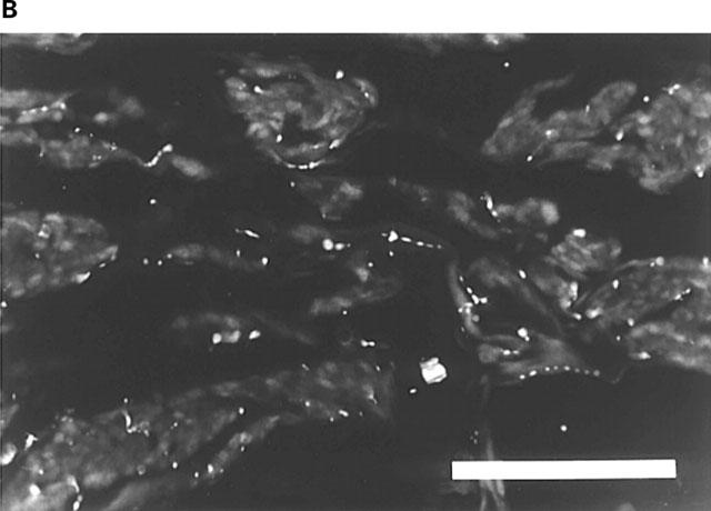
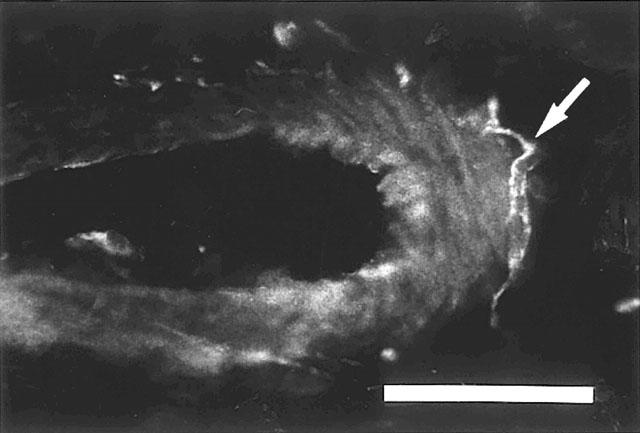
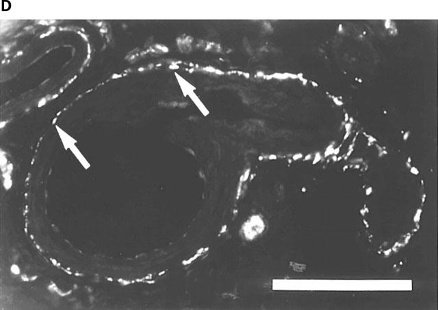
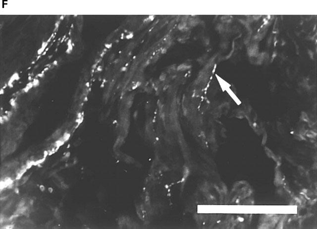
1. In the rat corpus cavernosum (CC), the distribution of immunoreactivity for neuronal and endothelial NO synthase (nNOS and eNOS), and the pattern of NOS-immunoreactive (-IR) nerves in relation to some other nerve populations, were investigated. Cholinergic nerves were specifically immunolabelled with antibodies to the vesicular acetylcholine transporter protein (VAChT). 2. In the smooth muscle septa surrounding the cavernous spaces, and around the central and helicine arteries, the numbers of PGP- and tyrosine hydroxylase (TH)-IR terminals were large, whereas neuropeptide Y (NPY)-, VAChT-, nNOS-, and vasoactive intestinal polypeptide (VIP)-IR terminals were found in few to moderate numbers. 3. Double immunolabelling revealed that VAChT- and nNOS-IR terminals, VAChT- and VIP-IR terminals, nNOS-IR and VIP-IR terminals, and TH- and NPY-IR terminals showed coinciding profiles, and co-existence was verified by confocal laser scanning microscopy. TH immunoreactivity was not found in VAChT-, nNOS-, or VIP-IR nerve fibres or terminals. 4. An isolated strip preparation of the rat CC was developed, and characterized. In this preparation, cumulative addition of NO to noradrenaline (NA)-contracted strips, produced concentration-dependent, rapid, and almost complete relaxations. Electrical field stimulation of endothelin-1-contracted preparations produced frequency-dependent responses: a contractile twitch followed by a fast relaxant response. After cessation of stimulation, there was a slow relaxant phase. Inhibition of NO synthesis, or blockade of guanylate cyclase, abolished the first relaxant phase, whereas the second relaxation was unaffected. 5. The results suggest that in the rat CC, nNOS, VAChT- and VIP-immunoreactivities can be found in the same parasympathetic cholinergic neurons. Inhibitory neurotransmission involves activation of the NO-system, and the release of other, as yet unknown, transmitters.


Figures

Figure 1
Figure 1
Rat corpus cavernosum. (A) PGP-IR terminals accompanying smooth muscle bundles. FITC-immunofluorescence. (B) Reduced number of PGP-IR terminals following chemical sympathectomy. FITC-immunofluorescence. (C) AChE-positive terminals along smooth muscle bundles and forming plexuses around branches of central arteries. Bars=100 μm.
Figure 1
Figure 1
Rat corpus cavernosum. (A) PGP-IR terminals accompanying smooth muscle bundles. FITC-immunofluorescence. (B) Reduced number of PGP-IR terminals following chemical sympathectomy. FITC-immunofluorescence. (C) AChE-positive terminals along smooth muscle bundles and forming plexuses around branches of central arteries. Bars=100 μm.
Figure 2
Figure 2
Rat corpus cavemosum. Branch of a central artery with HO-1-IR nerve fibres (arrow) at the outer margins of the vessel. Texas Red-immunofluorescence. Bar=50 μm.
Figure 3
Figure 3
Rat corpus cavernosum. (A) VAChT-IR terminals along smooth muscle bundles. FITC-immunofluorescence. (B) same section as in (A). NOS-IR terminals displaying profiles (arrows) coinciding with VAChT-IR terminals in (A). Texas Red (TR)-immunofluorescence. (C) Branch of a central artery surrounded by a plexus of VAChT-IR terminals. FITC-immunofluorescence. (D) Same section as in (C). VIP-IR terminals with profiles (arrows) coinciding with VAChT-IR terminals in (C). TR-immunofluorescence. (E) Branch of a central artery and smooth muscle bundles with NOS-IR terminals. FITC-immunofluorescence. (F) Same section as in (E). VIP-IR terminals with profiles (arrows) coinciding with NOS-IR terminals in (E). TR-immunofluorescence. Bars=100 μm.
Figure 3
Figure 3
Rat corpus cavernosum. (A) VAChT-IR terminals along smooth muscle bundles. FITC-immunofluorescence. (B) same section as in (A). NOS-IR terminals displaying profiles (arrows) coinciding with VAChT-IR terminals in (A). Texas Red (TR)-immunofluorescence. (C) Branch of a central artery surrounded by a plexus of VAChT-IR terminals. FITC-immunofluorescence. (D) Same section as in (C). VIP-IR terminals with profiles (arrows) coinciding with VAChT-IR terminals in (C). TR-immunofluorescence. (E) Branch of a central artery and smooth muscle bundles with NOS-IR terminals. FITC-immunofluorescence. (F) Same section as in (E). VIP-IR terminals with profiles (arrows) coinciding with NOS-IR terminals in (E). TR-immunofluorescence. Bars=100 μm.
Figure 4
Figure 4
Rat corpus cavernosum. (A) Branch of a central artery surrounded by VAChT-IR terminals. FITC-immunofluorescence. (B) Same section as in (A). TH-IR terminals displaying profiles which are similar, but not coinciding with VAChT-IR terminals in (A). TR-immunofluorescence. (C) Branch of a central artery surrounded by a plexus of VAChT-IR terminals. TR-immunofluorescence. (D) Same section as in (C). NPY-IR terminals displaying profiles, some of which are similar to VAChT-IR terminals in (C). FITC-immunofluorescence. (E) Branch of a central artery surrounded by VAChT-IR terminals following chemical sympathectomy. TR-immunofluorescence. (F) Same section as in (E). Single remaining NPY-IR terminals (compare (D)), which are similar to VAChT-IR terminals in (E). FITC-immunofluorescence. Bars=100 μm.
Figure 4
Figure 4
Rat corpus cavernosum. (A) Branch of a central artery surrounded by VAChT-IR terminals. FITC-immunofluorescence. (B) Same section as in (A). TH-IR terminals displaying profiles which are similar, but not coinciding with VAChT-IR terminals in (A). TR-immunofluorescence. (C) Branch of a central artery surrounded by a plexus of VAChT-IR terminals. TR-immunofluorescence. (D) Same section as in (C). NPY-IR terminals displaying profiles, some of which are similar to VAChT-IR terminals in (C). FITC-immunofluorescence. (E) Branch of a central artery surrounded by VAChT-IR terminals following chemical sympathectomy. TR-immunofluorescence. (F) Same section as in (E). Single remaining NPY-IR terminals (compare (D)), which are similar to VAChT-IR terminals in (E). FITC-immunofluorescence. Bars=100 μm.
Figure 5
Figure 5
Rat corpus cavernosum. Confocal microscopy. (A) VAChT-IR nerve varicosities. FITC-immunofluorescence. (B) same sections as in (A). NOS-IR nerve varicosities co-existing with VAChT-IR varicosities in (A). Texas Red (TR)-immunofluorescence. (C) VAChT-IR nerve varicosities. FITC-immunofluorescence. (D) same sections as in (C). VIP-IR nerve varicosities co-existing with VAChT-IR varicosities in (C). Texas Red (TR)-immunofluorescence. (E) NOS-IR nerve varicosities. FITC-immunofluorescence. (F) same sections as in (E). Separate TH-IR nerve varicosities. TR-immunofluorescence. Each image consists of 22 consecutive sections with 0.30 μm between adjacent sections. Bars=10 μm.
Figure 5
Figure 5
Rat corpus cavernosum. Confocal microscopy. (A) VAChT-IR nerve varicosities. FITC-immunofluorescence. (B) same sections as in (A). NOS-IR nerve varicosities co-existing with VAChT-IR varicosities in (A). Texas Red (TR)-immunofluorescence. (C) VAChT-IR nerve varicosities. FITC-immunofluorescence. (D) same sections as in (C). VIP-IR nerve varicosities co-existing with VAChT-IR varicosities in (C). Texas Red (TR)-immunofluorescence. (E) NOS-IR nerve varicosities. FITC-immunofluorescence. (F) same sections as in (E). Separate TH-IR nerve varicosities. TR-immunofluorescence. Each image consists of 22 consecutive sections with 0.30 μm between adjacent sections. Bars=10 μm.
Figure 6
Figure 6
(A) Relaxant effect of nitric oxide (NO; n=13, N=12) on isolated preparations of rat corpus cavernosum (CC) contracted with l-noradrenaline (NA; 10−5M), before and after pretreatment with ODQ (10−6M; n=6, N=6). Values are given as mean±s.e.mean. (B) Tracing describing the relaxations by NO (M), added cumulatively, in one NA-contracted rat CC preparation. (C) Same preparation as in (B). Tracing showing the relaxant effect of NO after pretreatment with ODQ (10−6M). Unfilled circles: NO; filled circles NO+ODQ. **P<0.01; ***P<0.001.
Figure 7
Figure 7
(A) Inhibitory effect of electrical field stimulation (EFS; 5 s) in rat isolated corpus cavernosum (CC; n=6, N=6) contracted with endothelin1 (ET; 3×10⁻⁸M), before and after pretreatment with Nω-nitro-L-arginine (L-NNA; 10−4M). Values are given as mean±s.e.mean. (B) Tracing showing the effect of EFS in one ET-contracted preparation of rat CC. Unfilled circles: phase I (first relaxant phase); filled circles: phase I+L-NNA; unfilled triangles: phase II (second relaxant phase); filled triangles: phase II+L-NNA. *P<0.05; ***P<0.001.
Figure 8
Figure 8
(A and C) Tracing describing the effect of 15 s continuous electrical field stimulation (EFS) in two separate preparations of rat corpus cavernosum CC contracted with endothelin-1 (ET; 3×10⁻⁸M). (B) Same preparation as in (A). Effect of 15 s EFS after pretreatment with L-NNA (10−4M). (D) Same preparation as in (C). Effect of 15 s EFS after pretreatment with ODQ (10−6M). Incomplete lines represents resting tension level before addition of endothelin-1. Thick complete lines correspond to the duration of stimulation. Roman numerals represent the first (I) and second (II) relaxant phases.

Similar articles

Cited by

References

    1. ALM P., LARSSON B., EKBLAD E., SUNDLER F., ANDERSSON K.E. Immunohistochemical localization of peripheral nitric oxide synthase-containing nerves using antibodies raised against synthetized C- and N-terminal fragments of a cloned enzyme from rat brain. Acta Physiol. Scand. 1993;148:421–429. - PubMed
    1. ANDERSSON K.-E., WAGNER G. Physiology of penile erection. Physiol. Revs. 1995;75:191–236. - PubMed
    1. ARVIDSSON U., REIDL M., ELDE R., MEISTER B. Vesicular acetylcholine transporter (VAChT) protein: A novel and unique marker for cholinergic neurons in the central and peripheral nervous systems. J. Comp. Neurol. 1997;378:454–467. - PubMed
    1. BURNETT A.L., LOWENSTEIN C.J., BREDT D.S., CHANG T.S.K., SNYDER S.H. Nitric oxide: A physiologic mediator of penile erection. Science. 1992;257:401–403. - PubMed
    1. BURNETT A.L., NELSON R.J., CALVIN D.C., LIU J.-L., DEMAS D.E., KLEIN S.L., KRIEGFELD L.J., DAWSON V.L., DAWSON T.M., SNYDER S.H. Nitric oxide-dependent penile erection in mice lacking neuronal nitric oxide synthase. Molec. Med. 1996;2:288–296. - PMC - PubMed

Publication types

MeSH terms

LinkOut - more resources