. 1999 Jun 28;145(7):1419-33.
doi: 10.1083/jcb.145.7.1419.

A complex web of signal-dependent trafficking underlies the triorganellar distribution of P-selectin in neuroendocrine PC12 cells

Affiliations

A complex web of signal-dependent trafficking underlies the triorganellar distribution of P-selectin in neuroendocrine PC12 cells

A D Blagoveshchenskaya et al. J Cell Biol. .

Abstract

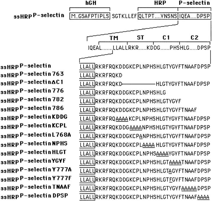
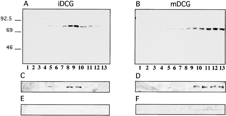
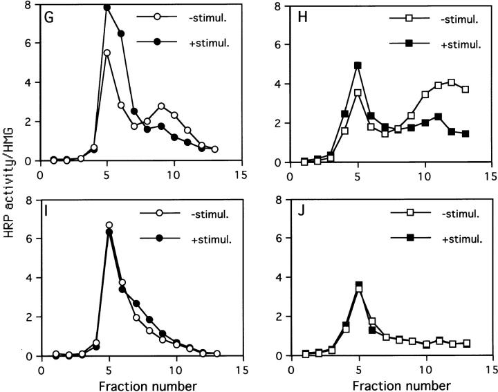
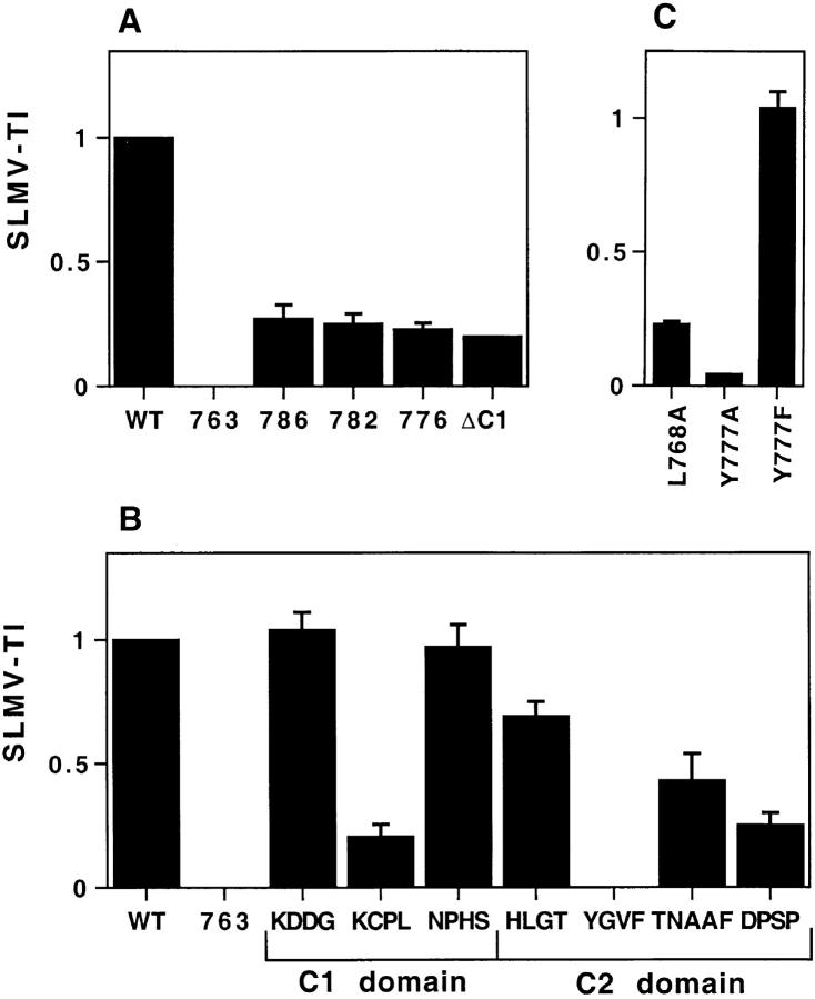
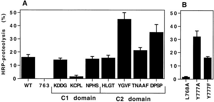
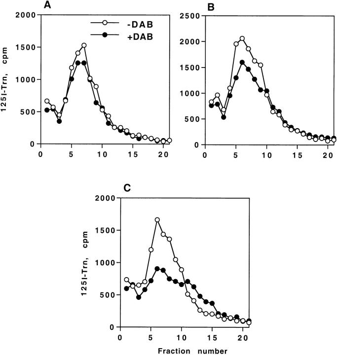
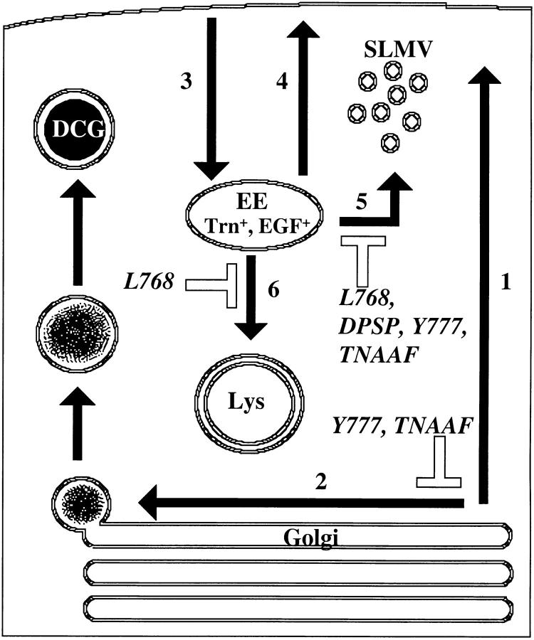
By analyzing the trafficking of HRP-P-selectin chimeras in which the lumenal domain of P-selectin was replaced with horseradish peroxidase, we determined the sequences needed for targeting to synaptic-like microvesicles (SLMV), dense core granules (DCG), and lysosomes in neuroendocrine PC12 cells. Within the cytoplasmic domain of P-selectin, Tyr777 is needed for the appearance of P-selectin in immature and mature DCG, as well as for targeting to SLMV. The latter destination also requires additional sequences (Leu768 and 786DPSP789) which are responsible for movement through endosomes en route to the SLMV. Leu768 also mediates transfer from early transferrin (Trn)-positive endosomes to the lysosomes; i.e., operates as a lysosomal targeting signal. Furthermore, SLMV targeting of HRP-P-selectin chimeras, but not the endogenous SLMV protein synaptophysin/p38, previously shown to be delivered to SLMV directly from the plasma membrane, is a Brefeldin A-sensitive process. Together, these data are consistent with a model of SLMV biogenesis which involves an endosomal intermediate in PC12 cells. In addition, we have discovered that impairment of SLMV or DCG targeting results in a concomitant increase in lysosomal delivery, illustrating the entwined relationships between routes leading to regulated secretory organelles (RSO) and to lysosomes.


Figures

Figure 2
Figure 2
Schematic illustration of HRP–P-selectin chimeras. The top line shows the components used for construction: boxes represent the individual components; sequences outside boxes were added during construction. hGH, human growth hormone signal sequence; HRP, enzymatically active domain of HRP; P-selectin, transmembrane (TM); and cytoplasmic domains of P-selectin. The 35 residues of the wild-type cytoplasmic domain have been assigned to the stop transfer (ST), C1 and C2 domains according to exon–intron boundaries. The bottom part shows the full amino acid sequences of the cytoplasmic domains of the chimeras starting with the wild-type P-selectin tail, ssHRPP-selectin. The carboxy-terminal end of the TM domain shown is boxed. The name of each chimera is listed to the left of the diagram. The number at the end of the name indicates the amino acid changed to a stop signal (numbering from the human P-selectin sequence). The tetrapeptide sequence(s) or number of a single amino acid at the end of each chimera's name show the residues that have been replaced by alanine.
Figure 5
Figure 5
Distribution of HRP activity, 125I-Trn radioactivity and synaptophysin/p38 immunoreactivity after subcellular fractionation on 5–25% Glycerol gradients. (A) PC12 cells expressing wild-type ssHRPP-selectin were fed with 125I-Trn (▵) for 1 h at 37°C, homogenized, and then PNS was fractionated on 5–25% Glycerol gradients to isolate SLMV. HRP activity for ssHRPP-selectin (•) is expressed in arbitrary units corresponding to the amount of HRP activity in each fraction divided by that in the homogenate. (B) Aliquots from each fraction across the gradient were subjected to 10% SDS-PAGE and Western blotted with polyclonal antibody against p38.
Figure 1
Figure 1
A two-step subcellular fractionation procedure for separation of late endosomes and DCG. (A) Distribution of intracellular compartments from PC12 cells on 1–16% Ficoll gradients. PC12 cells expressing ssHRPP-selectin were loaded with 3H-Dopamine, or labeled with 125I-Trn or125I-EGF as described in Materials and Methods. Cells were homogenized in HB and the PNS was centrifuged on 1–16% Ficoll gradients and fractionated. The early/recycling endosomes are shown by the distribution of 125I-Trn endocytosed for 60 min at 37°C (▵). Late endosomes are shown by the distribution of 125I-EGF internalized for 20 min at 37°C (▪). The distribution of NAGA activity (OD420 nm; ○), HRP activity (OD450 nm; ▪) and 3H-Dopamine radioactivity (filled plus signs) along 1–16% Ficoll gradients are shown. (B) Separation of DCG and late endosomes on a secondary 0.9–1.85 M sucrose gradient. Fractions 14–20 from the 1–16% Ficoll gradients were pooled, diluted with HB, and recentrifuged on 0.9–1.85 M sucrose gradients to equilibrium. The distributions of NAGA activity (○), HRP activity (•), 3H-Dopamine radioactivity (filled plus signs),125I-EGF internalized for 20 min at 37°C (▪) and 125I-Trn (▵) after centrifugation on this gradient are shown.
Figure 3
Figure 3
Targeting of HRP–P-selectin chimeras to DCG in PC12 cells. PC12 cells expressing the HRP–P-selectin chimeras shown in Fig. 2 were labeled with 3H-Dopamine and fractionated as described in the legend for Fig. 1. Targeting to DCG was quantitated by calculating a GTI for each chimera. Each GTI is expressed on a scale related to the WT chimera (GTI = 1) and the tail-less chimera (GTI = 0). Each bar represents the mean ± SE of 3–10 independent determinations for each chimera. Deviations of <0.015 are not displayed. (A) GTIs for the deletion mutants; (B) GTIs for the tetra-alanine substitutions; (C) GTIs for the point mutations.
Figure 4
Figure 4
Sorting of SgII, ssHRPP-selectin, and ssHRPP-selectinY777A to iDCG and mDCG. (A and B) PC12 cells expressing ssHRPP-selectin were pulsed for 10 min at 37°C with 35S-labeling mix and chased for 20 min at 37°C (A) or for 16 h (B) at 37°C to label iDCG or mDCG, respectively. The PNS obtained from these cells was centrifuged on initial 0.3–1.2 M sucrose velocity gradients followed by recentrifugation of fractions corresponding to iDCG or mDCG on 0.9–1.7 M sucrose equilibrium gradients as described in Materials and Methods. After fractionation, 200 μl of each fraction was diluted with NDET buffer and immunoprecipitated with anti-SgII. The samples were then separated by 10% SDS-PAGE under reducing conditions, dried, and exposed to a PhosphorImager screen (Bio-Rad Laboratories). Alternatively, cells expressing ssHRPP-selectin (C–H) or ssHRPP-selectinY777A (I and J) were stimulated with 10 mM Carbachol for 30 min at 37°C (E, F, G •, H ▪, I •, and J ▪), or incubated without secretagogue (C, D, G ○, H □, I ○, and J □) and subjected to the two-step fractionation procedure for separation of iDCG and mDCG, as described in the legend for A and B. After fractionation, aliquots of each fraction were separated by SDS-PAGE followed by Western blotting using anti-SgII followed by 125I–protein A (C–F) or used for measurement of HRP activity normalized to that in the homogenate (G–J).
Figure 4
Figure 4
Sorting of SgII, ssHRPP-selectin, and ssHRPP-selectinY777A to iDCG and mDCG. (A and B) PC12 cells expressing ssHRPP-selectin were pulsed for 10 min at 37°C with 35S-labeling mix and chased for 20 min at 37°C (A) or for 16 h (B) at 37°C to label iDCG or mDCG, respectively. The PNS obtained from these cells was centrifuged on initial 0.3–1.2 M sucrose velocity gradients followed by recentrifugation of fractions corresponding to iDCG or mDCG on 0.9–1.7 M sucrose equilibrium gradients as described in Materials and Methods. After fractionation, 200 μl of each fraction was diluted with NDET buffer and immunoprecipitated with anti-SgII. The samples were then separated by 10% SDS-PAGE under reducing conditions, dried, and exposed to a PhosphorImager screen (Bio-Rad Laboratories). Alternatively, cells expressing ssHRPP-selectin (C–H) or ssHRPP-selectinY777A (I and J) were stimulated with 10 mM Carbachol for 30 min at 37°C (E, F, G •, H ▪, I •, and J ▪), or incubated without secretagogue (C, D, G ○, H □, I ○, and J □) and subjected to the two-step fractionation procedure for separation of iDCG and mDCG, as described in the legend for A and B. After fractionation, aliquots of each fraction were separated by SDS-PAGE followed by Western blotting using anti-SgII followed by 125I–protein A (C–F) or used for measurement of HRP activity normalized to that in the homogenate (G–J).
Figure 6
Figure 6
Targeting of HRP–P-selectin chimeras to SLMV in PC12 cells. PC12 cells expressing the HRP–P-selectin chimeras indicated in Fig. 2 were homogenized and fractionated using 5–25% Glycerol gradients to isolate SLMV. SLMV targeting indexes (SLMV-TI) were then calculated. Each bar represents the mean ± SE of 3–10 independent determinations for each chimera. Each SLMV-TI is expressed on a scale related to the WT chimera (SLMV-TI = 1) and the tail-less chimera (SLMV-TI = 0). Deviations of <0.015 are not displayed. (A) SLMV-TIs for the deletion mutants; (B) SLMV-TIs for the tetra-alanine substitutions; (C) SLMV-TIs for the point mutations.
Figure 7
Figure 7
HRP proteolysis of HRP–P-selectin chimeras in PC12 cells. Cells expressing the HRP–P-selectin chimeras indicated were subjected to a Triton X-114 partitioning assay. HRP activity was measured both in the upper hydrophilic phase and in the lower hydrophobic phase to determine the fraction of soluble HRP activity found in the lysate. Each bar represents the mean ± SE of 3–10 independent determinations. (A) The tetra-alanine substitutions; (B) the point mutations.
Figure 8
Figure 8
Targeting of internalized 125I-2H11 to lysosomes. PC12 cells expressing the chimeras indicated were fed with 125I-2H11 for 1 h at 37°C and subjected to a two-step fractionation procedure for isolation of lysosomal compartments (see Materials and Methods). A shows the distribution of NAGA (○) compared with HRP activity for ssHRPP-selectin (▪) and tail-less ssHRPP-selectin763 (□). HRP activity was determined in each fraction and normalized to the total HRP activity in the homogenate. B shows the distribution of NAGA activity (○) compared with 125I-2H11 radioactivity for ssHRPP-selectin (filled plus signs) and for ssHRPP-selectin763 (empty plus signs). 125I-2H11 radioactivity was counted in each fraction across the gradient and normalized to the total radioactivity in the homogenate. (C) the LTI for a variety of chimeras. Each bar represents the mean ± SE of four independent experiments.
Figure 9
Figure 9
DAB-induced density shift of internalized 125I-Trn in PC12 cells expressing HRP–P-selectin chimeras. Cells expressing ssHRPTrnR(tail−) (A), ssHRPP-selectin (B), or ssHRPP-selectinL768A (C) were fed with 125I-Trn for 60 min at 37°C. Ligand present on the cell surface was removed by MES buffer treatment. Cells were homogenized in HB and the PNS was split into two parts. One half was incubated with DAB and H2O2 (•) and the other half with HB alone (○). After the DAB reaction, the samples were centrifuged on 1–16% Ficoll gradients. The distribution of 125I-Trn along these gradients is shown for DAB-incubated or control samples. Data from one typical experiment (out of two) is shown.
Figure 10
Figure 10
Effect of BFA on the targeting of ssHRPP-selectin and synaptophysin/p38 to SLMV. (A) PC12 cells expressing ssHRPP-selectin were incubated in the presence (○) or absence of 10 μg/ml BFA (•) for 1 h at 37°C and a PNS was obtained for centrifugation on 5–25% glycerol gradients. HRP activity was determined in each fraction. Data are expressed as the amount of HRP activity in each fraction divided by that in the homogenate. (B) Aliquots from pooled SLMV peaks, as defined by the distribution of HRP activity shown in A, and from homogenates from the cells treated or mock-treated with BFA were collected, proteins were separated by 10% SDS-PAGE and Western blotted with a polyclonal antibody against p38. Tracks 1 and 3 show p38 in the SLMV peak. Tracks 2 and 4 show p38 in the homogenates. Samples were from the cells treated or mock-treated with BFA, as indicated.
Figure 11
Figure 11
A schematic model for post-Golgi trafficking of P-selectin in PC12 cells. Newly synthesized P-selectin reaching the Golgi is either constitutively transported to the cell surface (1) or is sorted to the DCG in a signal-dependent (YGVF/ TNAAF with a critical contribution from Tyr777) fashion (2). After appearance on the cell surface, it is rapidly internalized into the early, Trn-positive, EGF-positive endosomes (3). From here, some chimera recycles to the plasma membrane (4), whereas the rest is delivered to SLMV (5) or to lysosomes (6). Steps 5 and 6 are mediated by Leu768. Trafficking to SLMV (5) from endosomes is also mediated by Tyr777, TNAAF, and DPSP.

References

    1. Arvan P, Castle D. Sorting and storage during secretory granule biogenesis: looking backward and looking forward. Biochem J. 1998;332:593–610. - PMC - PubMed
    1. Barr FA, Huttner WB. A role for ADP-ribosylation factor 1, but not COP I, in secretory vesicles biogenesis from the trans-Golgi network. FEBS Lett. 1996;384:65–70. - PubMed
    1. Bauerfeind R, Huttner WB. Biogenesis of constitutive secretory vesicles, secretory granules and synaptic vesicles. Curr Opin Cell Biol. 1993;5:628–635. - PubMed
    1. Blagoveshchenskaya AD, Norcott JP, Cutler DF. Lysosomal targeting of P-selectin is mediated by a novel sequence within its cytoplasmic tail. J Biol Chem. 1998a;273:2729–2737. - PubMed
    1. Blagoveshchenskaya AD, Hewitt EW, Cutler DF. A balance of opposing signals within the cytoplasmic tail controls the lysosomal targeting of P-selectin. J Biol Chem. 1998b;273:27896–27903. - PubMed

Publication types

MeSH terms