

doi: 10.1101/gad.13.12.1524.
Targeting genes for self-excision in the germ line
Affiliations
- PMID: 10385621
- PMCID: PMC316811
- DOI: 10.1101/gad.13.12.1524
Item in Clipboard
Targeting genes for self-excision in the germ line
Genes Dev.
.
Abstract
A procedure is described that directs the self-induced deletion of DNA sequences as they pass through the male germ line of mice. The testes-specific promoter from the angiotensin-converting enzyme gene was used to drive expression of the Cre-recombinase gene. Cre was linked to the selectable marker Neor, and the two genes flanked with loxP elements. This cassette was targeted to the Hoxa3 gene in mouse ES cells that were in turn used to generate chimeric mice. In these chimeras, somatic cells derived from the ES cells retained the cassette, but self-excision occurred in all ES-cell-derived sperm.
Figures
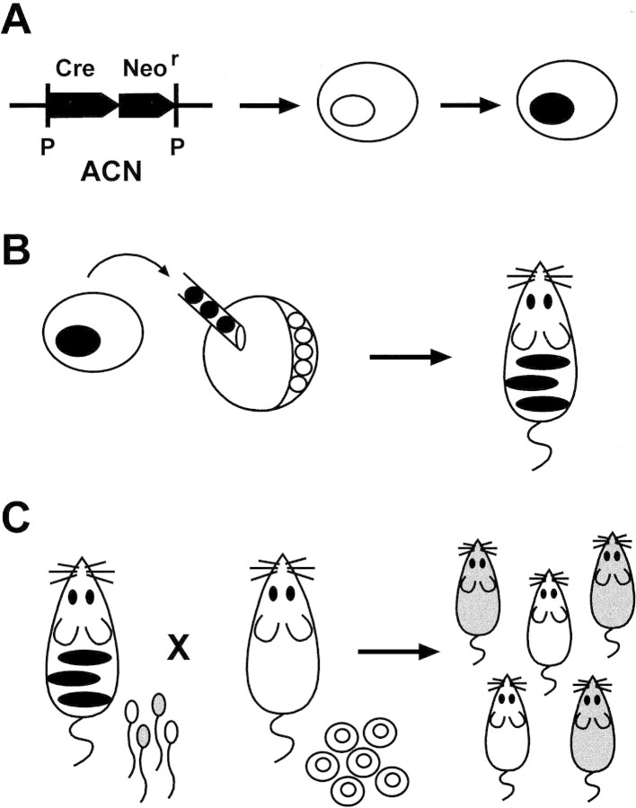
Testes-specific self-excision. (A) A
selectable marker gene, Neor, with a constitutive
promoter, is transferred by homologous recombination to a specific
locus in a mouse ES cell. The Neor gene is linked to
a Cre gene that is under transcriptional control of the tACE
promoter, and the two genes are flanked with loxP sites (P);
the entire cassette, ACN, is introduced by gene targeting to a specific
locus in a mouse ES cell. (B) ES cells, heterozygous for an
allele containing the integrated cassette, are injected into wild-type
mouse blastocysts and the blastocysts allowed to develop; the resulting
animals are chimeric for wild-type (host-derived) cells (white) and
ES-derived cells (black). (C) Male chimeric animals will
transmit through their sperm one of two alleles of the locus of
interest: wild-type (white) or mutant (gray); if self-excision has
occurred the mutant allele will be marked only by a loxP site,
the final product of the testes-specific self-excision reaction.
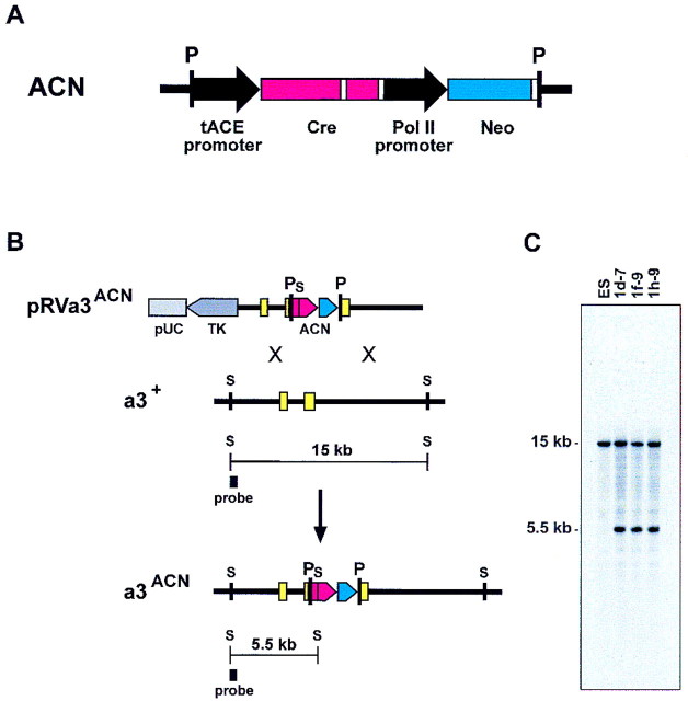
Targeting of a self-excision cassette to
Hoxa3. (A) Self-excision cassette, ACN: The
testes-specific elements from the mouse ACE gene (black arrow)
are placed 5′ of the modified Cre structural gene (Gu et
al. 1993) (red), followed, 3′, with the minimal polyadenylation
signal from HSV–TK (Thomas and Capecchi 1987) (white box); an
intron, derived from the SV40 t-antigen gene (white box), is
inserted into the Cre gene; the Neor gene
(blue) is controlled by a promoter from the mouse RNA polymerase II
gene (black arrow) and followed also by the HSV–TK poly(A)
site (white box). The 5′ and 3′ ends of this cassette contain
loxP sites (P). (B) Gene targeting at Hoxa3:
(Top line) The targeting vector pRVa3ACN; the vector
contains 11 kb of mouse genomic DNA into which the self-excision
cassette ACN has been inserted in the homeodomain of Hoxa3
(McGinnis et al. 1984); the genomic sequences are linked to the
HSV–TK gene (dark gray) and all are contained on a pUC-based
plasmid backbone (light gray); the ACN cassette contains at its 5′
end an SstI site (S), used as a marker for homologous
integration of the cassette at the Hoxa3 gene;
(second line) the wild-type Hoxa3 locus;
(bottom line) the predicted structure of the recombinant
Hoxa3ACN allele. The 5′ flanking probe used to
detect recombination is indicated, and the diagnostic
SstI-generated DNA fragments delineated beneath each locus.
Yellow boxes designate Hoxa3 exons; other SstI sites
in the vector are not indicated. (C) Southern transfer
analysis: DNA from the parental cell line (ES) and the homologous
recombinant ES lines used to generate mice was restricted with
SstI; radiolabeled DNA probe is depicted in B.
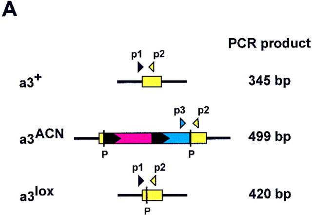
Genetic transmission of Hoxa3 alleles.
(A) The PCR-based genotyping of the three Hoxa3
alleles: Primer 1 (p1) is from the Hoxa3 intron; primer 2 (p2)
is from coding exon 2-derived sequences (antisense); primer 3 (p3) is
from the Neor gene. Predicted sizes are indicated;
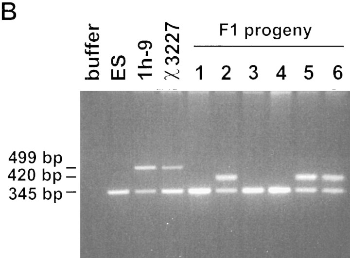
color coding is as in Fig. 2. (B) Genotyping of DNA: DNA is
from wild-type ES cells (ES), recombinant ES cell line, 1h-9, tail
biopsies from a chimeric male, χ3227, generated from 1h-9, and tail
tissue from F1 progeny of the chimera; amplified DNA was
electrophoresed through agarose and stained with ethidium bromide.
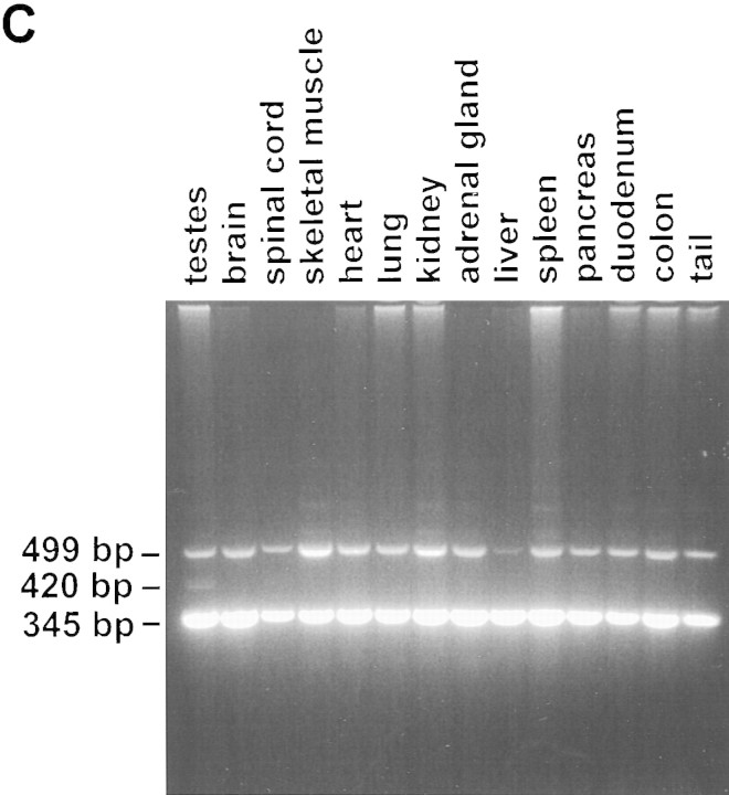
Sizes correspond to those listed in a. (C) Absence of
excision in somatic tissue. A single chimeric male derived from cell
line 1h-9 was sacrificed at 8 weeks of age. DNA extracted from each of
the indicated tissues was analyzed by PCR as in B. Analysis of
a second chimera showed an identical result.
Genetic transmission of Hoxa3 alleles.
(A) The PCR-based genotyping of the three Hoxa3
alleles: Primer 1 (p1) is from the Hoxa3 intron; primer 2 (p2)
is from coding exon 2-derived sequences (antisense); primer 3 (p3) is
from the Neor gene. Predicted sizes are indicated;
color coding is as in Fig. 2. (B) Genotyping of DNA: DNA is
from wild-type ES cells (ES), recombinant ES cell line, 1h-9, tail
biopsies from a chimeric male, χ3227, generated from 1h-9, and tail
tissue from F1 progeny of the chimera; amplified DNA was
electrophoresed through agarose and stained with ethidium bromide.
Sizes correspond to those listed in a. (C) Absence of
excision in somatic tissue. A single chimeric male derived from cell
line 1h-9 was sacrificed at 8 weeks of age. DNA extracted from each of
the indicated tissues was analyzed by PCR as in B. Analysis of
a second chimera showed an identical result.
Similar articles
-
Efficient removal of loxP-flanked DNA sequences in a gene-targeted locus by transient expression of Cre recombinase in fertilized eggs.Mol Reprod Dev. 1997 Feb;46(2):109-13. doi: 10.1002/(SICI)1098-2795(199702)46:2<109::AID-MRD1>3.0.CO;2-U. Mol Reprod Dev. 1997. PMID: 9021742
-
Cre-loxP-mediated gene replacement: a mouse strain producing humanized antibodies.Curr Biol. 1994 Dec 1;4(12):1099-103. doi: 10.1016/s0960-9822(00)00248-7. Curr Biol. 1994. PMID: 7704573
-
Targeting collecting tubules using the aquaporin-2 promoter.Exp Nephrol. 1999 Jan-Feb;7(1):67-74. doi: 10.1159/000020587. Exp Nephrol. 1999. PMID: 9892817
-
Targeted viral delivery of Cre recombinase induces conditional gene deletion in cardiovascular circuits of the mouse brain.Physiol Genomics. 2004 Jun 17;18(1):25-32. doi: 10.1152/physiolgenomics.00048.2004. Epub 2004 Jun 17. Physiol Genomics. 2004. PMID: 15069166
-
Newer approaches to genetic modeling in mice: tissue-specific protein expression as studied using angiotensin-converting enzyme (ACE).Am J Pathol. 2003 Sep;163(3):807-17. doi: 10.1016/S0002-9440(10)63441-4. Am J Pathol. 2003. PMID: 12937122 Free PMC article. Review. No abstract available.
Cited by
-
Ptbp2 represses adult-specific splicing to regulate the generation of neuronal precursors in the embryonic brain.Genes Dev. 2012 Jul 15;26(14):1626-42. doi: 10.1101/gad.191338.112. Genes Dev. 2012. PMID: 22802532 Free PMC article.
-
Analysis of kinesin-2 function in photoreceptor cells using synchronous Cre-loxP knockout of Kif3a with RHO-Cre.Invest Ophthalmol Vis Sci. 2006 Nov;47(11):5039-46. doi: 10.1167/iovs.06-0032. Invest Ophthalmol Vis Sci. 2006. PMID: 17065525 Free PMC article.
-
Targeted mutagenesis of the murine transferrin receptor-2 gene produces hemochromatosis.Proc Natl Acad Sci U S A. 2002 Aug 6;99(16):10653-8. doi: 10.1073/pnas.162360699. Epub 2002 Jul 19. Proc Natl Acad Sci U S A. 2002. PMID: 12134060 Free PMC article.
-
Thyroid hormone action in the absence of thyroid hormone receptor DNA-binding in vivo.J Clin Invest. 2003 Aug;112(4):588-97. doi: 10.1172/JCI18377. J Clin Invest. 2003. PMID: 12925699 Free PMC article.
-
Characterization of Nkx6-2-derived neocortical interneuron lineages.Cereb Cortex. 2009 Jul;19 Suppl 1(Suppl 1):i1-10. doi: 10.1093/cercor/bhp038. Epub 2009 Apr 10. Cereb Cortex. 2009. PMID: 19363146 Free PMC article.
References
-
- Broach JR, Hicks JB. Replication and recombination functions associated with the yeast plasmid, 2 μ circle. Cell. 1980;21:501–508. - PubMed
-
- Capecchi MR. Human germline gene therapy: A discussion on how and why. In: Stock G, Campbell J, editors. Engineering the human germline. New York, NY: Oxford University Press; 1999. . (In press.)
-
- Colledge WH, Abella BS, Southern KW, Ratcliff R, Jiang C, Cheng SH, MacVinish LJ, Anderson JR, Cuthbert AW, Evans MJ. Generation and characterization of a ΔF508 cystic fibrosis mouse model. Nat Genet. 1995;10:445–452. - PubMed
Publication types
MeSH terms
Substances
Associated data
- Actions
LinkOut - more resources
Full Text Sources
Other Literature Sources
Molecular Biology Databases