. 1999 Jun 15;13(12):1529-39.
doi: 10.1101/gad.13.12.1529.

ACF consists of two subunits, Acf1 and ISWI, that function cooperatively in the ATP-dependent catalysis of chromatin assembly

Affiliations

ACF consists of two subunits, Acf1 and ISWI, that function cooperatively in the ATP-dependent catalysis of chromatin assembly

T Ito et al. Genes Dev. .

Abstract

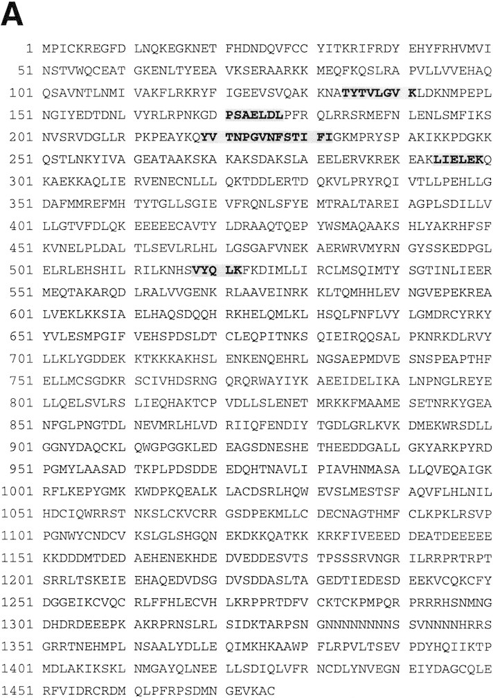
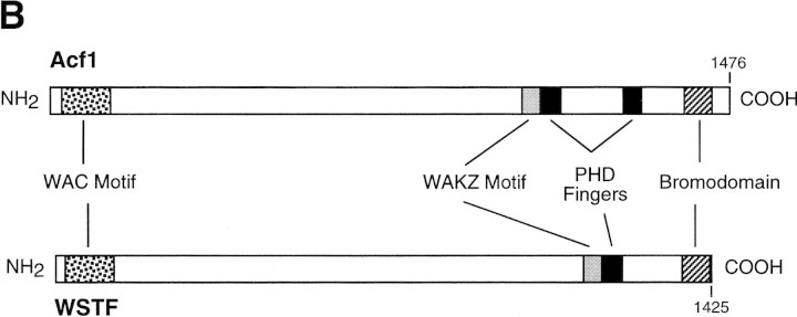
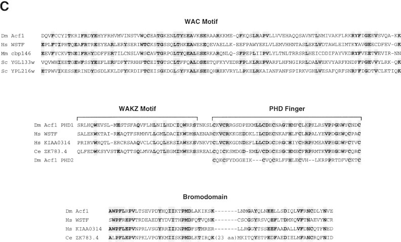
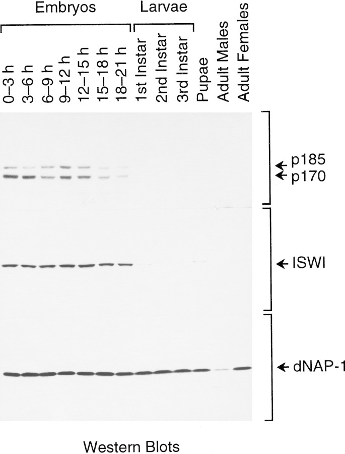
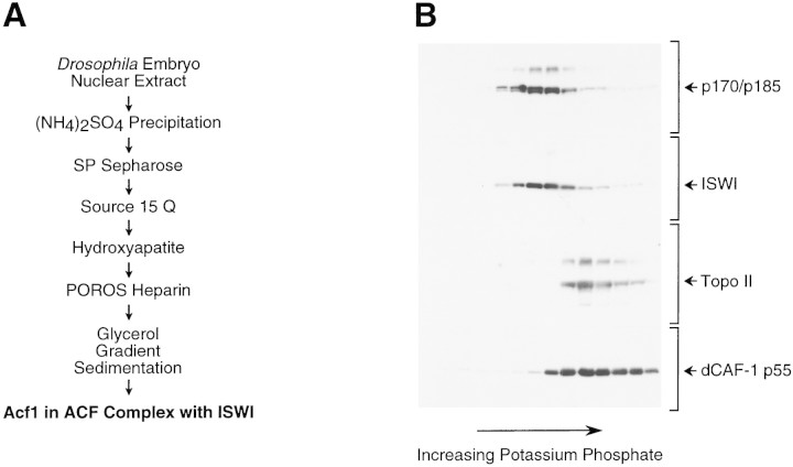
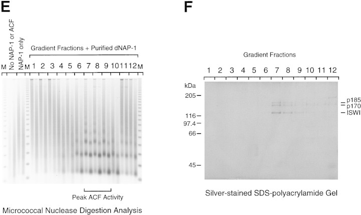
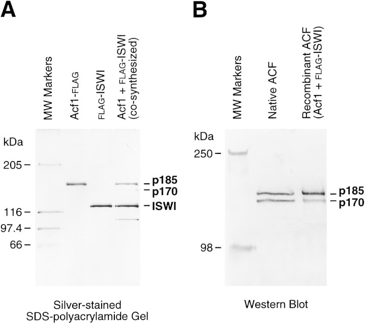
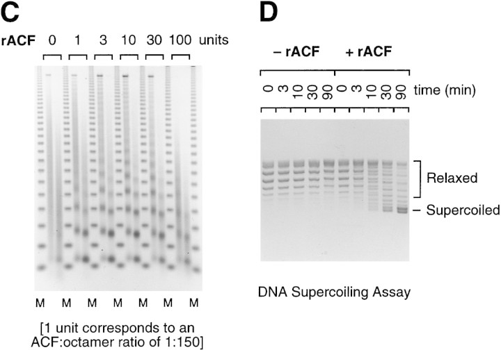
The assembly of core histones and DNA into periodic nucleosome arrays is mediated by ACF, an ISWI-containing factor, and NAP-1, a core histone chaperone, in an ATP-dependent process. We describe the isolation of Drosophila acf1 cDNA, which encodes the p170 and p185 forms of the Acf1 protein in ACF. Acf1 is a novel protein that contains two PHD fingers, one bromodomain, and two new conserved regions. Human WSTF, which is encoded by one of multiple genes that is deleted in Williams syndrome individuals, is the only currently known mammalian protein with each of the conserved motifs in Acf1. Purification of the native form of Acf1 led to the isolation of ACF comprising Acf1 (both p170 and p185 forms) and ISWI. Native Acf1 did not copurify with components of NURF or CHRAC, which are other ISWI-containing complexes in Drosophila. Purified recombinant ACF, consisting of Acf1 (either p185 alone or both p170 and p185) and ISWI, catalyzes the deposition of histones into extended periodic nucleosome arrays. Notably, the Acf1 and ISWI subunits function synergistically in the assembly of chromatin. ISWI alone exhibits a weak activity that is approximately 3% that of ACF. These results indicate that both Acf1 and ISWI participate in the chromatin assembly process and suggest further that the Acf1 subunit confers additional functionality to the general 'motor' activity of ISWI.


Figures

Figure 1
Figure 1
Sequence and features of Acf1 protein. (A) Predicted amino acid sequence of Acf1, as deduced from the cDNA sequence. The shaded regions indicate amino acid residues of native ACF that were determined by protein microsequencing. The acf1 cDNA sequence is in GenBank (accession no.AF148962). (B) Schematic diagram of Acf1. In addition to two PHD fingers (Schindler et al. 1993; Aasland et al. 1995) and a bromodomain (Haynes et al. 1992; Jeanmougin et al. 1997), Acf1 has two novel conserved regions termed WAC and WAKZ motifs. WSTF (Lu et al. 1998) is a human protein that appears to be related to Drosophila Acf1. (C) Alignment of sequence motifs in Acf1 with some of their counterparts in other proteins. cbp146 was identified in a gene trap screen for chromosomal and nuclear proteins in mice (Tate et al. 1998; CT146 cell line). YGL133w and YPL216w are from S. cerevisiae. KIAA0314 was identified in a screen for novel human cDNAs (Nagase et al. 1997) and appears to be identical to TTF-I-interacting peptide 5, which was identified in a yeast two-hybrid screen (also see Jansa et al. 1998). ZK783.4 is from C. elegans.
Figure 1
Figure 1
Sequence and features of Acf1 protein. (A) Predicted amino acid sequence of Acf1, as deduced from the cDNA sequence. The shaded regions indicate amino acid residues of native ACF that were determined by protein microsequencing. The acf1 cDNA sequence is in GenBank (accession no.AF148962). (B) Schematic diagram of Acf1. In addition to two PHD fingers (Schindler et al. 1993; Aasland et al. 1995) and a bromodomain (Haynes et al. 1992; Jeanmougin et al. 1997), Acf1 has two novel conserved regions termed WAC and WAKZ motifs. WSTF (Lu et al. 1998) is a human protein that appears to be related to Drosophila Acf1. (C) Alignment of sequence motifs in Acf1 with some of their counterparts in other proteins. cbp146 was identified in a gene trap screen for chromosomal and nuclear proteins in mice (Tate et al. 1998; CT146 cell line). YGL133w and YPL216w are from S. cerevisiae. KIAA0314 was identified in a screen for novel human cDNAs (Nagase et al. 1997) and appears to be identical to TTF-I-interacting peptide 5, which was identified in a yeast two-hybrid screen (also see Jansa et al. 1998). ZK783.4 is from C. elegans.
Figure 2
Figure 2
Western blot analysis of Acf1 at different stages of Drosophila development. Equivalent amounts of total soluble protein derived from Drosophila at the indicated stages of development were subjected to polyacrylamide–SDS gel electrophoresis and Western blot analysis.
Figure 3
Figure 3
Purification of the native form of Acf1 leads to the isolation of ACF comprising Acf1 (p170 and p185) and ISWI. (A) Scheme for the purification of native form of Acf1 from Drosophila embryos. (B) Hydroxyapatite chromatography. The peak gradient fractions from the Source 15Q (Pharmacia Biotech) column were applied to a Bio-Gel HT hydroxyapatite (Bio-Rad) column, and protein was eluted with a linear potassium phosphate gradient. The column fractions were subjected to Western blot analysis with antibodies against Drosophila Acf1 (p170/p185), ISWI, topoisomerase II, and dCAF-1 p55 in conjunction with 125I-labeled protein A. With the Acf1 Western blot, the p170 and p185 forms of Acf1 were not clearly resolved. Also, the slower migrating species that cross-reacts with the Acf1 antiserum is not recognized by the affinity-purified antibodies (e.g., see Fig. 2). (C) POROS heparin chromatography. The peak hydroxyapatite fractions were applied to a POROS heparin (PerSeptive Biosystems) column, and protein was eluted with a linear NaCl gradient. The column fractions were subjected to Western blot analysis, as in B. The control sample is an ACF-containing fraction from the Source 15Q chromatography step. The p170 and p185 forms of Acf1 were not clearly resolved. (D) Glycerol gradient sedimentation. The peak POROS heparin fractions were subjected to 15%–40% (vol/vol) glycerol gradient sedimentation. The glycerol gradient fractions were subjected to Western blot analysis, as in B and C. The p170 and p185 forms of Acf1 were not clearly resolved. (E) Micrococcal nuclease digestion analysis. ACF activity in the glycerol gradient fractions was tested by micrococcal nuclease digestion analysis. Chromatin assembly reactions contained 10 μl of each 400 μl fraction and were carried out as described in Materials and Methods. The samples were then partially digested with two different concentrations of micrococcal nuclease. The resulting DNA fragments were deproteinized, resolved by 1.5% agarose gel electrophoresis, and visualized by staining with ethidium bromide. The mass markers (M) are the 123-bp DNA ladder (GIBCO-BRL). The peak of ACF activity is seen in fractions 7–9. (F) Native ACF consists of Acf1 (p185 and p170) and ISWI. Glycerol gradient fractions were subjected to 6% polyacrylamide–SDS gel electrophoresis, and proteins were visualized by silver staining. The sizes of molecular mass markers and the ACF subunits are indicated. The traces of dCAF-1 p55/NURF-55 that were seen in Western blots of the glycerol gradient fractions (D) could not be detected in these silver-stained SDS–polyacrylamide gels.
Figure 3
Figure 3
Purification of the native form of Acf1 leads to the isolation of ACF comprising Acf1 (p170 and p185) and ISWI. (A) Scheme for the purification of native form of Acf1 from Drosophila embryos. (B) Hydroxyapatite chromatography. The peak gradient fractions from the Source 15Q (Pharmacia Biotech) column were applied to a Bio-Gel HT hydroxyapatite (Bio-Rad) column, and protein was eluted with a linear potassium phosphate gradient. The column fractions were subjected to Western blot analysis with antibodies against Drosophila Acf1 (p170/p185), ISWI, topoisomerase II, and dCAF-1 p55 in conjunction with 125I-labeled protein A. With the Acf1 Western blot, the p170 and p185 forms of Acf1 were not clearly resolved. Also, the slower migrating species that cross-reacts with the Acf1 antiserum is not recognized by the affinity-purified antibodies (e.g., see Fig. 2). (C) POROS heparin chromatography. The peak hydroxyapatite fractions were applied to a POROS heparin (PerSeptive Biosystems) column, and protein was eluted with a linear NaCl gradient. The column fractions were subjected to Western blot analysis, as in B. The control sample is an ACF-containing fraction from the Source 15Q chromatography step. The p170 and p185 forms of Acf1 were not clearly resolved. (D) Glycerol gradient sedimentation. The peak POROS heparin fractions were subjected to 15%–40% (vol/vol) glycerol gradient sedimentation. The glycerol gradient fractions were subjected to Western blot analysis, as in B and C. The p170 and p185 forms of Acf1 were not clearly resolved. (E) Micrococcal nuclease digestion analysis. ACF activity in the glycerol gradient fractions was tested by micrococcal nuclease digestion analysis. Chromatin assembly reactions contained 10 μl of each 400 μl fraction and were carried out as described in Materials and Methods. The samples were then partially digested with two different concentrations of micrococcal nuclease. The resulting DNA fragments were deproteinized, resolved by 1.5% agarose gel electrophoresis, and visualized by staining with ethidium bromide. The mass markers (M) are the 123-bp DNA ladder (GIBCO-BRL). The peak of ACF activity is seen in fractions 7–9. (F) Native ACF consists of Acf1 (p185 and p170) and ISWI. Glycerol gradient fractions were subjected to 6% polyacrylamide–SDS gel electrophoresis, and proteins were visualized by silver staining. The sizes of molecular mass markers and the ACF subunits are indicated. The traces of dCAF-1 p55/NURF-55 that were seen in Western blots of the glycerol gradient fractions (D) could not be detected in these silver-stained SDS–polyacrylamide gels.
Figure 4
Figure 4
Chromatin assembly by purified recombinant Acf1 and ISWI. (A) Purification of recombinant Acf1 and ISWI. Acf1–Flag alone, Flag–ISWI alone, or Acf1 + Flag–ISWI together were synthesized with a baculovirus expression system and then purified by using the Flag affinity tag. The proteins (185 ng of Acf1–Flag or Acf1; 140 ng of Flag–ISWI) were analyzed by 6% polyacrylamide–SDS gel electrophoresis and silver staining. The polypeptide that migrates below Flag–ISWI in the cosynthesized Acf1 + Flag–ISWI preparation is an unknown contaminant. (B) Both p170 and p185 are synthesized from the acf1 cDNA. Native ACF (after hydroxyapatite chromatography, as shown in Fig. 3B) and recombinant ACF, which was obtained by cosynthesis of Acf1 and Flag–ISWI and purification with the Flag epitope tag, were subjected to 6% polyacrylamide–SDS gel electrophoresis and Western blot analysis with affinity-purified antibodies that recognize the TYE peptide of Acf1 (described in Materials and Methods). (C) Recombinant ACF can function catalytically in the assembly of chromatin. Chromatin assembly and micrococcal nuclease digestion assays were performed with varying concentrations of purified, recombinant ACF (rACF). The resulting DNA samples were resolved by 1.2% agarose gel electrophoresis and detected by staining with ethidium bromide. One unit of ACF is defined to be 22 fmoles, which corresponds to one ACF protomer per 150 core histone octamers in this assay. (D) DNA supercoiling analysis of chromatin assembly by recombinant ACF. Standard chromatin assembly reactions were carried out in the presence or absence of purified recombinant ACF (rACF, 10 U/reaction) with recombinant dNAP-1 and purified Drosophila core histones. The assembly reactions were terminated at the indicated time points by the addition of EDTA to 50 mm final concentration. The samples were then deproteinized and subjected to 1.0% agarose gel electrophoresis. The DNA was detected by staining with ethidium bromide.
Figure 4
Figure 4
Chromatin assembly by purified recombinant Acf1 and ISWI. (A) Purification of recombinant Acf1 and ISWI. Acf1–Flag alone, Flag–ISWI alone, or Acf1 + Flag–ISWI together were synthesized with a baculovirus expression system and then purified by using the Flag affinity tag. The proteins (185 ng of Acf1–Flag or Acf1; 140 ng of Flag–ISWI) were analyzed by 6% polyacrylamide–SDS gel electrophoresis and silver staining. The polypeptide that migrates below Flag–ISWI in the cosynthesized Acf1 + Flag–ISWI preparation is an unknown contaminant. (B) Both p170 and p185 are synthesized from the acf1 cDNA. Native ACF (after hydroxyapatite chromatography, as shown in Fig. 3B) and recombinant ACF, which was obtained by cosynthesis of Acf1 and Flag–ISWI and purification with the Flag epitope tag, were subjected to 6% polyacrylamide–SDS gel electrophoresis and Western blot analysis with affinity-purified antibodies that recognize the TYE peptide of Acf1 (described in Materials and Methods). (C) Recombinant ACF can function catalytically in the assembly of chromatin. Chromatin assembly and micrococcal nuclease digestion assays were performed with varying concentrations of purified, recombinant ACF (rACF). The resulting DNA samples were resolved by 1.2% agarose gel electrophoresis and detected by staining with ethidium bromide. One unit of ACF is defined to be 22 fmoles, which corresponds to one ACF protomer per 150 core histone octamers in this assay. (D) DNA supercoiling analysis of chromatin assembly by recombinant ACF. Standard chromatin assembly reactions were carried out in the presence or absence of purified recombinant ACF (rACF, 10 U/reaction) with recombinant dNAP-1 and purified Drosophila core histones. The assembly reactions were terminated at the indicated time points by the addition of EDTA to 50 mm final concentration. The samples were then deproteinized and subjected to 1.0% agarose gel electrophoresis. The DNA was detected by staining with ethidium bromide.
Figure 5
Figure 5
Acf1 and ISWI function cooperatively in the catalysis of chromatin assembly. (A) Chromatin assembly reactions were carried out with the indicated amounts of individual recombinant ACF subunits (Acf1–Flag or Flag–ISWI) or ACF (cosynthesized Acf1 + Flag–ISWI). In addition, where noted, ATP was not included in the reaction with ACF. These and other related experiments (see, e.g., Fig. 4C) indicate that ACF assembles chromatin ∼30-fold more efficiently than either subunit alone. One unit corresponds to 22 fmoles of protein. (B) Acf1 and ISWI can be combined postsynthetically to yield active ACF. Chromatin was assembled with either cosynthesized Acf1 + Flag–ISWI [which consists of p185, p170, and ISWI, as seen in Fig. 4A] or individual Acf1–Flag [which consists of p185 only, as shown in Fig. 4A] and Flag–ISWI polypeptides that had been combined after purification (postsynthetic combination of subunits). One unit corresponds to 22 fmoles of protein.
Figure 5
Figure 5
Acf1 and ISWI function cooperatively in the catalysis of chromatin assembly. (A) Chromatin assembly reactions were carried out with the indicated amounts of individual recombinant ACF subunits (Acf1–Flag or Flag–ISWI) or ACF (cosynthesized Acf1 + Flag–ISWI). In addition, where noted, ATP was not included in the reaction with ACF. These and other related experiments (see, e.g., Fig. 4C) indicate that ACF assembles chromatin ∼30-fold more efficiently than either subunit alone. One unit corresponds to 22 fmoles of protein. (B) Acf1 and ISWI can be combined postsynthetically to yield active ACF. Chromatin was assembled with either cosynthesized Acf1 + Flag–ISWI [which consists of p185, p170, and ISWI, as seen in Fig. 4A] or individual Acf1–Flag [which consists of p185 only, as shown in Fig. 4A] and Flag–ISWI polypeptides that had been combined after purification (postsynthetic combination of subunits). One unit corresponds to 22 fmoles of protein.

References

    1. Aasland R, Gibson TJ, Stewart AF. The PHD finger: Implications for chromatin-mediated transcriptional regulation. Trends Biochem Sci. 1995;20:56–59. - PubMed
    1. Adams CR, Kamakaka RT. Chromatin assembly: Biochemical identities and genetic redundancy. Curr Opin Genet Dev. 1999;9:185–190. - PubMed
    1. Annunziato AT. Histone acetylation during chromatin replication and nucleosome assembly. In: Wolffe AP, editor. The nucleus. Vol. 1. Greenwich, CT: JAI Press; 1995. pp. 31–56.
    1. Bellugi U, Bihrle A, Jernigan T, Trauner D, Doherty S. Neuropsychological, neurological, and neuroanatomical profile of Williams syndrome. Am J Hum Genet (Suppl.) 1990;6:115–125. - PubMed
    1. Bulger M, Kadonaga JT. Biochemical reconstitution of chromatin with physiological nucleosome spacing. Methods Mol Genet. 1994;5:241–262.

Publication types

MeSH terms

Associated data