

Determination of left/right asymmetric expression of nodal by a left side-specific enhancer with sequence similarity to a lefty-2 enhancer
- PMID: 10385627
- PMCID: PMC316797
- DOI: 10.1101/gad.13.12.1589
Determination of left/right asymmetric expression of nodal by a left side-specific enhancer with sequence similarity to a lefty-2 enhancer
Abstract
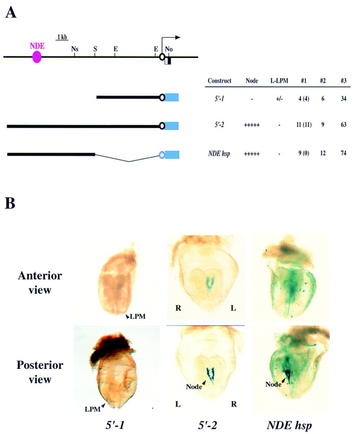
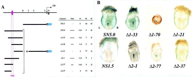
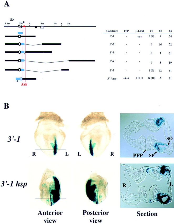
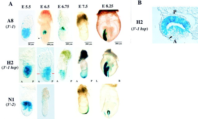
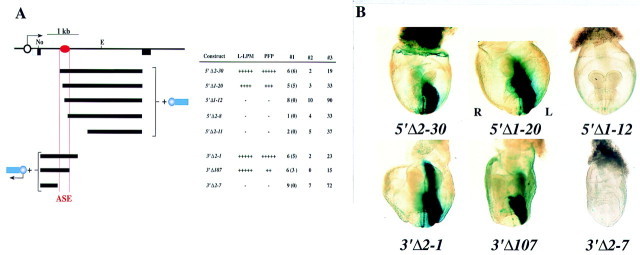
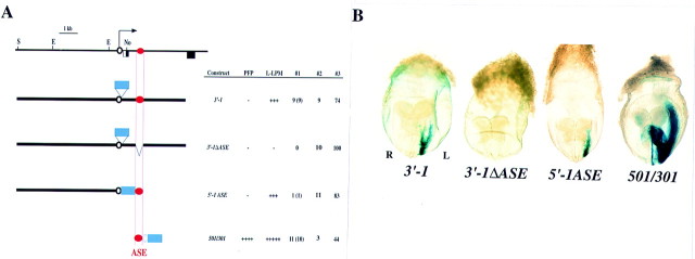
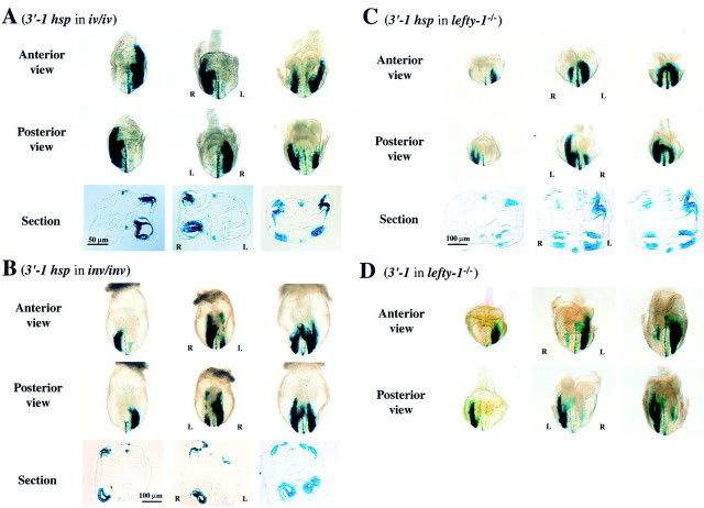
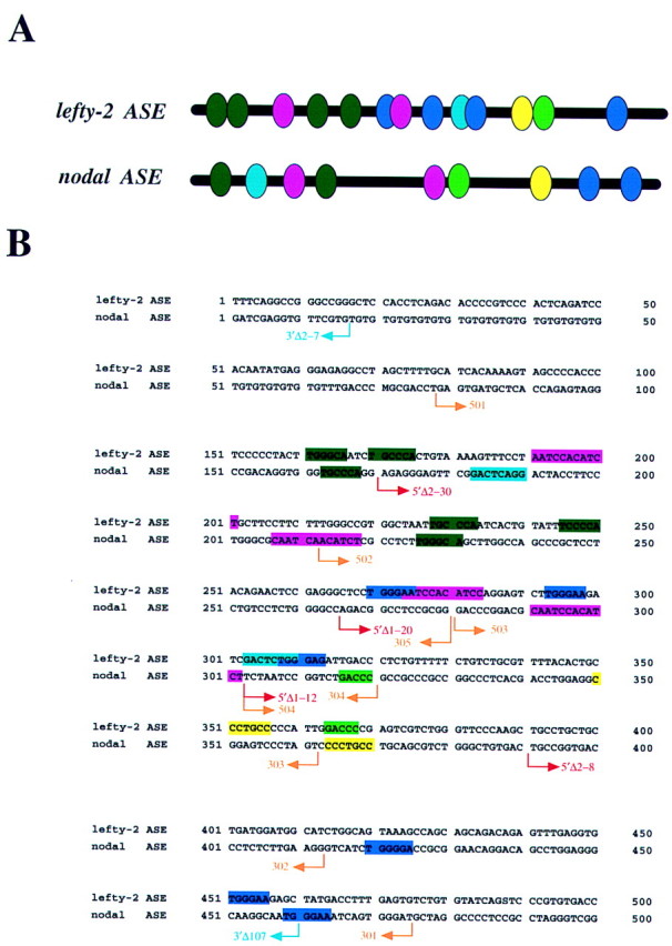
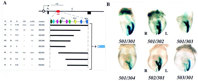
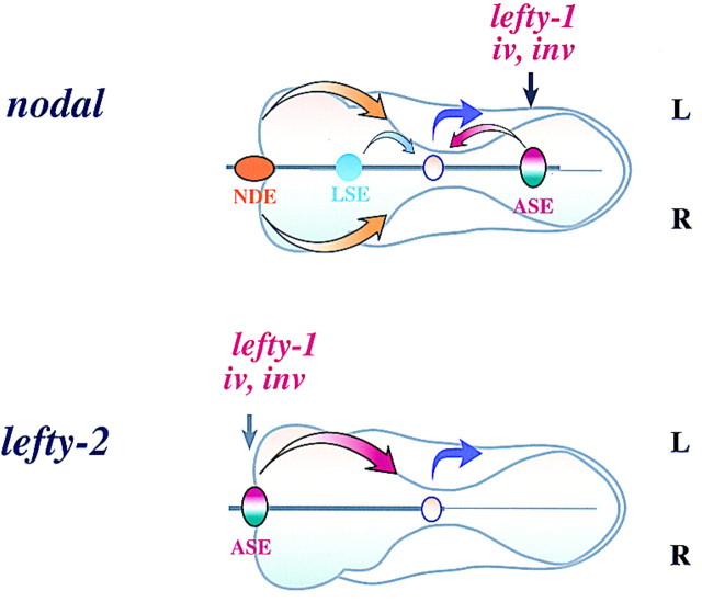
The nodal gene is expressed on the left side of developing mouse embryos and is implicated in left/right (L-R) axis formation. The transcriptional regulatory regions of nodal have now been investigated by transgenic analysis. A node-specific enhancer was detected in the upstream region (-9.5 to -8.7 kb) of the gene. Intron 1 was also shown to contain a left side-specific enhancer (ASE) that was able to direct transgene expression in the lateral plate mesoderm and prospective floor plate on the left side. A 3. 5-kb region of nodal that contained ASE responded to mutations in iv, inv, and lefty-1, all genes that act upstream of nodal. The same 3. 5- kb region also directed expression in the epiblast and visceral endoderm at earlier stages of development. Characterization of deletion constructs delineated ASE to a 340-bp region that was both essential and sufficient for asymmetric expression of nodal. Several sequence motifs were found to be conserved between the nodal ASE and the lefty-2 ASE, some of which appeared to be essential for nodal ASE activity. These results suggest that similar transcriptional mechanisms underlie the asymmetric expression of nodal and of lefty-2 as well as the earlier expression of nodal in the epiblast and endoderm.
Figures
Similar articles
-
Distinct transcriptional regulatory mechanisms underlie left-right asymmetric expression of lefty-1 and lefty-2.Genes Dev. 1999 Feb 1;13(3):259-69. doi: 10.1101/gad.13.3.259. Genes Dev. 1999. PMID: 9990851 Free PMC article.
-
Asymmetric and node-specific nodal expression patterns are controlled by two distinct cis-acting regulatory elements.Genes Dev. 1999 Jun 15;13(12):1575-88. doi: 10.1101/gad.13.12.1575. Genes Dev. 1999. PMID: 10385626 Free PMC article.
-
Two nodal-responsive enhancers control left-right asymmetric expression of Nodal.Dev Dyn. 2005 Apr;232(4):1031-6. doi: 10.1002/dvdy.20192. Dev Dyn. 2005. PMID: 15736223
-
TGFβ signaling in establishing left-right asymmetry.Semin Cell Dev Biol. 2014 Aug;32:80-4. doi: 10.1016/j.semcdb.2014.03.029. Epub 2014 Apr 2. Semin Cell Dev Biol. 2014. PMID: 24704359 Review.
-
Roles of nodal-lefty regulatory loops in embryonic patterning of vertebrates.Genes Cells. 2001 Nov;6(11):923-30. doi: 10.1046/j.1365-2443.2001.00481.x. Genes Cells. 2001. PMID: 11733030 Review.
Cited by
-
Functions of cilia in cardiac development and disease.Ann Hum Genet. 2024 Jan;88(1):4-26. doi: 10.1111/ahg.12534. Epub 2023 Oct 23. Ann Hum Genet. 2024. PMID: 37872827 Free PMC article. Review.
-
Removal of maternal retinoic acid by embryonic CYP26 is required for correct Nodal expression during early embryonic patterning.Genes Dev. 2009 Jul 15;23(14):1689-98. doi: 10.1101/gad.1776209. Genes Dev. 2009. PMID: 19605690 Free PMC article.
-
Graded Nodal/Activin signaling titrates conversion of quantitative phospho-Smad2 levels into qualitative embryonic stem cell fate decisions.PLoS Genet. 2011 Jun;7(6):e1002130. doi: 10.1371/journal.pgen.1002130. Epub 2011 Jun 23. PLoS Genet. 2011. PMID: 21731500 Free PMC article.
-
Arkadia enhances Nodal/TGF-beta signaling by coupling phospho-Smad2/3 activity and turnover.PLoS Biol. 2007 Mar;5(3):e67. doi: 10.1371/journal.pbio.0050067. PLoS Biol. 2007. PMID: 17341133 Free PMC article.
-
Dual roles of Cripto as a ligand and coreceptor in the nodal signaling pathway.Mol Cell Biol. 2002 Jul;22(13):4439-49. doi: 10.1128/MCB.22.13.4439-4449.2002. Mol Cell Biol. 2002. PMID: 12052855 Free PMC article.
References
-
- Beddington R, Robertson EJ. Axis development and early asymmetry in mammals. Cell. 1999;96:195–209. - PubMed
-
- Brown NA, Wolpert L. The development of handedness in left/right asymmetry. Development. 1990;109:1–9. - PubMed
-
- Brown NA, McCarthy A, Wolpert L. Development of handed body asymmetry in mammals. Ciba Found Symp. 1991;162:182–201. - PubMed
-
- Collignon J, Varlet I, Robertson EJ. Relationship between asymmetric nodal expression and the direction of embryonic turning. Nature. 1996;381:155–158. - PubMed
Publication types
MeSH terms
Substances
LinkOut - more resources
Full Text Sources
Molecular Biology Databases